. 2019 May 16;14(5):e0216709.
doi: 10.1371/journal.pone.0216709. eCollection 2019.

Reading canonical and modified nucleobases in 16S ribosomal RNA using nanopore native RNA sequencing

Affiliations

Reading canonical and modified nucleobases in 16S ribosomal RNA using nanopore native RNA sequencing

Andrew M Smith et al. PLoS One. .

Abstract

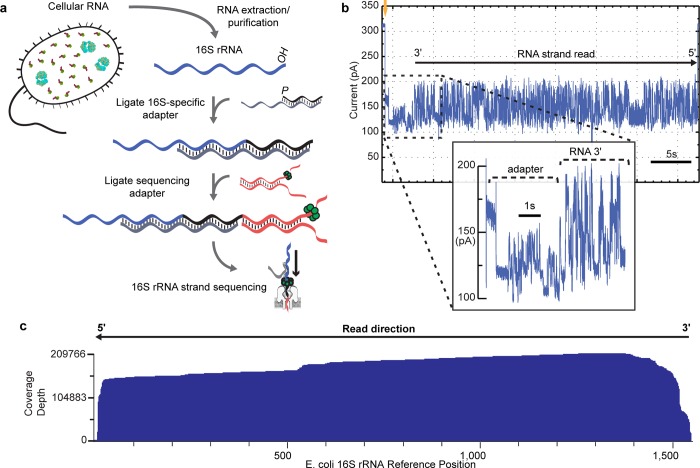
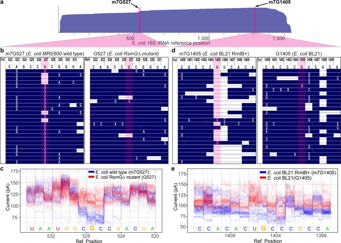
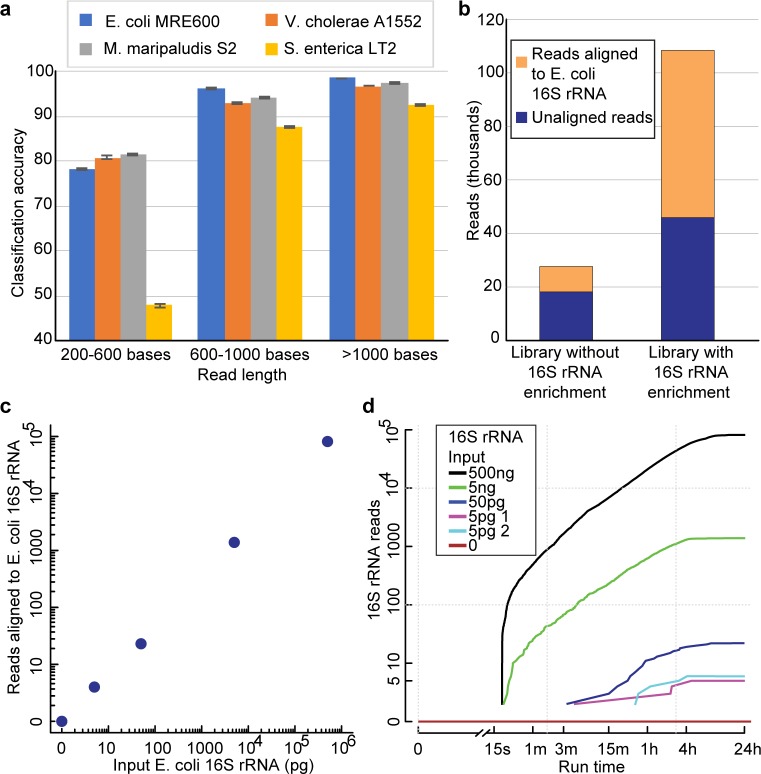
The ribosome small subunit is expressed in all living cells. It performs numerous essential functions during translation, including formation of the initiation complex and proofreading of base-pairs between mRNA codons and tRNA anticodons. The core constituent of the small ribosomal subunit is a ~1.5 kb RNA strand in prokaryotes (16S rRNA) and a homologous ~1.8 kb RNA strand in eukaryotes (18S rRNA). Traditional sequencing-by-synthesis (SBS) of rRNA genes or rRNA cDNA copies has achieved wide use as a 'molecular chronometer' for phylogenetic studies, and as a tool for identifying infectious organisms in the clinic. However, epigenetic modifications on rRNA are erased by SBS methods. Here we describe direct MinION nanopore sequencing of individual, full-length 16S rRNA absent reverse transcription or amplification. As little as 5 picograms (~10 attomole) of purified E. coli 16S rRNA was detected in 4.5 micrograms of total human RNA. Nanopore ionic current traces that deviated from canonical patterns revealed conserved E. coli 16S rRNA 7-methylguanosine and pseudouridine modifications, and a 7-methylguanosine modification that confers aminoglycoside resistance to some pathological E. coli strains.


Conflict of interest statement

MA holds options in Oxford Nanopore Technologies (ONT). MA is a paid consultant to ONT. MA is an inventor on 11 University of California patents licensed to ONT (6,267,872, 6,465,193, 6,746,594, 6,936,433, 7,060,50, 8,500,982, 8,679,747, 9,481,908, 9,797,013, 10,059,988, and 10,081,835). DRG, who contributed to each facet of the paper, is an employee of Oxford Nanopore Technologies. This does not alter our adherence to PLOS ONE policies on sharing data and materials.

Figures

Fig 1
Fig 1. Nanopore sequencing of individual E. coli 16S ribosomal RNA strands.
(a) Library preparation for MinION sequencing. Following RNA extraction, a 16S rRNA-specific adapter is hybridized and ligated to the 16S rRNA 3′ end. Next, a sequencing adapter bearing a RNA motor protein is hybridized and ligated to the 3′ overhang of the 16S rRNA adapter. The sample is then loaded into the MinION flowcell for sequencing. (b) Representative ionic current trace during translocation of a 16S rRNA strand from E. coli str. MRE600 through a nanopore. Upon capture of the 3′ end of an adapted 16S rRNA, the ionic current transitions from open channel (310 pA; gold arrow) to a series of discrete segments characteristic of the adapters (inset). This is followed by ionic current segments corresponding to base-by-base translocation of the 16S rRNA. The trace is representative of thousands of reads collected for individual 16S rRNA strands from E. coli. (c) Alignment of 200,000+ 16S rRNA reads to E. coli str MRE600 16S rRNA rrnD gene reference sequence. Reads are aligned in 5′ to 3′ orientation, after being reversed by the base-calling software. Numbering is according to canonical E. coli 16S sequence. Coverage across reference is plotted as a smoothed curve. In this experiment, 94.6% of reads that passed quality filters aligned to the reference sequence. Data presented here are from a single flow cell.
Fig 2
Fig 2. Detection of 7mG modifications in E. coli 16S rRNA.
(a) Diagram showing the positions along E. coli 16S rRNA that correspond to the expanded sequence alignments in panels b-e. Arrows indicate the positions of G527 and G1405 in the E. coli reference. (b) Alignment of nanopore RNA sequence reads proximal to position 527 of E. coli 16S rRNA. Numbered letters at the top represent DNA bases in the reference 16S rRNA gene. Blue regions in the body of the panel denote agreement between reference DNA bases and nanopore RNA strand base-calls. White letters denote base call differences between the reference and the nanopore reads, and horizontal white bars represent base deletions in the nanopore RNA reads. Columns highlighted in red correspond to position 527. The left inset is E. coli str. MRE600 (wild type) 16S rRNA (m7G527), and the right inset is RsmG mutant strain 16S rRNA (canonical G527). (c) Nanopore ionic current traces proximal to position 527 of the E. coli 16S rRNA reference. Blue traces are for wild type E. coli 16S rRNA translocation events bearing m7G at position 527. Red traces are for mutant strain 16S rRNA translocation events bearing a canonical G at position 527. (d) Alignment of nanopore RNA sequence reads proximal to position 1405 of E. coli 16S rRNA. Use of colors, shapes, and letters are as described for panel (b). The left inset is engineered mutant E. coli str. BL21 (RmtB+) 16S rRNA (m7G1405); the right inset is E. coli str. BL21 16S rRNA (G1405). (e) Nanopore ionic current traces proximal to position 1405 of the E. coli 16S rRNA reference. Blue traces are for mutant strain 16S rRNA translocation events bearing m7G at position 1405. Red traces are for wild type 16S rRNA translocation events bearing a canonical G at position 1405.
Fig 3
Fig 3. Nanopore 16S rRNA sequencing discriminates among microbes and detects E. coli 16S rRNA at low concentration.
(a) Classification accuracy from an in silico mixture of 16S rRNA reads from four microbes. Reads were binned based on length and 10 iterations of classification using 10,000 randomly sampled reads from at least 15,000 reads per microbe were performed. A read was called as correctly classified if it aligned to one of the 16S rRNA reference sequences for that microbe. The error bars indicate one standard deviation for the 10 iterations. (b) 16S rRNA sequencing yield for libraries prepared from E. coli str. K12 total RNA with and without enrichment. Sequencing libraries were prepared from 5 μg total RNA. The enrichment library used a desthiobiotinylated version of the 16S rRNA-specific adapter, which was hybridized and selected for using magnetic streptavidin beads (see Methods). The two 16S rRNA sequencing libraries were then prepared essentially the same way. (c) 16S rRNA reads from sequencing libraries prepared with E. coli str. MRE600 16S rRNA titered into 4.5 μg total RNA from HEK 293T cells. (d) 16S read accumulation over time in titration sequencing runs. The lines correspond to libraries shown in c.

References

    1. Garalde DR, Snell EA, Jachimowicz D, Sipos B, Lloyd JH, Bruce M, et al. Highly parallel direct RNA sequencing on an array of nanopores. Nat Methods. 2018;15: 201–206. 10.1038/nmeth.4577 - DOI - PubMed
    1. Kasianowicz JJ, Brandin E, Branton D, Deamer DW. Characterization of individual polynucleotide molecules using a membrane channel. Proc Natl Acad Sci U S A. 1996;93: 13770–13773. 10.1073/pnas.93.24.13770 - DOI - PMC - PubMed
    1. Lieberman KR, Cherf GM, Doody MJ, Olasagasti F, Kolodji Y, Akeson M. Processive replication of single DNA molecules in a nanopore catalyzed by phi29 DNA polymerase. J Am Chem Soc. 2010;132: 17961–17972. 10.1021/ja1087612 - DOI - PMC - PubMed
    1. Cherf GM, Lieberman KR, Rashid H, Lam CE, Karplus K, Akeson M. Automated forward and reverse ratcheting of DNA in a nanopore at 5-Å precision. Nat Biotechnol. 2012;30: 344–348. 10.1038/nbt.2147 - DOI - PMC - PubMed
    1. Manrao EA, Derrington IM, Laszlo AH, Langford KW, Hopper MK, Gillgren N, et al. Reading DNA at single-nucleotide resolution with a mutant MspA nanopore and phi29 DNA polymerase. Nat Biotechnol. 2012;30: 349–353. 10.1038/nbt.2171 - DOI - PMC - PubMed

Publication types

MeSH terms