. 2019 May 15;11(5):438.
doi: 10.3390/v11050438.

Virion-Associated Cholesterol Regulates the Infection of Human Parainfluenza Virus Type 3

Affiliations

Virion-Associated Cholesterol Regulates the Infection of Human Parainfluenza Virus Type 3

Qiaopeng Tang et al. Viruses. .

Abstract

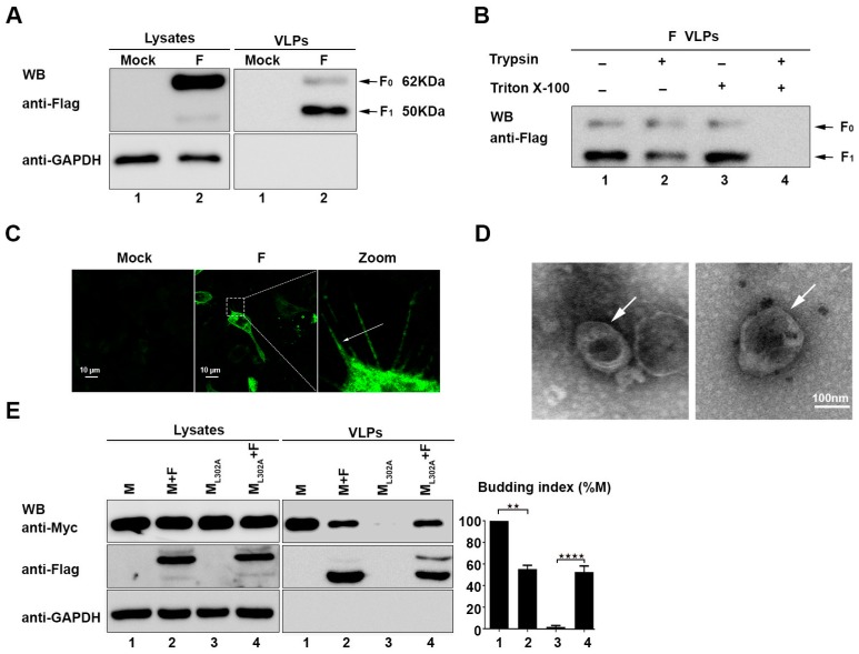
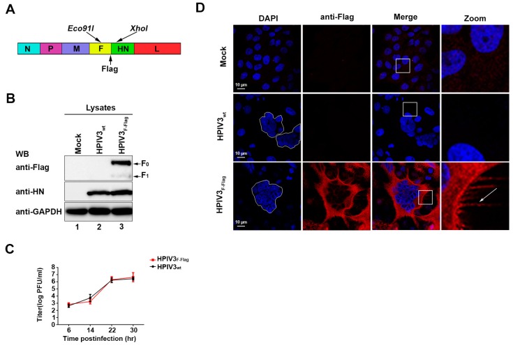
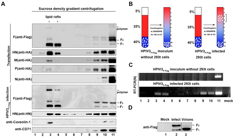
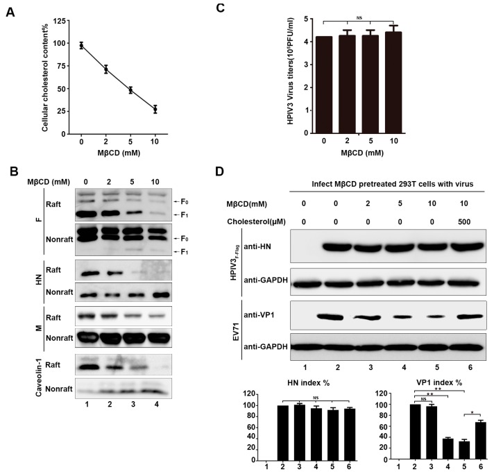
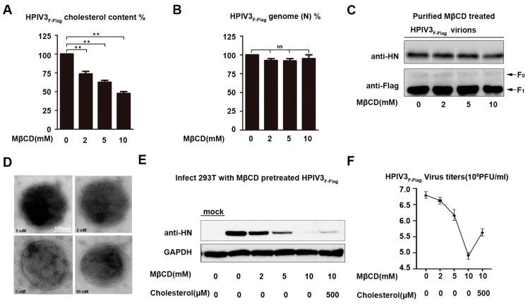
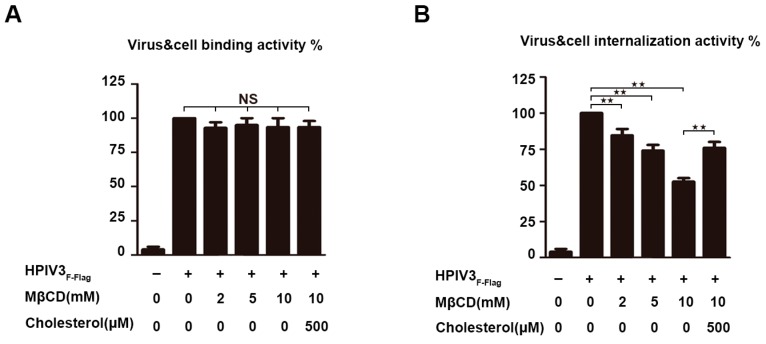
The matrix (M) proteins of paramyxoviruses bind to the nucleocapsids and cytoplasmic tails of glycoproteins, thus mediating the assembly and budding of virions. We first determined the budding characterization of the HPIV3 Fusion (F) protein to investigate the assembly mechanism of human parainfluenza virus type 3 (HPIV3). Our results show that expression of the HPIV3 F protein alone is sufficient to initiate the release of virus-like particles (VLPs), and the F protein can regulate the VLP-forming ability of the M protein. Furthermore, HPIV3F-Flag, which is a recombinant HPIV3 with a Flag tag at the C-terminus of the F protein, was constructed and recovered. We found that the M, F, and hemagglutinin-neuraminidase (HN) proteins and the viral genome can accumulate in lipid rafts in HPIV3F-Flag-infected cells, and the F protein mainly exists in the form of F1 in VLPs, lipid rafts, and purified virions. Furthermore, the function of cholesterol in the viral envelope and cell membrane was assessed via the elimination of cholesterol by methyl-β-cyclodextrin (MβCD). Our results suggest that the infectivity of HPIV3 was markedly reduced, due to defective internalization ability in the absence of cholesterol. These results reveal that HPIV3 might assemble in the lipid rafts to acquire cholesterol for the envelope of HPIV3, which suggests the that disruption of the cholesterol composition of HPIV3 virions might be a useful method for the design of anti-HPIV3 therapy.

Keywords: HPIV3; cholesterol; fusion protein; internalization; lipid rafts; viral assembly.


Conflict of interest statement

The authors declare no conflict of interest.

Figures

Figure 1
Figure 1
The human parainfluenza virus type 3 (HPIV3) Fusion (F) protein alone can secrete into the supernatant from transfected cells and form virus-like particles (VLPs). (A) 293T cells were transfected with the indicated plasmids for 36 h. The cell lysates and VLPs were prepared as described in Section 2, and then, the samples were subjected to western blot analysis (WB) by using anti-Flag and anti-GAPDH antibodies (Abs). Arrows indicate the bands of the detected proteins. (B) A protease protection assay of the HPIV3 F protein VLPs was performed, as described in Section 2 and then analyzed via WB with an anti-Flag antibody. (C) Cellular localization of the HPIV3 F protein. Hela cells were transfected with the plasmid encoding wild-type F, fixed 24 h after transfection, stained with an anti-Flag Ab, and visualized via confocal microscopy as describe in Section 2. Arrows indicate the filamentous structure formed by the F protein. (D) Representative TEM graphs of the F protein VLPs. VLP samples were prepared as previously described and then visualized by transmission electron microscopy. (E) The F protein was transfected with M/ML302A in 293T cells, and 36 h post-transfection, the cell lysates and VLPs were prepared as described in Section 2. Then, the samples were subjected to western blot analysis by using anti-Flag, anti-GAPDH, and anti-Myc Abs. Arrows indicate the bands of the detected proteins. Error bars, mean ± SD of three experiments. Student’s t test; * p < 0.05; ** p < 0.01; *** p < 0.001; **** p < 0.0001; NS, non-significant.
Figure 2
Figure 2
Recovery of recombinant HPIV3F-Flag. (A) Recombinant virus HPIV3F-Flag construction mode. (B) Hela cells were mock-infected or infected with wild-type HPIV3 or HPIV3F-Flag. At 24 h post-infection, cell lysates were subjected to WB as described in Section 2 with anti-Flag, anti- hemagglutinin-neuraminidase (HN), and anti-GAPDH Abs. (C) The growth properties of HPIV3 and HPIV3F-Flag. MK2 cells were infected with HPIV3 or HPIV3F-Flag at a multiplicity of infection (MOI) of 1; titers of supernatants that were harvested at 6, 14, 22, and 30 h after virus infection were examined. (D) Cellular localization of the HPIV3F-Flag F protein. Hela cells were mock-infected or infected with wild-type HPIV3 or HPIV3F-Flag. At 24 h post-infection, the cells were stained with anti-Flag and visualized via confocal microscopy, as described in Section 2. The arrows indicate the filamentous structure formed by HPIV3F-Flag.
Figure 3
Figure 3
The F, HN, and matrix (M) proteins and virus genome accumulate in lipid rafts. (A) 293T cells were transfected with F, HN, M, N, and P proteins or infected with HPIV3F-Flag. Afterwards, 36 h post-transfection or 24 h post-infection, the cells were extracted with 1% Triton X-100 at 4 °C. The lysates were loaded at the bottom of a flotation sucrose density gradient and subjected to equilibrium centrifugation. The gradient was fractionated from the top, and samples were analyzed by WB with anti-Flag, anti-HA, anti-Myc, anti-HN, anti-Caveolin-1, and anti-CD71 Abs. (B) The mode that purified HPIV3F-Flag virions or HPIV3F-Flag-infected cells were loaded at the bottom of a flotation sucrose density gradient. (C) Extracted RNA from each fraction was amplified targeting the HPIV3 N gene by RT-PCR and then examined. (D) The F protein in mock-infected, HPIV3F-Flag-infected cells, and purified virions. MK2 cells were mock-infected or HPIV3F-Flag-infected at a MOI of 0.1. At 36 h post-infection, cells supernatants were subjected to ultracentrifugation through a 20% sucrose cushion to pellet the virions, and cells were lysed, as described in Section 2.
Figure 4
Figure 4
Destruction of lipid rafts of cellular membranes did not prevent HPIV3 infection and budding. (A) 293T cells were incubated with various concentrations of methyl-β-cyclodextrin (MβCD) at 37 °C. Subsequently, 30 min. after treatment with MβCD, cellular cholesterol was measured by an AmplexTM Red Cholesterol Assay Kit according to the manufacturer’s protocol. (B) 293T cells were transfected with the indicated plasmids. Then, 36 h post-transfection, the cells were incubated with various concentrations of MβCD at 37 °C. Next, 30 min. after treatment with MβCD, the cells were extracted with 1% Triton X-100 at 4 °C. The lysate was loaded at the bottom of a flotation sucrose density gradient and subjected to equilibrium centrifugation. Fractions of lipid rafts and non-lipid rafts were analyzed by WB with anti-Flag, anti-HA, anti-Myc, and anti-Caveolin-1 Abs. (C) 293T cells were infected with HPIV3 at a MOI of 0.1. Afterwards, 12 h post-infection, the cells were treated with the indicated MβCD for 30 min, and 12 h after treatment with MβCD, the supernatant of the cells was collected, and the virus titers were measured. (D) 293T cells were incubated with MβCD and cholesterol as indicated. Next, the cells were infected with HPIV3F-Flag or EV71. Then, 12 h post-infection, cell lysates were subjected to WB as described in Section 2 with anti-HN, anti-VP1, and anti-GAPDH Abs. Error bars, mean ± SD of three experiments. Student’s t test; * p < 0.05; ** p < 0.01; *** p < 0.001; **** p < 0.0001; NS, non-significant.
Figure 5
Figure 5
Infectivity of cholesterol-depleted HPIV3 was markedly reduced. (A) HPIV3F-Flag virus was pretreated with various concentrations of MβCD or left untreated, and then, the virions were repurified using a sucrose cushion in order to analyze the cholesterol content using an AmplexTM Red Cholesterol Assay Kit according to the manufacturer’s protocol. (B) The relative number of the genomes of the sucrose-cushion-repurified virus that was treated with MβCD and cholesterol were detected by Q-PCR. (C) The number of the HN and F proteins on the repurified MβCD-treated HPIV3 virions were detected by WB. (D) Electron microscope image of MβCD-treated virus. The sucrose-cushion-repurified HPIV3 virus that was treated with MβCD was negative stained and observed by electron microscope. (E) 293T cells were infected with sucrose-cushion-repurified virus that was treated with various concentrations of MβCD and cholesterol, and at 24 h post-infection, the cell lysate was analyzed by WB with anti-HN and anti-GAPDH Abs. (F) Viral titers of MβCD-treated or untreated HPIV3. Error bars, mean ± SD of three experiments. Student’s t test; * p < 0.05; ** p < 0.01; *** p < 0.001; **** p < 0.0001; NS, non-significant.
Figure 6
Figure 6
Cholesterol-depleted HPIV3 particles are defective for internalization. (A) Virus-cell binding assay. HPIV3F-Flag virus was treated with MβCD and cholesterol as indicated and was bound to the surfaces of 293T cells on ice at a MOI of 1. Then, the cells were washed with cold PBS to remove un-adsorbed virus, and total RNA was extracted from cells by using TRIzol according to the manufacturer’s instructions. Then, RT-PCR assay was conducted to quantify the amount of cell-bound viral RNA. (B) Internalization assay. Cells were warmed to 37 °C and maintained for 2 h, after which they were treated with 0.25% trypsin for 10 min. at 37 °C, total RNA were extracted by TRIzol, and then RT-PCR assay was conducted to quantify the amount of internalized viral RNA. Error bars, mean ± SD of three experiments. Student’s t test; * p < 0.05; ** p < 0.01; *** p < 0.001; **** p < 0.0001; NS, non-significant.

Similar articles

Cited by

References

    1. Porotto M., Murrell M., Greengard O., Doctor L., Moscona A. Influence of the human parainfluenza virus 3 attachment protein’s neuraminidase activity on its capacity to activate the fusion protein. J. Virol. 2005;79:2383–2392. doi: 10.1128/JVI.79.4.2383-2392.2005. - DOI - PMC - PubMed
    1. Simons K., Toomre D. Lipid rafts and signal transduction. Nat. Rev. Mol. Cell Biol. 2000;1:31–39. doi: 10.1038/35036052. - DOI - PubMed
    1. Rajendran L., Simons K. Lipid rafts and membrane dynamics. J. Cell Sci. 2005;118:1099–1102. doi: 10.1242/jcs.01681. - DOI - PubMed
    1. Viard M., Parolini I., Sargiacomo M., Fecchi K., Ramoni C., Ablan S., Ruscetti F.W., Wang J.M., Blumenthal R. Role of cholesterol in human immunodeficiency virus type 1 envelope protein-mediated fusion with host cells. J. Virol. 2002;76:11584–11595. doi: 10.1128/JVI.76.22.11584-11595.2002. - DOI - PMC - PubMed
    1. Norkin L.C., Anderson H.A., Wolfrom S.A., Oppenheim A. Caveolar endocytosis of simian virus 40 is followed by brefeldin A-sensitive transport to the endoplasmic reticulum, where the virus disassembles. J. Virol. 2002;76:5156–5166. doi: 10.1128/JVI.76.10.5156-5166.2002. - DOI - PMC - PubMed

Publication types