. 2019 May 15;11(5):1077.
doi: 10.3390/nu11051077.

Polyphenol-Enriched Plum Extract Enhances Myotubule Formation and Anabolism while Attenuating Colon Cancer-induced Cellular Damage in C2C12 Cells

Affiliations

Polyphenol-Enriched Plum Extract Enhances Myotubule Formation and Anabolism while Attenuating Colon Cancer-induced Cellular Damage in C2C12 Cells

Faten A Alsolmei et al. Nutrients. .

Abstract

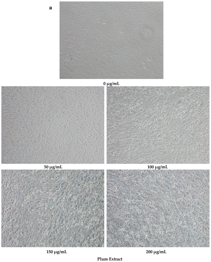
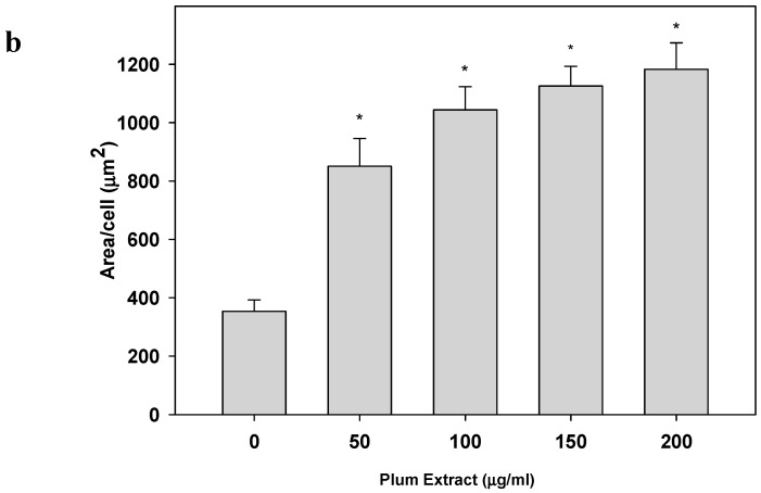
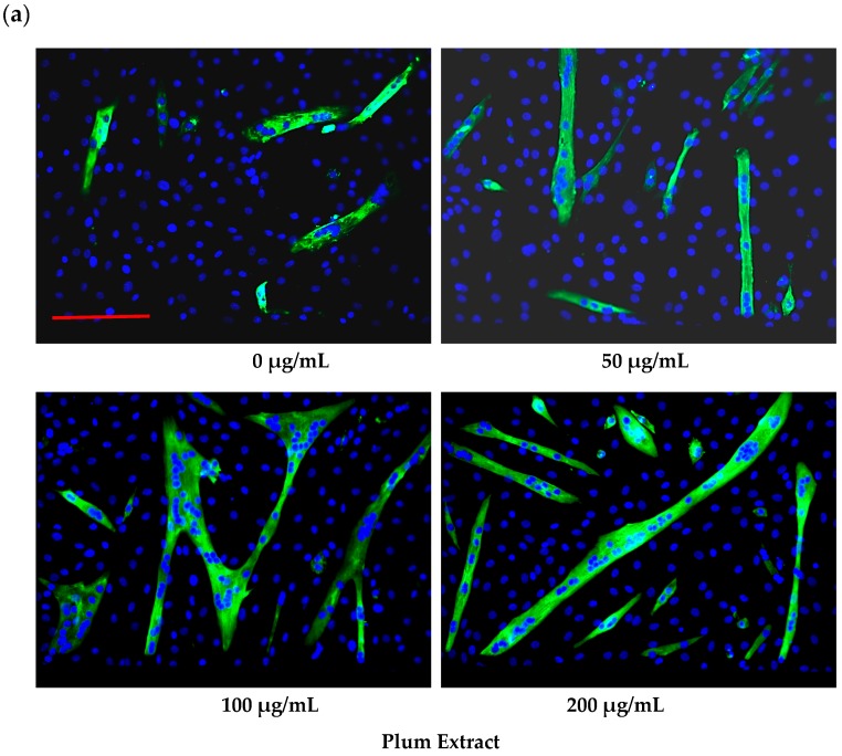
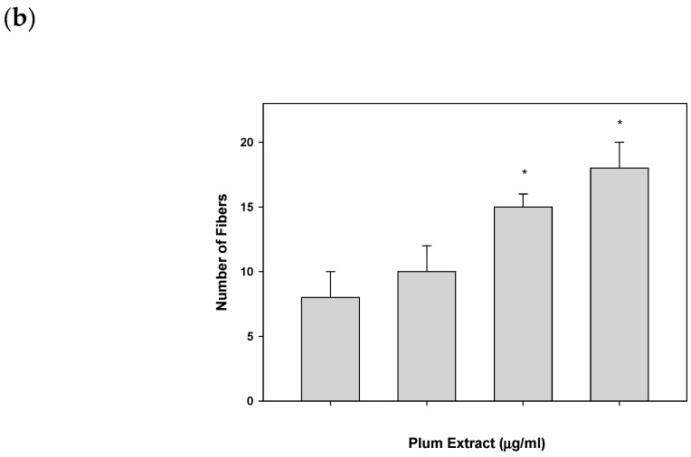
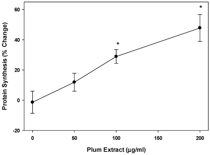
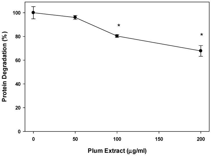
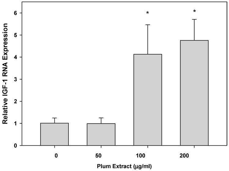
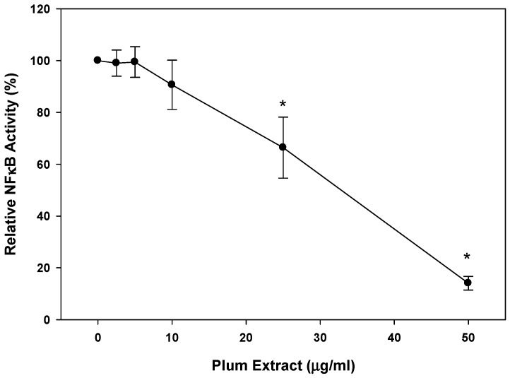
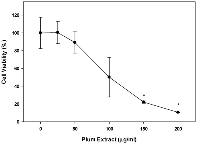
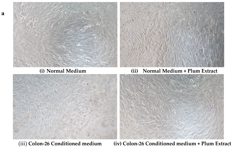
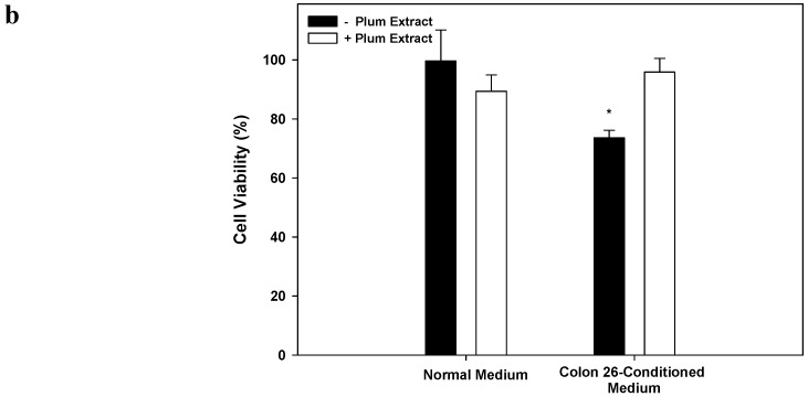
Preventing muscle wasting in certain chronic diseases including cancer is an ongoing challenge. Studies have shown that polyphenols derived from fruits and vegetables shows promise in reducing muscle loss in cellular and animal models of muscle wasting. We hypothesized that polyphenols derived from plums (Prunus domestica) could have anabolic and anti-catabolic benefits on skeletal muscle. The effects of a polyphenol-enriched plum extract (PE60) were evaluated in vitro on C2C12 and Colon-26 cancer cells. Data were analyzed using a one-way ANOVA and we found that treatment of myocytes with plum extract increased the cell size by ~3-fold (p < 0.05) and stimulated myoblast differentiation by ~2-fold (p < 0.05). Plum extract induced total protein synthesis by ~50% (p < 0.05), reduced serum deprivation-induced total protein degradation by ~30% (p < 0.05), and increased expression of Insulin-Like Growth Factor-1 (IGF-1) by ~2-fold (p < 0.05). Plum extract also reduced tumor necrosis factor α (TNFα)-induced nuclear factor κB (NFκB) activation by 80% (p < 0.05) in A549/NF-κB-luc cells. In addition, plum extract inhibited the growth of Colon-26 cancer cells, and attenuated cytotoxicity in C2C12 myoblasts induced by soluble factors released from Colon-26 cells. In conclusion, our data suggests that plum extract may have pluripotent health benefits on muscle, due to its demonstrated ability to promote myogenesis, stimulate muscle protein synthesis, and inhibit protein degradation. It also appears to protect muscle cell from tumor-induced cytotoxicity.

Keywords: cachexia; cancer; muscle wasting; myoblasts; plum; protein synthesis.


Conflict of interest statement

Suzette Pereira and Paul Johns are currently employed by Abbott. Padmavathy Krishnan is an ex-Abbott employee. Rafat Siddiqui, Haiwen Li, and Faten Alsolmei have no conflict of interest.

Figures

Figure 1
Figure 1
The effect of plum extract on C2C12 myoblast cell size. (a) The representative pictures of myoblast after treatment with varying concentration of plum extract (100 × magnified images) taken by a Nikon Microscope. The bar represents a length of 500 μm. (b) The size of myoblast was determined using Element-BR software as described in “Materials and Methods”. The data are expressed as mean ± SD for at least three experiments. All comparisons were made to control (untreated cells) using one-way ANOVA; significant differences are reported at * p < 0.05.
Figure 1
Figure 1
The effect of plum extract on C2C12 myoblast cell size. (a) The representative pictures of myoblast after treatment with varying concentration of plum extract (100 × magnified images) taken by a Nikon Microscope. The bar represents a length of 500 μm. (b) The size of myoblast was determined using Element-BR software as described in “Materials and Methods”. The data are expressed as mean ± SD for at least three experiments. All comparisons were made to control (untreated cells) using one-way ANOVA; significant differences are reported at * p < 0.05.
Figure 2
Figure 2
The effect of plum extract on C2C12 myoblast differentiation. (a) Images of differentiated cells after treatment with varying concentration of plum extract showing nuclei stained in blue (Hoechst 33342) and myofibers stained in green (Alexa 488). Pictures were taken at 200× magnification using a Nikon Fluorescent Microscope. The bar represents a length of 300 μm. (b) Fused cells from five random fields were counted manually under 200× as described in “Materials and Methods”. The data are expressed as mean ± SD for at least three experiments. All comparisons were made to control (untreated cells) using one-way ANOVA; significant differences are reported at * p < 0.05.
Figure 2
Figure 2
The effect of plum extract on C2C12 myoblast differentiation. (a) Images of differentiated cells after treatment with varying concentration of plum extract showing nuclei stained in blue (Hoechst 33342) and myofibers stained in green (Alexa 488). Pictures were taken at 200× magnification using a Nikon Fluorescent Microscope. The bar represents a length of 300 μm. (b) Fused cells from five random fields were counted manually under 200× as described in “Materials and Methods”. The data are expressed as mean ± SD for at least three experiments. All comparisons were made to control (untreated cells) using one-way ANOVA; significant differences are reported at * p < 0.05.
Figure 3
Figure 3
The effect of plum extract on myotubule protein synthesis. Protein synthesis was measure by the incorporation of labeled phenylalanine into total myotubule proteins in response to various levels of plum extract. Data were computed as cpm/mg of proteins followed by calculation of % change over control. The data were expressed as mean ± SD for at least three experiments. All comparisons were made to control (untreated cells) using one-way ANOVA; significant differences are reported at * p < 0.05.
Figure 4
Figure 4
The effect of plum extract on myotubule protein degradation. Proteolysis was induced by 48 h-serum starvation in the presence or absence of plum extract, and monitored by release of radioactive tyrosine from pre-labelled cells. Data were computed as cpm/mg of proteins and then % change over control was calculated. The data were expressed as mean ± SD for at least three experiments. All comparisons were made to control (untreated cells) using one-way ANOVA; significant differences are reported at * p < 0.05.
Figure 5
Figure 5
The effect of plum extract of IGF-1 gene expression. Total RNA was extracted from C2C12 myotubules treated with various concentrations of plum extract and compared to untreated control. All results were obtained from at least three independent biological repeats. Data were analyzed using the ΔΔCT method. Glyceraldehyde-3-phosphate dehydrogenase (GAPDH) genes were used as house-keeping genes for expression calculation. All comparisons were made to control (untreated cells) using one-way ANOVA; significant differences are reported at * p < 0.05.
Figure 6
Figure 6
Effects of plum extract on NFkB activation. The effect of plum extract on TNFα-mediated NFkB activation was measured in the A549/NFκB-luc reporter stable cell line. Activity was measured in terms of luciferase activity. The data are reported as the relative percent inhibition of TNFα-mediated NFkB activation. The data are expressed as mean ± SD for at least three experiments. All comparisons were made to control (untreated cells) using one-way ANOVA; significant differences are reported at * p < 0.05.
Figure 7
Figure 7
Effect of plum extract on Colon-26 adenocarcinoma cells. Data were calculated as % inhibition of cell growth in response to various concentrations of plum extract. The data are expressed as mean ± SD for at least three replicates. All comparisons were made to control (untreated cells) using one-way ANOVA; the significant differences are reported at * p < 0.05.
Figure 8
Figure 8
The effect of plum extract on C2C12 viability in response to Colon-26-induced cytotoxicity. (a) Differentiated C2C12 myotubes were treated with normal medium (i & ii) or Colon-26-conditioned medium (iii & iv) in the absence (i & iii) or presence (ii & iv) of plum extract (50 μg/mL). (b) The viability of C2C12 myotubules were determined using WST-1 assay. The data is expressed as mean ± SD for at least three experiments. All comparisons were made to control (untreated cells) using one-way ANOVA; significant differences are reported at * p < 0.05.
Figure 8
Figure 8
The effect of plum extract on C2C12 viability in response to Colon-26-induced cytotoxicity. (a) Differentiated C2C12 myotubes were treated with normal medium (i & ii) or Colon-26-conditioned medium (iii & iv) in the absence (i & iii) or presence (ii & iv) of plum extract (50 μg/mL). (b) The viability of C2C12 myotubules were determined using WST-1 assay. The data is expressed as mean ± SD for at least three experiments. All comparisons were made to control (untreated cells) using one-way ANOVA; significant differences are reported at * p < 0.05.

References

    1. Nixon D.W., Heymsfield S.B., Cohen A.E., Lutne M.H., Ansley J., Lawson D.H., Rudman D. Protein calorie under-nutrition in hospitalized cancer patients. Am. J. Med. 1980;68:683–690. doi: 10.1016/0002-9343(80)90254-5. - DOI - PubMed
    1. Warren S. The immediate causes of death in cancer. Am. J. Med. Sci. 1932;184:610–616. doi: 10.1097/00000441-193211000-00002. - DOI
    1. Evans W.J., Morley J.E., Argiles J., Bales C., Baracos V., Guttridge D., Jatoi A., Kalantar-Zadeh K., Lochs H., Mantovani G., et al. Cachexia: A new definition. Clin. Nutr. 2008;27:793–799. doi: 10.1016/j.clnu.2008.06.013. - DOI - PubMed
    1. Fearon K.C., Glass D.J., Guttridge D.C. Cancer cachexia: Mediators, signaling, and metabolic pathways. Cell Metab. 2012;16:153–166. doi: 10.1016/j.cmet.2012.06.011. - DOI - PubMed
    1. Fearon K., Strasser F., Anker S.D., Bosaeus I., Bruera E., Fainsinger R.L., Jatoi A., Loprinzi C., MacDonald N., Mantovani G., et al. Definition and classification of cancer cachexia: An international consensus. Lancet Oncol. 2011;12:489–495. doi: 10.1016/S1470-2045(10)70218-7. - DOI - PubMed

Grants and funding

LinkOut - more resources