. 2019 Sep;78(9):1205-1214.
doi: 10.1136/annrheumdis-2018-214783. Epub 2019 May 16.

TNF-induced inflammatory genes escape repression in fibroblast-like synoviocytes: transcriptomic and epigenomic analysis

Affiliations

TNF-induced inflammatory genes escape repression in fibroblast-like synoviocytes: transcriptomic and epigenomic analysis

Christopher Loh et al. Ann Rheum Dis. 2019 Sep.

Abstract

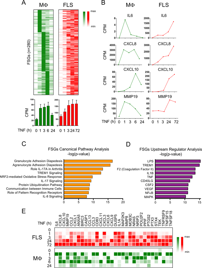
Objective: We investigated genome-wide changes in gene expression and chromatin remodelling induced by tumour necrosis factor (TNF) in fibroblast-like synoviocytes (FLS) and macrophages to better understand the contribution of FLS to the pathogenesis of rheumatoid arthritis (RA).

Methods: FLS were purified from patients with RA and CD14+ human monocyte-derived macrophages were obtained from healthy donors. RNA-sequencing, histone 3 lysine 27 acetylation (H3K27ac), chromatin immunoprecipitation-sequencing (ChIP-seq) and assay for transposable accessible chromatin by high throughput sequencing (ATAC-seq) were performed in control and TNF-stimulated cells.

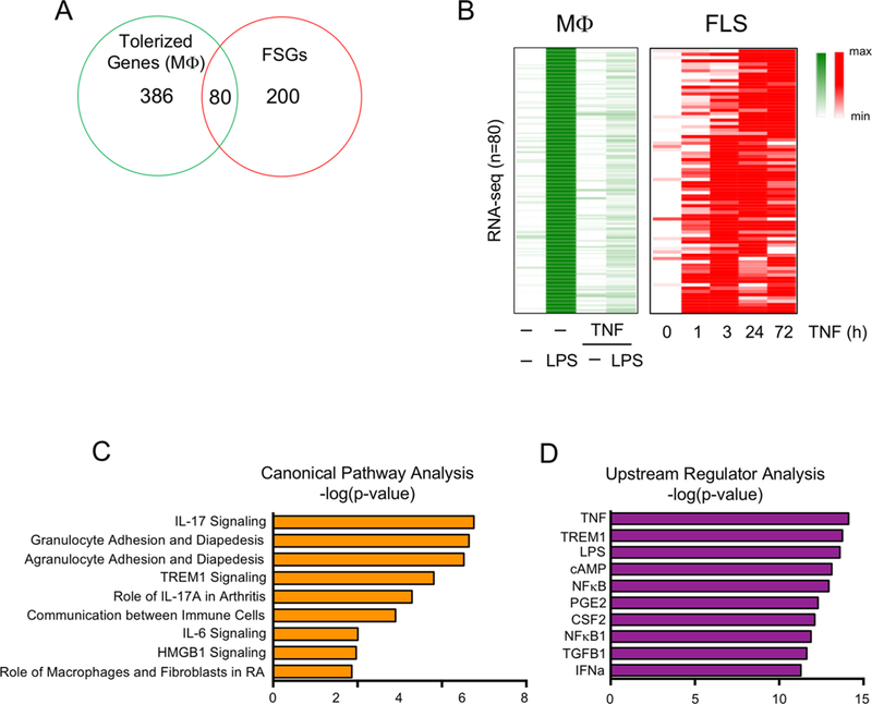
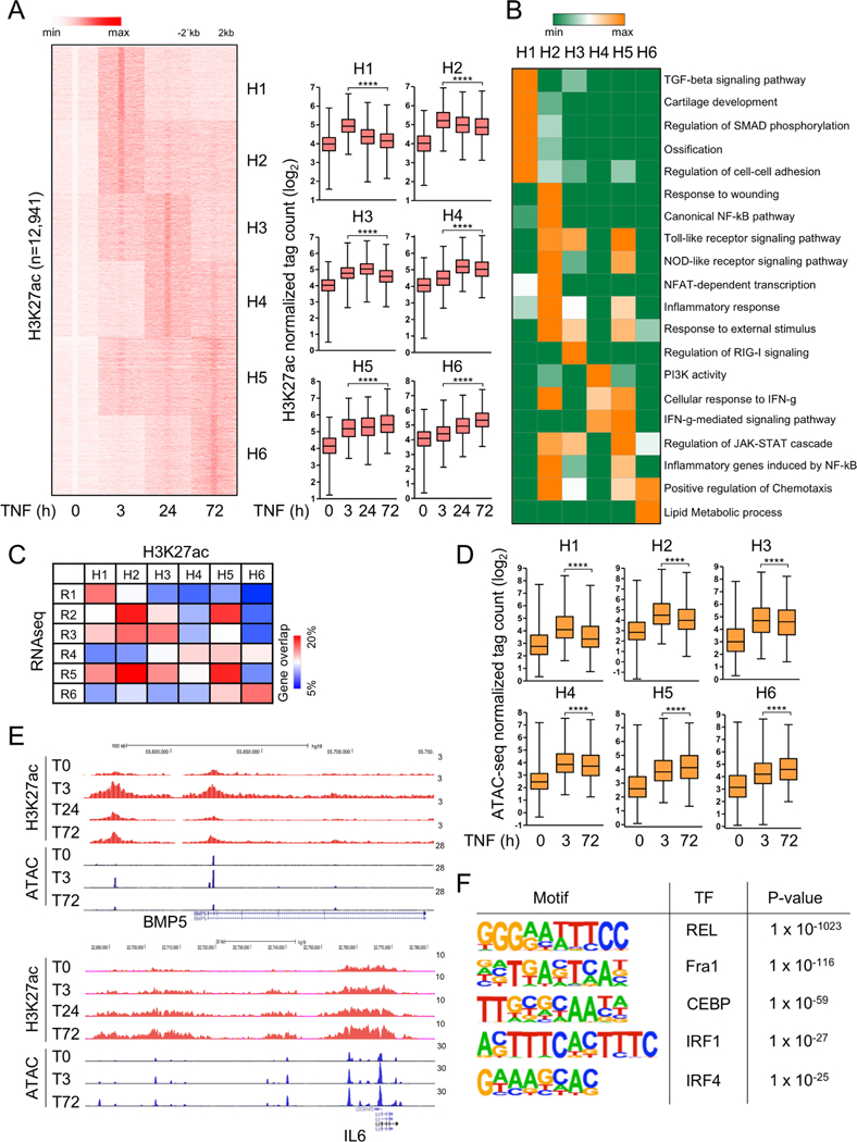
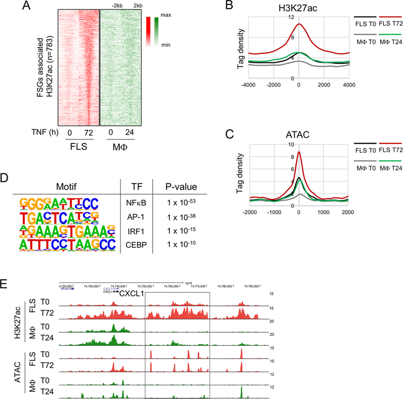
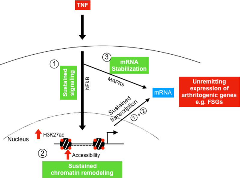
Results: We discovered 280 TNF-inducible arthritogenic genes which are transiently expressed and subsequently repressed in macrophages, but in RA, FLS are expressed with prolonged kinetics that parallel the unremitting kinetics of RA synovitis. 80 out of these 280 fibroblast-sustained genes (FSGs) that escape repression in FLS relative to macrophages were desensitised (tolerised) in macrophages. Epigenomic analysis revealed persistent H3K27 acetylation and increased chromatin accessibility in regulatory elements associated with FSGs in TNF-stimulated FLS. The accessible regulatory elements of FSGs were enriched in binding motifs for nuclear factor kappa-light-chain-enhancer of activated B cells (NF-κB), interferon-regulatory factors (IRFs) and activating protein-1 (AP-1). Inhibition of bromodomain and extra-terminal motif (BET) proteins, which interact with histone acetylation, suppressed sustained induction of FSGs by TNF.

Conclusion: Our genome-wide analysis has identified the escape of genes from transcriptional repression in FLS as a novel mechanism potentially contributing to the chronic unremitting synovitis observed in RA. Our finding that TNF induces sustained chromatin activation in regulatory elements of the genes that escape repression in RA FLS suggests that altering or targeting chromatin states in FLS (eg, with inhibitors of BET proteins) is an attractive therapeutic strategy.

Keywords: fibroblasts; rheumatoid arthritis; synovitis; tnf-alpha.


Conflict of interest statement

Competing interests: None declared. GDK is at the time of publication of this study an employee at Regeneron Pharmaceuticals Inc. and declares no conflict of interest related to the content of this manuscript.

Figures

Figure 1
Figure 1
Genes that are transiently induced by TNF in macrophages exhibit sustained expression in FLS. (A) Kinetic analysis of TNF-induced mRNA transcripts whose expression is transient in macrophages but sustained in FLS. RNA-seq analysis was performed in a time course of TNF stimulation (10 ng/mL) using macrophages (three replicates from independent blood donors) and FLS (two replicates from independent patients with RA). Two hundred and eighty genes (FSGs) transiently expressed in macrophages but sustained in FLS are displayed on a heatmap (upper panels). Bar graphs (lower panels) represent CPM values for the FSGs. Error bars indicate SEM. (B) Representative genes from the gene set of the FSGs are presented in CPM values. (C and D) Ingenuity pathway analysis of the FSGs defined in (A). (E) Expression of a subset of gene transcripts from the FSGs defined in (A) is presented relative to the maximum expression. CPM, counts per million; FLS, fibroblast-like synoviocytes; FSG, fibroblast-sustained genes; IL, interleukin; LPS, lipopolysaccharide; Mφ, macrophages; mRNA, messenger RNA; MAPK, mitogen-activated protein kinases; NF-κB, nuclear factor kappa-light-chain-enhancer of activated B cells; RA, rheumatoid arthritis; RNA-seq, RNA-sequencing; TNF, tumour necrosis factor.
Figure 2
Figure 2
Tolerised genes in macrophage exhibit sustained expression in FLS after TNF exposure. (A) Venn diagram showing the overlap between 466 macrophage tolerised genes (robustly LPS-inducible genes that are minimally induced by secondary LPS in macrophages pretreated with TNF) and the FSGs identified in figure 1A. (B) Heatmap depicting expression of the 80 genes in the overlap region in (A) presented relative to the maximum expression. (C and D) Ingenuity pathway analysis of the 80 genes that were tolerised in macrophages but whose expression was sustained in RA FLS. FLS, fibroblast-like synoviocytes; FSG, fibroblast-sustained genes; IFN, interferron; IL, interleukin; IFN, interferron; LPS, lipopolysaccharide; Mφ, macrophages; NF-κB, nuclear factor kappa-light-chain-enhancer of activated B cells; RA, rheumatoid arthritis; TGFB1, transforming growth factor beta 1; TNF, tumour necrosis factor.
Figure 3
Figure 3
Epigenomic landscape changes in FLS after TNF exposure. (A) Genome-wide H3K27ac ChIP-seq analysis (two biological replicates from independent patients with RA) of differentially induced peaks at each TNF-stimulated time points (3, 24 and 72 hours). The left panel depicts K-means clustering of 12 941 peaks (one per row) that were induced more than fourfold by TNF (p<0.0001) into six clusters (H1–H6); expression is presented relative to the maximum (set as 1). Box graphs represent H3K27ac ChIP-seq normalised tag densities (log2) at enhancers of a given cluster under the indicated conditions (right). ****P<0.0001, Wilcoxon matched-pairs signed rank test. (B) GREAT gene ontology analysis using the peaks in each cluster defined in (A). (C) Heatmap presentation of the percentage of genes (percentage of gene overlap) in each gene expression clusters R1–R6 (online supplementary figure S1A) that overlap with genes associated with H3K27ac enhancer clusters H1–H6 identified in (A). (D) Box graphs represent ATAC-seq normalised tag densities (log2) at H3K27ac peaks defined in (A). This represents quantitation of the data shown as a heatmap in online supplementary figure S3D. ****P<0.0001, Wilcoxon matched-pairs signed rank test. (E) Representative University of California Santa Cruz Genome Browser tracks displaying normalised profiles for H3K27ac ChIP-seq and ATAC-seq signals at BMP5 (cluster 1) and IL6 (cluster 6) locus. (F) Motifs enriched under inducible ATAC-seq peaks at 72 hours of TNF stimulation. ChIP-seq and ATAC-seq analyses were performed with two FLS replicates from independent patients with RA that yielded similar results (online supplementary figure S3A). ATAC-seq, assay for transposable accessible chromatin by high throughput sequencing; ChIP-seq, chromatin immunoprecipitation-sequencing; FLS, fibroblast-like synoviocytes; FSG, fibroblast-sustained genes; H3K27ac, histone 3 lysine 27 acetylation; IFN, interferron; IL, interleukin; IRF, interferon regulatory factors; NF-κB, nuclear factor kappa-light-chain-enhancer of activated B cells; C/EBP, CCAAT/enhancer-binding protein; RA, rheumatoid arthritis; TGF, transforming growth factor; TNF, tumour necrosis factor.
Figure 4
Figure 4
Persistent H3K27 acetylation is associated with sustained gene expression in FLS. (A) Heatmaps of normalised tag densities for H3K27ac ChIP-seq peaks associated with the FSGs defined in figure 1A in FLS and macrophages. (B) Histogram of H3K27ac-seq normalized tag densities for peaks defined in (A). (C) Histogram of ATAC-seq normalised tag densities at H3K27ac peaks defined in (A). (D) Motifs enriched under inducible ATAC-seq peaks associated with the FSGs at 72 hours of TNF stimulation. (E) Representative University of California Santa Cruz Genome Browser tracks displaying normalised profiles for H3K27ac ChIP-seq and ATAC-seq signals at the CXCL1 locus. Box enclose genomic regions that are differentially regulated across conditions. ATAC-seq, assay for transposable accessible chromatin by high throughput sequencing; ChIP-seq, chromatin immunoprecipitation-sequencing; FLS, fibroblast-like synoviocytes; FSG, fibroblast-sustained genes; H3K27ac, histone 3 lysine 27 acetylation; IRF, interferon regulatory factors; Mφ, macrophages; NF-κB, nuclear factor kappa-light-chain-enhancer of activated B cells; RA, rheumatoid arthritis; TNF, tumour necrosis factor.
Figure 5
Figure 5
Transcriptomic analysis of I-BET effects on TNF-induced inflammatory gene expression in FLS. (A) Volcano plot of transcriptomic changes in RA FLS stimulated with TNF (10 ng/mL) in the presence or absence of I-BET (10 M) for 24 hours; red dots correspond to genes with significant (p<0.05) and greater than twofold expression changes. RNA sequencing was performed in three independent biological replicates (FLS derived from three different patients with RA). (B) REACTOME pathway analysis of 1697 genes significantly suppressed by I-BET (greater than twofold and p<0.05) in TNF-stimulated FLS. (C) Heatmap depicting expression of genes that are TNF upregulated (greater than twofold and p<0.05) and I-BET suppressed (greater than twofold and p<0.05). Out of 911 TNF upregulated genes, 617 genes are suppressed by I-BET. (D) One hundred and ten genes out 280 FSGs were significantly suppressed by I-BET. (E) Representative TNF-inducible genes which were suppressed by I-BET (gene expression presented in CPM values). CPM, counts per million; FLS, fibroblast-like synoviocytes; FSG, fibroblast-sustained genes; I-BET, bromodomain and extra-terminal protein inhibitor; IL, interleukin; NF-κB, nuclear factor kappa-light-chain-enhancer of activated B cells; RA, rheumatoid arthritis; TNF, tumour necrosis factor.
Figure 6
Figure 6
Three-component model that explains the sustained expression of arthritogenic genes (FSGs) in RA FLS. TNF triggers in RA FLS: (1) prolonged activation of NF-κB, (2) chromatin remodelling (increased H3K27ac and chromatin accessibility) and (3) MAPK-dependent mRNA stabilisation. Prolonged activation of NF-κB together with chromatin remodelling maintain continuous transcription of arthritogenic genes in RA FLS. Continuous transcription and mRNA stabilisation contribute to the sustained expression of these genes (FSGs) in RA FLS, ultimately perpetuating synovitis. FLS, fibroblast-like synoviocytes; FSG, fibroblast-sustained genes; H3K27ac, histone 3 lysine 27 acetylation; mRNA, messenger RNA; MAPK, mitogen-activated protein kinases; NF-κB, nuclear factor kappa-light-chain-enhancer of activated B cells; RA, rheumatoid arthritis; TNF, tumour necrosis factor.

References

    1. Burmester GR, Pope JE. Novel treatment strategies in rheumatoid arthritis. The Lancet 2017;389:2338–48. - PubMed
    1. Prince FHM, Bykerk VP, Shadick NA, et al. Sustained rheumatoid arthritis remission is uncommon in clinical practice. Arthritis Res Ther 2012;14. - PMC - PubMed
    1. Mclnnes IB, Schett G. Pathogenetic insights from the treatment of rheumatoid arthritis. The Lancet 2017;389:2328–37. - PubMed
    1. Bottini N, Firestein GS. Duality of fibroblast-like synoviocytes in RA: passive responders and imprinted aggressors. Nat Rev Rheumatol 2013;9:24–33. - PMC - PubMed
    1. Donlin LT, Jayatilleke A, Giannopoulou EG, et al. Modulation of TNF-induced macrophage polarization by synovial fibroblasts. J Immunol 2014;193:2373–83. - PMC - PubMed

Publication types

MeSH terms

Substances