. 2019 May 28;116(22):10943-10951.
doi: 10.1073/pnas.1901777116. Epub 2019 May 16.

Long-term neurocognitive benefits of FLASH radiotherapy driven by reduced reactive oxygen species

Affiliations

Long-term neurocognitive benefits of FLASH radiotherapy driven by reduced reactive oxygen species

Pierre Montay-Gruel et al. Proc Natl Acad Sci U S A. .

Erratum in

Abstract

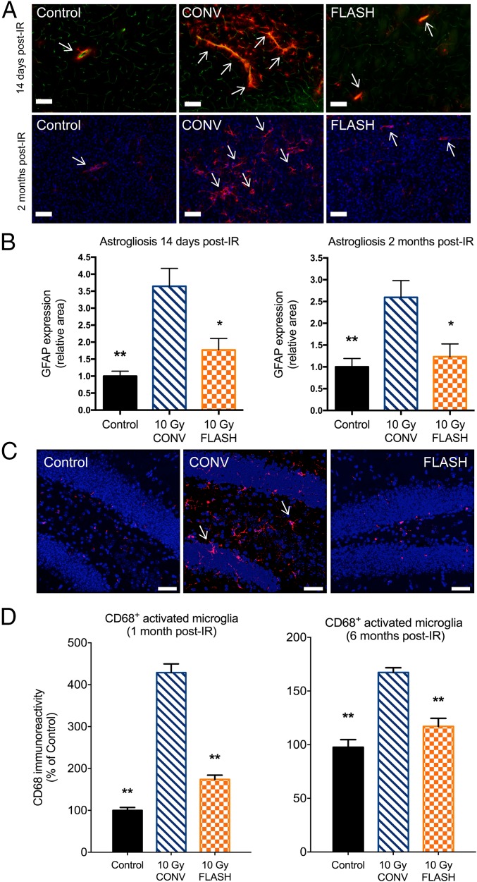
Here, we highlight the potential translational benefits of delivering FLASH radiotherapy using ultra-high dose rates (>100 Gy⋅s-1). Compared with conventional dose-rate (CONV; 0.07-0.1 Gy⋅s-1) modalities, we showed that FLASH did not cause radiation-induced deficits in learning and memory in mice. Moreover, 6 months after exposure, CONV caused permanent alterations in neurocognitive end points, whereas FLASH did not induce behaviors characteristic of anxiety and depression and did not impair extinction memory. Mechanistic investigations showed that increasing the oxygen tension in the brain through carbogen breathing reversed the neuroprotective effects of FLASH, while radiochemical studies confirmed that FLASH produced lower levels of the toxic reactive oxygen species hydrogen peroxide. In addition, FLASH did not induce neuroinflammation, a process described as oxidative stress-dependent, and was also associated with a marked preservation of neuronal morphology and dendritic spine density. The remarkable normal tissue sparing afforded by FLASH may someday provide heretofore unrealized opportunities for dose escalation to the tumor bed, capabilities that promise to hasten the translation of this groundbreaking irradiation modality into clinical practice.

Keywords: cognitive dysfunction; neuroinflammation; neuronal morphology; reactive oxygen species; ultra-high dose-rate irradiation.


Conflict of interest statement

The authors declare no conflict of interest.

Figures

Fig. 1.
Fig. 1.
FLASH-RT minimizes radiation-induced neurocognitive complications. WT mice were tested for cognitive function using the NOR (A), OIP (B), and TO (C) tasks. CONV irradiation (10 Gy) caused significant reductions in the DI on the NOR and TO tasks and similar trends on the OIP task compared with controls. In each instance, FLASH-RT prevented radiation-induced cognitive deficits. Mean ± SEM (n = 8–12 mice per group). P values were derived from ANOVA and the Bonferroni test. *P < 0.05, **P < 0.01 compared with the 10-Gy CONV group. At 6 mo post-RT, mice were subjected to the EPM test (D), LDB test (E), and FST (F). Mice subjected to CONV irradiation spent significantly less time in the open arms of the EPM and exhibited significantly fewer transitions between the light and dark regions of the LDB compared with controls. In contrast, FLASH cohorts showed a significant increase in the number of transitions between the light and dark compartments compared with CONV cohorts. Mice exposed to CONV irradiation spent significantly more time floating compared with either controls or the FLASH cohort. Mean ± SEM (n = 8–10 mice per group). P values were derived from ANOVA and the Bonferroni test. *P < 0.05, **P < 0.01 compared with the 10-Gy CONV group. (G) Exposure to either irradiation modality did not impair the acquisition of conditioned fear (three tone-shock pairings). All mice showed a gradual decrease in freezing behavior over the 20 extinction trials (tone only); however, the time spent freezing was significantly greater for the mice irradiated with CONV compared with controls or the FLASH cohort. (g1) Control and FLASH mice successfully abolished fear memory compared with the CONV group. Mean ± SEM (n = 8–10 mice per group). P values were derived from two-way repeated ANOVA followed by the Bonferroni test: *P < 0.05, **P < 0.01, ***P < 0.001.
Fig. 2.
Fig. 2.
FLASH effect depends on tissue partial pressure of oxygen (pO2) and reduced ROS production. WT mice anesthetized under normoxia or carbogen breathing (O2 groups) were subjected to the NOR test 2 mo postirradiation. (A) Increase in normal brain pO2 caused by carbogen breathing before and during the irradiation delivery reversed the neurocognitive sparing induced by FLASH found under normoxia to levels observed after CONV irradiation. Data are expressed as mean DI ± SD (n = 5–16 animals per group). P values were derived from the Mann–Whitney U test: ***P < 0.001 compared with the control nonirradiated group and +P < 0.05; ++P < 0.01 compared with the control + O2 group. ns, not significant. (B) Water equilibrated at a 4% O2 tension was irradiated with CONV and FLASH-RT, and H2O2 production was quantified by Amplex Red measurements. FLASH irradiation produces significantly less H2O2 than CONV at equivalent doses. Mean ± SD P values were derived from Mann–Whitney U test: ***P < 0.001.
Fig. 3.
Fig. 3.
FLASH-RT reduces indications of neuroinflammation. Micrographs show GFAP+ astrocytes (red) in the vicinity (arrows) of CD31+ endothelial cells (green). (Scale bars: 100 µm.) (A) CONV irradiation (IR) leads to a marked rise in GFAP+ cells, indicating an increase in reactive gliosis. FLASH did not elicit such increased levels of reactive gliosis and was comparable to controls. (B) Quantification of these data at 2 wk and 2 mo post-IR reveals qualitatively similar yet significant effects. For each post-IR time, CONV IR increased reactive gliosis significantly, whereas FLASH-RT did not, being statistically similar to controls. Mean ± SD (n = 5 animals per group), P values were derived from the nonparametric Mann–Whitney U test: *P < 0.05, **P < 0.01 compared with the 10-Gy CONV group. (C) Micrographs from the hippocampal dentate hilus (DH) and granule cell layer (GCL) show CD68+ activated microglia (red, arrows) against granule cell neurons (blue). (Scale bars: 40 µm.) (D) There was a marked increase in activated microglia (CD68+) at both 1 and 6 mo following CONV IR compared with controls. FLASH-RT prevented the increase in activated microglia and was statistically indistinguishable from controls. Mean ± SEM (n = 4 animals per group). P values were derived from ANOVA and the Bonferroni test: **P < 0.01 compared with the 10-Gy CONV group.
Fig. 4.
Fig. 4.
FLASH-RT preserves host neuronal morphology 2 and 6 mo postirradiation (post-IR). (A) Neuronal dendrites (green) along with major branch points (blue) are shown in each IR cohort. The neuronal arborization is reduced 1 mo post-IR by CONV IR (10-Gy CONV) compared with controls, an effect not apparent after FLASH. (B) Higher magnification view of dendritic spines (red) against the dendritic tree (green). Dendritic spine numbers are reduced following CONV IR compared with controls, an effect again not evident in the FLASH-irradiated brain. (Scale bars: A, 20 μm; B, 5 μm.) (C) Reductions in dendritic area, length, and branching following CONV IR compared with controls were evident, effects that were all significantly preserved in the FLASH-irradiated brain. (D) Similar findings were evident following quantification of dendritic spines, where reductions in spine numbers, density, and volume were found after CONV IR compared with controls. (E) Analyses of granule cell neurons at 6 mo post-IR reveals persistent reductions in dendritic area, length, and branching following CONV IR compared with controls, effects that were all ameliorated significantly in the FLASH-irradiated brain. (F) Similar findings were again evident following quantification of dendritic spines, where reductions in spine numbers, density, and volume were found after CONV IR compared with controls. With the exception of spine numbers, FLASH again preserved dendritic spine parameters significantly. Data are expressed as mean ± SEM (n = 4 animals per group). P values were derived from one-way ANOVA followed by Bonferroni multiple comparison post hoc analysis: *P < 0.05, **P < 0.01, ***P < 0.001.
Fig. 5.
Fig. 5.
Differential physicochemical events distinguish FLASH from CONV irradiation. For the delivery of a similar dose, FLASH irradiation is 1,000-fold more rapid than CONV irradiation. (A) While CONV irradiation transpires during ongoing chemical and biological responses, FLASH does not interact with these early radiation reactions. (B) FLASH induces the rapid depletion of oxygen and a transient local hypoxia, thereby reducing ROS levels and normal brain toxicity compared with CONV irradiation.

References

    1. Begg AC, Stewart FA, Vens C (2011) Strategies to improve radiotherapy with targeted drugs. Nat Rev Cancer 11:239–253. - PubMed
    1. Bentzen SM. (2006) Preventing or reducing late side effects of radiation therapy: Radiobiology meets molecular pathology. Nat Rev Cancer 6:702–713. - PubMed
    1. Yarnold J, Brotons MC (2010) Pathogenetic mechanisms in radiation fibrosis. Radiother Oncol 97:149–161. - PubMed
    1. Bortfeld T. (2006) IMRT: A review and preview. Phys Med Biol 51:R363–R379. - PubMed
    1. Butler JM, Rapp SR, Shaw EG (2006) Managing the cognitive effects of brain tumor radiation therapy. Curr Treat Options Oncol 7:517–523. - PubMed

Publication types

Substances