. 2019 Jun;30(6):1006-1019.
doi: 10.1681/ASN.2018040362. Epub 2019 May 16.

Nephrin Signaling Results in Integrin β 1 Activation

Affiliations

Nephrin Signaling Results in Integrin β 1 Activation

Christopher Philipp Dlugos et al. J Am Soc Nephrol. 2019 Jun.

Abstract

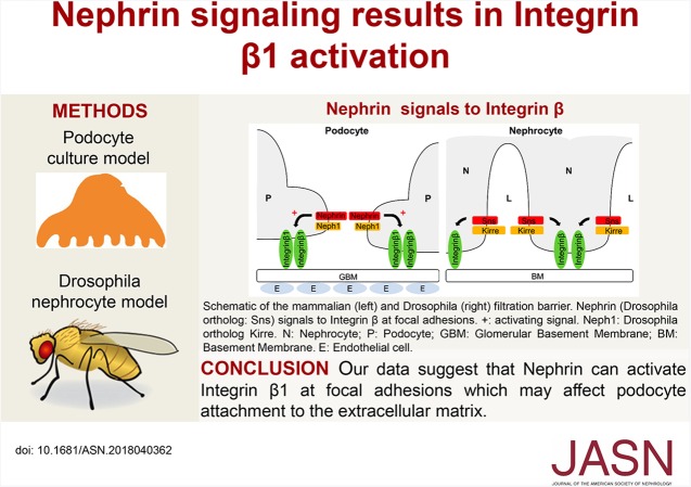
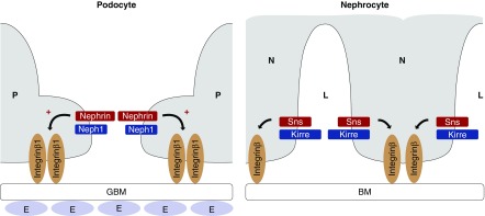
Background: Patients with certain mutations in the gene encoding the slit diaphragm protein Nephrin fail to develop functional slit diaphragms and display severe proteinuria. Many adult-onset glomerulopathies also feature alterations in Nephrin expression and function. Nephrin signals from the podocyte slit diaphragm to the Actin cytoskeleton by recruiting proteins that can interact with C3G, a guanine nucleotide exchange factor of the small GTPase Rap1. Because Rap activity affects formation of focal adhesions, we hypothesized that Nephrin transmits signals to the Integrin receptor complex, which mediates podocyte adhesion to the extracellular matrix.

Methods: To investigate Nephrin's role in transmitting signals to the Integrin receptor complex, we conducted genetic studies in Drosophila nephrocytes and validated findings from Drosophila in a cultured human podocyte model.

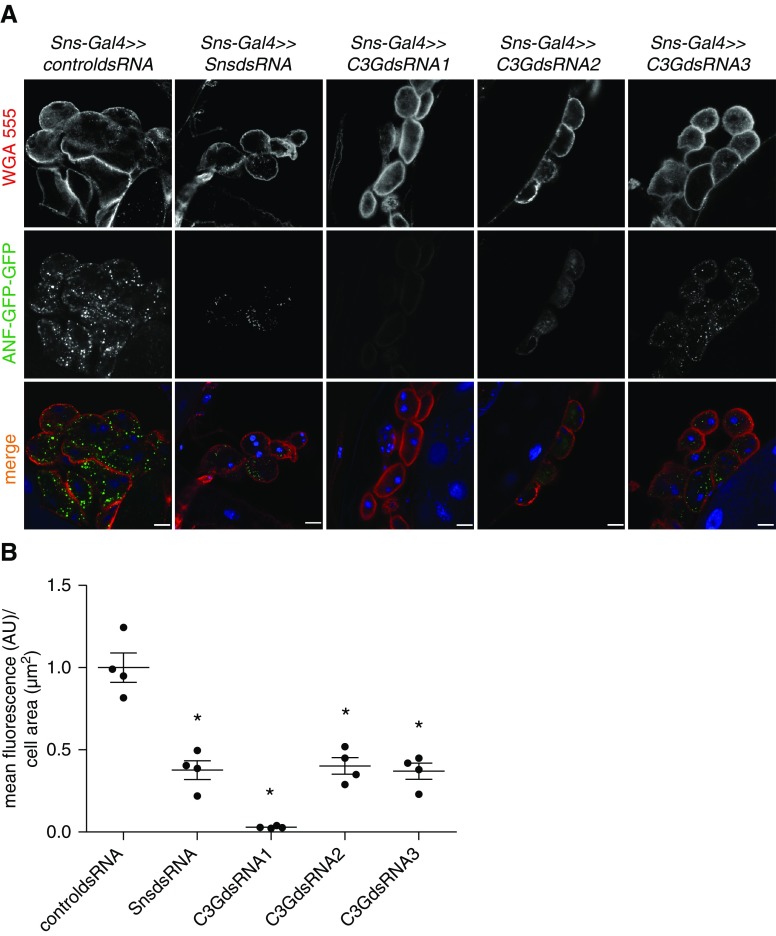
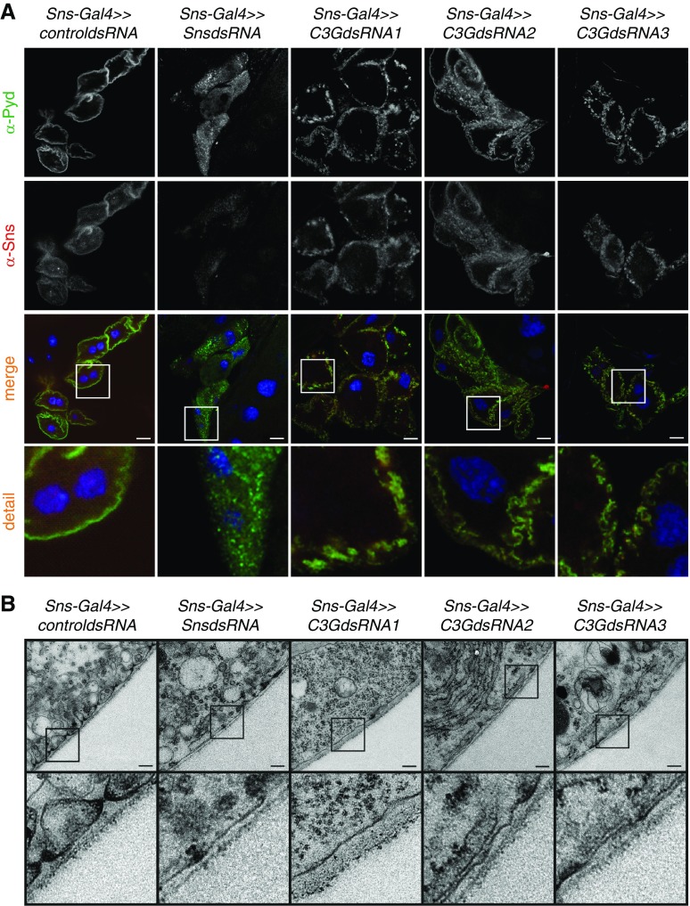
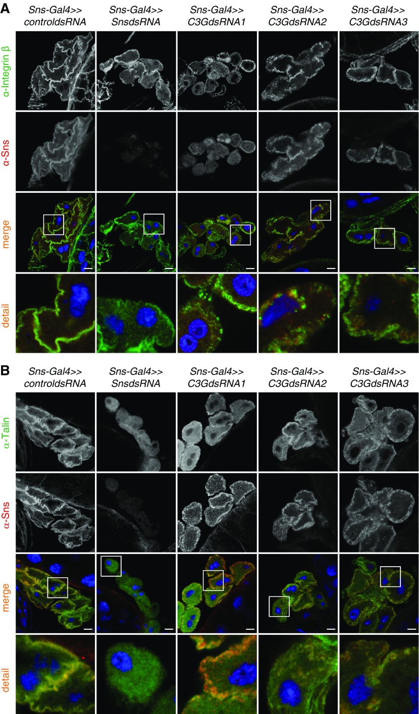
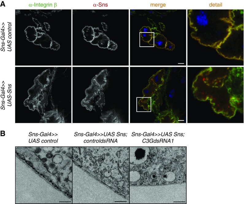
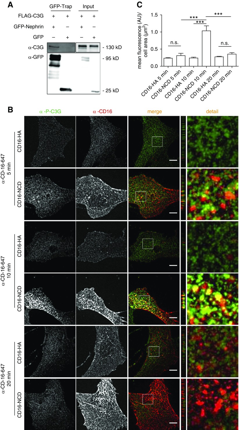
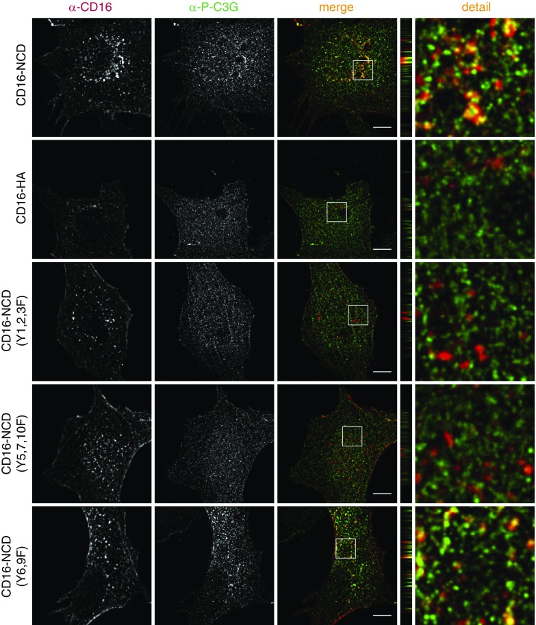
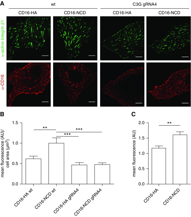
Results: Drosophila nephrocytes form a slit diaphragm-like filtration barrier and express the Nephrin ortholog Sticks and stones (Sns). A genetic screen identified c3g as necessary for nephrocyte function. In vivo, nephrocyte-specific gene silencing of sns or c3g compromised nephrocyte filtration and caused nephrocyte diaphragm defects. Nephrocytes with impaired Sns or C3G expression displayed an altered localization of Integrin and the Integrin-associated protein Talin. Furthermore, gene silencing of c3g partly rescued nephrocyte diaphragm defects of an sns overexpression phenotype, pointing to genetic interaction of sns and c3g in nephrocytes. We also found that activated Nephrin recruited phosphorylated C3G and resulted in activation of Integrin β1 in cultured podocytes.

Conclusions: Our findings suggest that Nephrin can mediate a signaling pathway that results in activation of Integrin β1 at focal adhesions, which may affect podocyte attachment to the extracellular matrix.

Keywords: cytoskeleton; nephrin; renal cell biology; signaling.


Figures

None
Graphical abstract
Figure 1.
Figure 1.
Knockdown of sns or c3g impairs nephrocyte function in vivo. (A) Uptake of secreted ANF-GFP-GFP into nephrocytes is shown in prepared control (RNAi for or83b), c3g, or sns knockdown nephrocytes. Knockdown in nephrocytes was accomplished using sns-Gal4. Samples were stained with wheat germ agglutinin-Alexa555 (WGA) to visualize membranes. Merged images are shown in the lower panel. Scale bar, 10 µm. (B) Accumulation of ANF-GFP-GFP in nephrocytes was quantified. Shown are means and SEM in arbitrary units (AU) per µm2 cell area normalized to control condition. *P<0.05 by unpaired two-tailed Mann–Whitney test. n=4.
Figure 2.
Figure 2.
Knockdown of sns or c3g leads to loss of nephrocyte diaphragms and lacunae. (A) Nephrocytes were prepared and immunofluorescence analysis was performed with antibody specific for Sns (red) and Pyd (green) (mammalian ortholog: ZO-1/2). Merged images and higher magnifications (×4) are shown in the bottom panels. Scale bar, 10 µm. (B) TEM analysis of control (RNAi for or83b), sns, or c3g knockdown nephrocytes is shown. Note that three different RNAi lines were used for c3g knockdown. Knockdown in nephrocytes was accomplished using sns-Gal4. Upper panel scale bar, 200 nm. Lower panel, zoom ×4 compared with the upper panel. (A and B) n=3.
Figure 3.
Figure 3.
Knockdown of sns or c3g results in altered targeting of the focal adhesion proteins Integrin and Talin in nephrocytes. Immunofluorescence analysis of control (RNAi for or83b), sns, or c3g knockdown nephrocytes is shown. Knockdown in nephrocytes was accomplished using sns-Gal4. Nephrocytes were prepared and immunofluorescence analysis was performed with antibody specific for Integrin (A), Talin (B), and Sns. Merged images and higher magnifications (×4) of the marked area are shown in the bottom panels. Third panel scale bar, 10 µm. (A and B) n=3.
Figure 4.
Figure 4.
sns and c3g genetically interact in nephrocytes. (A) Immunofluorescence analysis of nephrocytes overexpressing sns or control plasmid by sns-Gal4. Nephrocytes were stained with antibodies specific for Integrin β (green) and Sns (red). Merged images and higher magnifications (×4) of the marked area are shown in the columns to the right. Scale bar, 10 µm. (B) Shown are TEM images from Drosophila with overexpression of sns and knockdown of the control gene or83b, or flies with sns overexpression and c3g knockdown in nephrocytes. Knockdown in nephrocytes was accomplished using sns-Gal4. Scale bar, 200 nm. n=3.
Figure 5.
Figure 5.
phospho-C3G is recruited to activated Nephrin. (A) Flag-C3G and either GFP-Nephrin or GFP were transiently expressed in HEK293T cells. GFP-trap analysis revealed that Nephrin and C3G are part of one protein complex. (B) Podocytes were transiently transfected with CD16-NCD or CD16-HA and anti-CD16-Alexa647 antibody was added to the medium for the indicated time to induce Nephrin clustering. Cells were fixed, and immunofluorescence analysis was performed with antibody specific for p-C3G. (C) Statistical analysis of mean fluorescence of p-C3G in arbitrary units (AU) per µm2 cell area. Shown are means and SEM. ***P<0.001 by unpaired two-tailed t test; NS, not significant. One representative experiment of three is shown. Ten cells per condition were evaluated. Scale bar, 10 µm.
Figure 6.
Figure 6.
Recruitment of phospho-C3G to Nephrin depends on tyrosine residues also necessary for recruitment of the FAK-Cas-Crk complex to Nephrin. Human podocytes that transiently express CD16-NCD or Nephrin Y-to-F mutants as indicated were incubated with anti–CD16-Alexa647 antibody to induce Nephrin clustering. Podocytes were fixed and immunofluorescence analysis was performed with antibody specific for p-C3G. z-panel to the right. Zoom in to the far right. Scale bar, 10 µm; n=3.
Figure 7.
Figure 7.
Nephrin activation results in Integrin β1 activation. (A) Control podocytes or C3G KO podocytes (gRNA 4) transiently expressing CD16-NCD or CD16-HA were incubated with anti-CD16-Alexa647 antibody to initiate Nephrin clustering. Podocytes were fixed, and immunofluorescence analysis was performed using antibody specific for active Integrin β1. Scale bar, 10 µm. (B) Statistical analysis of mean fluorescence intensity of active Integrin β1 in arbitrary units (AU) per µm2 cell area. **P<0.01, ***P<0.001 by unpaired two-tailed t test. n=3; 20 cells per condition were evaluated. (C) Flow cytometry analysis of cultured podocytes inducibly expressing CD16-NCD or CD16-HA that were incubated with anti–CD16-Alexa647 antibody to initiate Nephrin clustering is shown. Podocytes were scraped from the dish, stained with antibody specific for active Integrin β1, and fixed. The statistical analysis by unpaired two-tailed t test shows the mean fluorescence intensity of active Integrin β1 in AU normalized to the control condition. n=3, **P<0.01. KO, knockout; wt, wild type.
Figure 8.
Figure 8.
Nephrin signals to Integrin β in podocytes and Drosophila nephrocytes. To the left, a schematic of the mammalian filtration barrier is shown, with the fenestrated endothelium (E), the glomerular basement membrane (GBM), and two mammalian podocyte (P) foot processes with the slit diaphragm in between. Slit diaphragm proteins Nephrin and Neph1 are depicted as well as Integrin β1, which connects the basal podocyte with the GBM. Nephrin activation in podocyte culture results in activation of Integrin β1 (arrow marked with +). To the right, a schematic of the Dm nephrocyte (N) filtration barrier is shown, which consists of the basement membrane (BM) and the intracellular junction that spans the gaps between invaginations in the nephrocyte plasma membrane. Nephrocyte slit diaphragm proteins that are orthologs of Nephrin (Sns in Dm) and Neph1 (Kirre in Dm) are shown. Drosophila Integrin β connects the nephrocyte with the basement membrane. Sns gain of function results in mislocalization of Integrin β (depicted by arrow). L, nephrocyte lacunae.

References

    1. Pavenstädt H, Kriz W, Kretzler M: Cell biology of the glomerular podocyte. Physiol Rev 83: 253–307, 2003 - PubMed
    1. Wiggins RC: The spectrum of podocytopathies: A unifying view of glomerular diseases. Kidney Int 71: 1205–1214, 2007 - PubMed
    1. Faul C, Asanuma K, Yanagida-Asanuma E, Kim K, Mundel P: Actin up: Regulation of podocyte structure and function by components of the actin cytoskeleton. Trends Cell Biol 17: 428–437, 2007 - PubMed
    1. Scott RP, Quaggin SE: Review series: The cell biology of renal filtration. J Cell Biol 209: 199–210, 2015 - PMC - PubMed
    1. Harburger DS, Calderwood DA: Integrin signalling at a glance. J Cell Sci 122:159–163, 2009 - PMC - PubMed

Publication types