. 2019 Jul 17;39(29):5728-5739.
doi: 10.1523/JNEUROSCI.2739-18.2019. Epub 2019 May 16.

Anterior Cingulate Cortex to Ventral Hippocampus Circuit Mediates Contextual Fear Generalization

Affiliations

Anterior Cingulate Cortex to Ventral Hippocampus Circuit Mediates Contextual Fear Generalization

Xin-Lan Bian et al. J Neurosci. .

Abstract

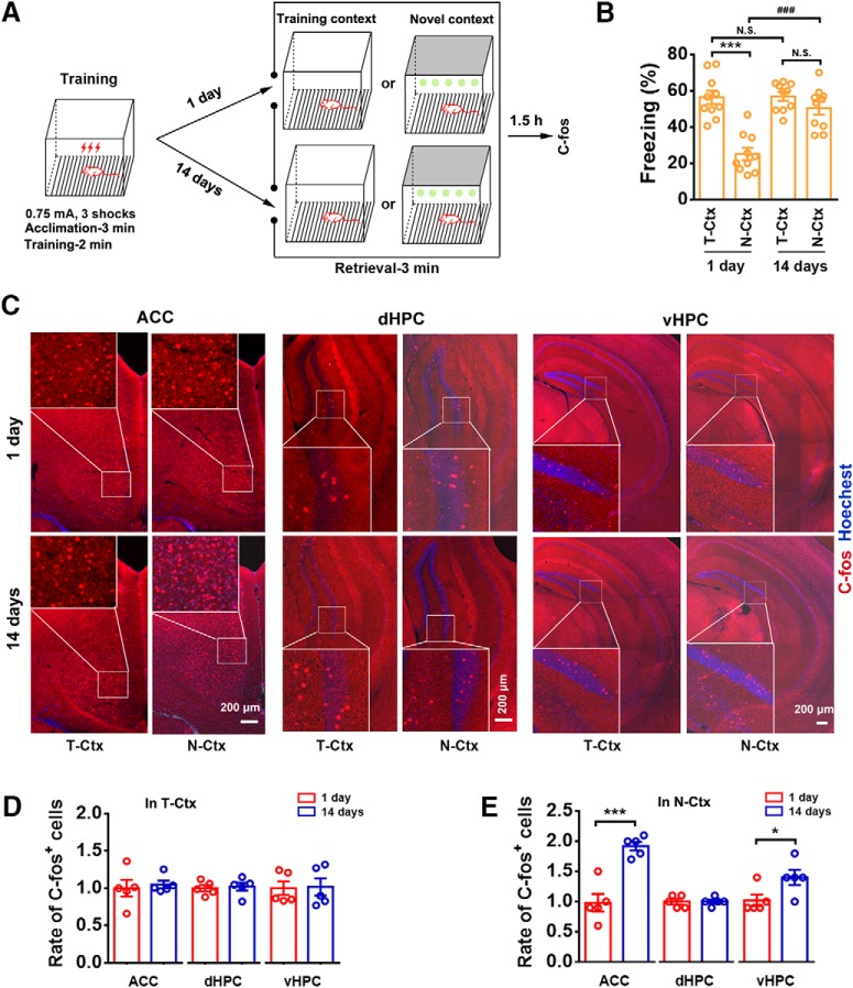
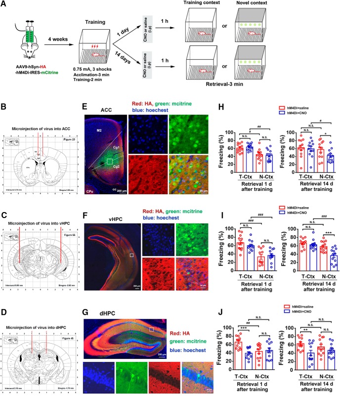
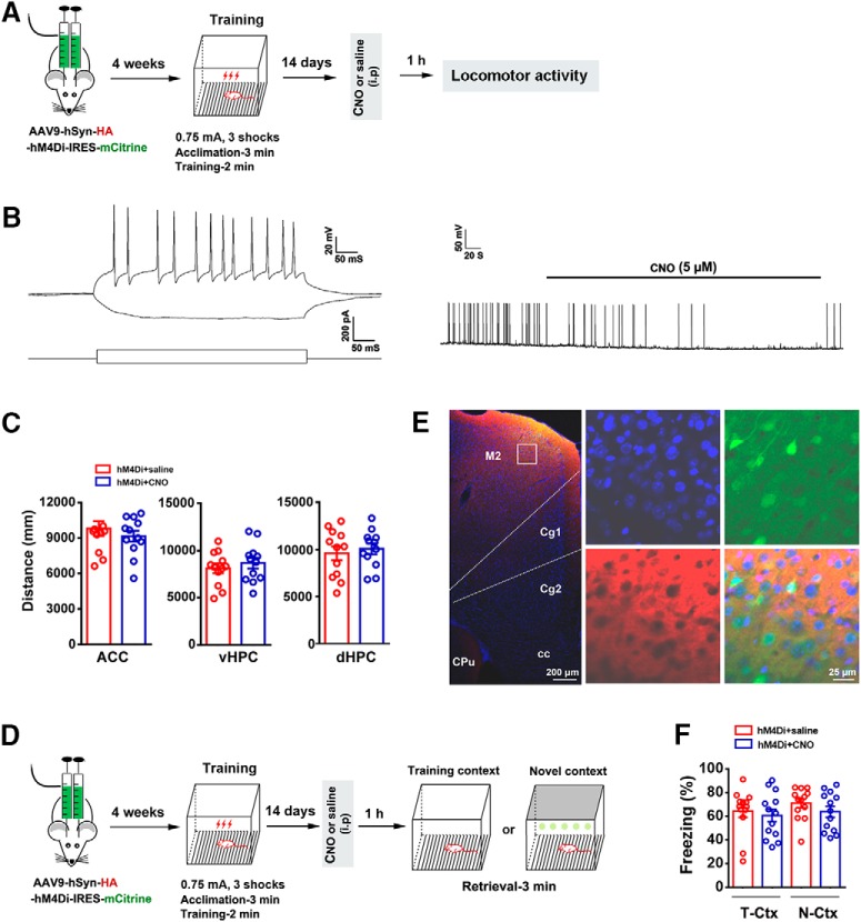
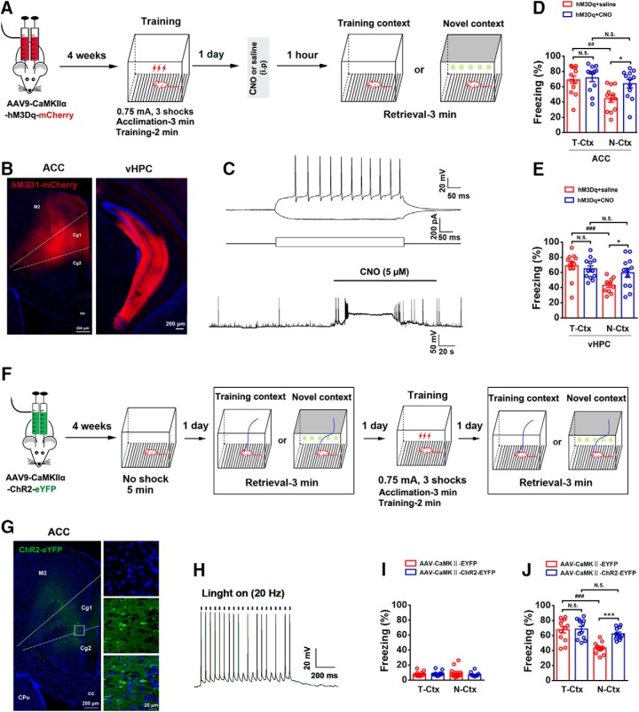
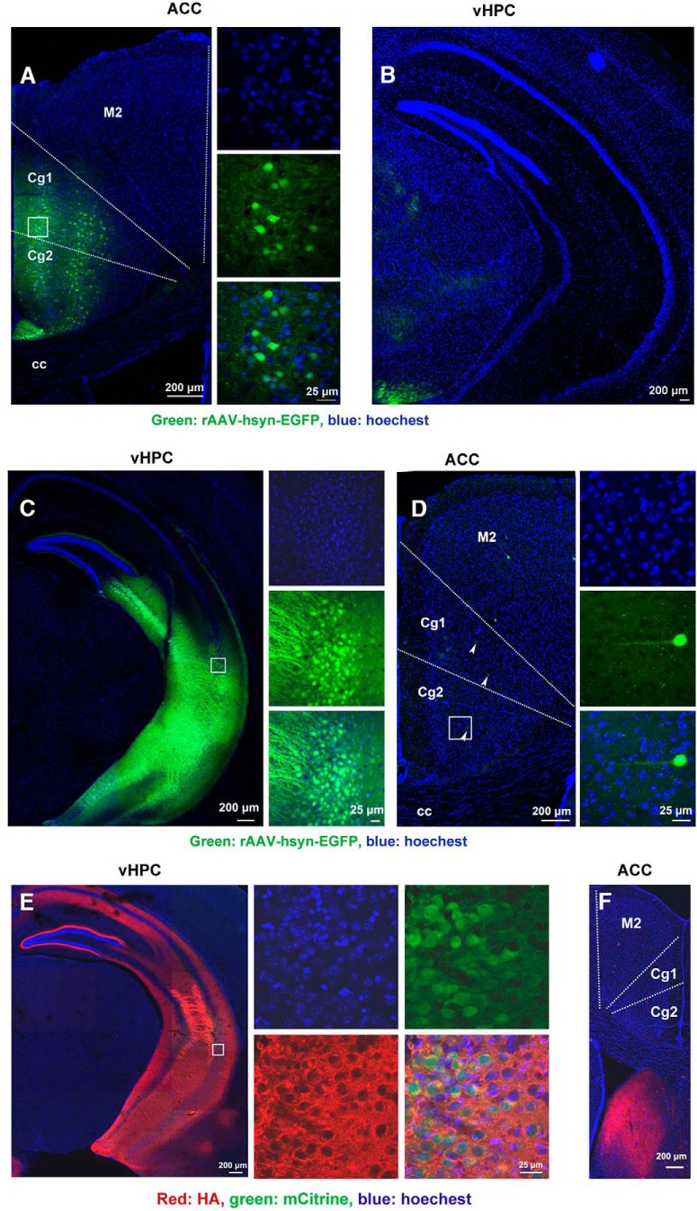
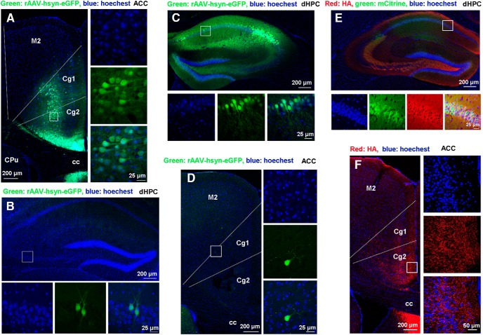
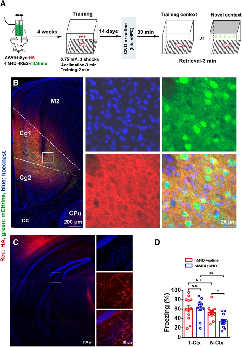
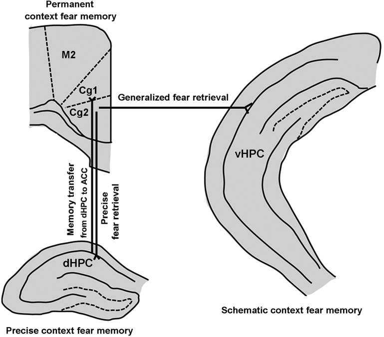
Contextual fear memory becomes less context-specific over time, a phenomenon referred to as contextual fear generalization. Overgeneralization of contextual fear memory is a core symptom of post-traumatic stress disorder (PTSD), but circuit mechanisms underlying the generalization remain unclear. We show here that neural projections from the anterior cingulate cortex (ACC) to ventral hippocampus (vHPC) mediate contextual fear generalization in male mice. Retrieval of contextual fear in a novel context at a remote time point activated cells in the ACC and vHPC, as indicated by significantly increased C-fos+ cells. Using chemogenetic or photogenetic manipulations, we observed that silencing the activity of ACC or vHPC neurons reduced contextual fear generalization at the remote time point, whereas stimulating the activity of ACC or vHPC neurons facilitated contextual fear generalization at a recent time point. We found that ACC neurons projected to the vHPC unidirectionally, and importantly, silencing the activity of projection fibers from the ACC to vHPC inhibited contextual fear generalization at the remote time point. Together, our findings reveal an ACC to vHPC circuit that controls expression of fear generalization and may offer new strategies to prevent or reverse contextual fear generalization in subjects with anxiety disorders, especially in PTSD.SIGNIFICANCE STATEMENT Overgeneralization of contextual fear memory is a cardinal feature of PTSD, but circuit mechanisms underlying it remain unclear. Our study indicates that neural projections from the anterior cingulate cortex to ventral hippocampus control the expression of contextual fear generalization. Thus, manipulating the circuit may prevent or reverse fear overgeneralization in subjects with PTSD.

Keywords: anterior cingulate cortex; anxiety disorders; contextual fear memory; memory generalization; neural circuit; ventral hippocampus.


Figures

Figure 1.
Figure 1.
Implication of ACC and vHPC in fear generalization. A, Design of the experiments for BE. B, Freezing levels of mice during recent and remote retrieval tests (1 and 14 d after training respectively) in T-Ctx or N-Ctx (two-way ANOVA followed by Bonferroni post hoc test; environment F(1,36) = 32.134, p < 0.001; time F(1,36) = 15.069, p < 0.001; interaction F(1,36) = 13.969, p = 0.001; n = 10 for each group; ***p < 0.001, ###p < 0.001). C, Representative Immunofluorescence images of ACC, dHPC, and vHPC showing c-fos+ cells and high-magnification images (top-left corner, ACC; lower-left corner, dHPC and vHPC) from selected areas. Red, C-fos; blue, Hoechst. D, Number of c-fos+ cells in ACC, dHPC, and vHPC of mice subjected to recent or remote fear retrieval in T-Ctx (n = 5; ACC: t(1/8) = 0.408, p = 0.694; dHPC: t(1/8) = 0.308, p = 0.766; vHPC: t(1/8) = 0.116, p = 0.910, two-tailed t test). E, Number of c-fos+ cells in ACC, dHPC, and vHPC of mice subjected to recent or remote fear retrieval in N-Ctx (n = 5; ACC: t(1/8) = 5.718, ***p < 0.001; dHPC: t(1/8) = 0.183, p = 0.860; vHPC: t(1/8) = 2.522, *p = 0.036, two-tailed t test). N.S., no statistics significance, p ≥ 0.05.
Figure 2.
Figure 2.
Activity of ACC or vHPC neurons is necessary for fear memory generalization. A, Design of the experiments for HJ. BD, AAV injection sites. E–G, Representative images (E, F, left; G, top) of the mice ACC, vHPC or dHPC infected by AAV9-hSyn-HA-hM4Di-IRES-mCitrine and high-magnification images (E, F, right; G, bottom) from selected areas on the left (E, F) of top (G) images. HJ, Freezing levels of mice retrieved 1 d (left) and 14 d (right) in T-Ctx or N-Ctx after training, in which the activity of neurons in the ACC, vHPC, or dHPC was silenced by CNO (i.p.) at 1 h before retrieval (three-way ANOVA followed by Bonferroni post hoc test. For ACC, treatment: F(1,88) = 1.712, p = 0.194; environment: F(1,88) = 19.276, p < 0.001; time: F(1,88) = 0.433, p = 0.512; interaction: F(1,88) = 0.299, p = 0.586; n = 12 for each group; *p < 0.05, #p < 0.05, ##p < 0.01. For vHPC, treatment: F(1,86) = 5.996, p = 0.016; environment: F(1,86) = 57.007, p < 0.001; time: F(1,86) = 5.363, p = 0.023; interaction: F(1,86) = 5.500, p = 0.021, n = 11 for two groups in N-Ctx at the recent time point and n = 12 for other groups; ***p < 0.001, ###p < 0.001. For dHPC, treatment: F(1,86) = 14.610, p < 0.001; environment: F(1,86) = 1.351, p = 0.248; time: F(1,86) = 0.563, p = 0.455; interaction: F(1,86) = 1.047, p = 0.309, n = 11 for two groups treated with CNO at the recent time point and n = 12 for other groups; **p < 0.01, ***p < 0.001, ##p < 0.01. M2: secondary motor cortex; Cg1: cingulate cortex, area 1; Cg2: cingulate cortex, area 2; CPu: caudate putamen (striatum); cc: corpus callosum. N.S., no statistics significance, p ≥ 0.05.
Figure 3.
Figure 3.
Silencing the activity of ACC, vHPC, or dHPC does not change locomotor activity and silencing the activity of M2 does not change freezing levels. A, Design of the experiments for C. B, The ability of the hM4D/CNO system to inhibit the firing of neurons in the ACC using in vitro whole-cell recordings. Left, Representative current-clamp traces of a neuron response to positive and negative 500 ms current injections (−40 and +120 pA, respectively). Right, In neurons expressing hM4-mCitrine, bath application of CNO completely abolished cell discharge of action potentials and this effect was reversed upon CNO washout. C, The locomotor activity in different groups (n = 12; ACC: t(1/22) = 0.837, p = 0.412; vHPC: t(1/22) = 0.723, p = 0.477; dHPC: t(1/22) = 0.509, p = 0.616, two-tailed t test). D, Design of the experiments for F. E, Representative image of the mice M2 infected by recombinant virus AAV9-hSyn-HA-hM4Di-IRES-mCitrine (left) and high-magnification images (right) from selected areas of the left image. F, Freezing levels of mice retrieved 14 d (right) in T-Ctx or N-Ctx after training, in which the activity of neurons in the M2 were silenced by CNO (i.p.) at 1 h before retrieval (two-way ANOVA followed by Bonferroni post hoc test; treatment: F(1,48) = 1.335, p = 0.254; environment: F(1,48) = 1.014, p = 0.319; interaction: F(1,48) = 0.121, p = 0.729; n = 13 for each group. M2: secondary motor cortex; Cg1: cingulate cortex, area 1; Cg2: cingulate cortex, area 2; CPu: caudate putamen (striatum); cc: corpus callosum.
Figure 4.
Figure 4.
Activity of ACC and vHPC neurons is sufficient for fear memory generalization. A, Design of the experiments for D and E. B, Representative images of the mice ACC or vHPC infected by AAV9-CaMKIIα-hM3Dq-mCherry. C, The ability of the hM3D/CNO system to activate the firing of neurons in the ACC using in vitro whole-cell recordings. Top, Representative current-clamp traces of a neuron response to positive and negative 500 ms current injections (−40 and +120 pA, respectively). Bottom, In neurons expressing hM3-mcherry, bath application of CNO elicited action potential firing and this effect was reversed upon CNO washout. D, E, Freezing levels of mice retrieved 1 d in T-Ctx or N-Ctx after training, in which ACC (D) or vHPC (E) neurons were activated by CNO at 1 h before retrieval (two-way ANOVA followed by Bonferroni post hoc test; ACC, treatment: F(1,44) = 4.697, p = 0.036; environment: F(1,44) = 9.958, p = 0.003; interaction: F(1,44) = 2.710, p = 0.107; n = 12 for each group; *p < 0.05, ##p < 0.01; vHPC, treatment: F(1,43) = 1.771, p = 0.190; environment: F(1,43) = 11.392, p = 0.002; interaction: F(1,43) = 4.960, p = 0.031; n = 11 for the group treated with saline in the N-Ctx and n = 12 for other groups; *p < 0.05, ###p < 0.001). F, Design of the experiments for I and J. G, Images showing ChR2-eYFP-expressing ACC neurons (left) and high-magnification images from a selected area in the leftward image. H, ChR2-mediated action potentials in ACC neuron were evoked by 5 ms pulses of light (465 nm) with 20 Hz in current-clamp mode. I, Freezing levels in T-Ctx or N-Ctx before training (two-way ANOVA followed by Bonferroni post hoc test; optogenetics: F(1,44) = 0.462, p = 0.500; environment: F(1,44) = 0.008, p = 0.930; interaction: F(1,44) = 1.186, p = 0.282; n = 12 for each group). J, Freezing levels in T-Ctx or N-Ctx after training (two-way ANOVA followed by Bonferroni post hoc test; optogenetics: F(1,44) = 9.016, p = 0.004; environment; F(1,44) = 22.042, p = 0.000; interaction: F(1,44) = 7.367, p = 0.009; n = 12 for each group; ***p < 0.001, ###p < 0.001. M2: secondary motor cortex; Cg1: cingulate cortex, area 1; Cg2: cingulate cortex, area 2; CPu: caudate putamen (striatum); cc: corpus callosum. N.S., no statistics significance, p ≥ 0.05.
Figure 5.
Figure 5.
Projection pattern between the ACC and vHPC. A, Representative images of the ACC infected by rAAV-hSyn-EGFP (left) and high-magnification images (right) from selected areas in the left image. B, Representative image of vHPC of mouse 6 weeks after receiving microinjection of rAAV-hSyn-EGFP into the ACC. C, Representative image of the vHPC infected by rAAV-hSyn-EGFP (left) and high-magnification images (right) from selected areas in the left image. D, Representative image of the ACC (left) of mouse 6 weeks after receiving microinjection of rAAV-hSyn-EGFP into the vHPC and high-magnification images (right) from selected areas in the left image. The white arrows indicate EGFP+. E, Representative image of the vHPC infected by AAV9-hSyn-HA-hM4Di-IRES-mCitrine (left) and high-magnification images (right) from selected areas in the left image. F, Representative image of ACC of mouse 6 weeks after receiving microinjection of AAV9-hSyn-HA-hM4Di-IRES-mCitrine into the vHPC. M2: secondary motor cortex; Cg1: cingulate cortex, area 1; Cg2: cingulate cortex, area 2; cc: corpus callosum.
Figure 6.
Figure 6.
Projection pattern between the ACC and dHPC. A, Representative image of the ACC infected by rAAV-hSyn-EGFP (left) and high-magnification images (right) from selected areas in the left image. B, Representative image of the dHPC (top) of mouse 6 weeks after receiving microinjection of rAAV-hSyn-EGFP into the ACC and high-magnification images (bottom) from selected areas in the top image. C, Representative image of the dHPC infected by rAAV-hSyn-EGFP (top) and high-magnification images (bottom) from selected areas in the top image. D, Representative image of the ACC (left) of mouse 6 weeks after receiving microinjection of rAAV-hSyn-EGFP into the dHPC and high-magnification images (right) from selected areas in the left image. E, Representative image of the dHPC infected by AAV9-hSyn-HA-hM4Di-IRES-mCitrine (top) and high-magnification images (bottom) from selected areas in the top image. F, Representative image of ACC (left) of mouse 4 weeks after receiving microinjection of AAV9-hSyn-HA-hM4Di-IRES-mCitrine into the dHPC and high-magnification images (right) from selected areas in the left image. M2: secondary motor cortex; Cg1: cingulate cortex, area 1; Cg2: cingulate cortex, area 2; CPu: caudate putamen (striatum); cc: corpus callosum.
Figure 7.
Figure 7.
Neural projections from the ACC to vHPC mediate fear memory generalization. A, Design of the experiments for D. B, Representative image of the ACC infected by AAV9-hSyn-HA-hM4Di-IRES-mCitrine (left) and high-magnification images (right) from selected areas in the left image. C, Representative image of vHPC (left) of mouse 4 weeks after receiving microinjection of AAV9-hSyn-HA-hM4Di-IRES-mCitrine into the ACC and high-magnification images (right) from selected areas in the left image. D, Freezing levels of mice in T-Ctx or N-Ctx retrieved 14 d after training in which vHPC neurons receiving neural projections from the ACC were silenced by CNO at 30 min before retrieval (two-way ANOVA followed by Bonferroni post hoc test; treatment: F(1,44) = 2.340, p = 0.133; environment: F(1,44) = 11.533, p = 0.001; interaction: F(1,44) = 3.521, p = 0.067; n = 12 for each group; *p < 0.05, ##p < 0.01). M2: secondary motor cortex; Cg1: cingulate cortex, area 1; Cg2: cingulate cortex, area 2; CPu: caudate putamen (striatum); cc: corpus callosum. N.S., no statistics significance, p ≥ 0.05.
Figure 8.
Figure 8.
Schematic diagram of the synaptic interactions between the hippocampus and the ACC in controlling precise and generalized context fear memory (see text). M2: secondary motor cortex; Cg1: cingulate cortex, area 1; Cg2: cingulate cortex, area 2.

References

    1. Allsop SA, Wichmann R, Mills F, Burgos-Robles A, Chang CJ, Felix-Ortiz AC, Vienne A, Beyeler A, Izadmehr EM, Glober G, Cum MI, Stergiadou J, Anandalingam KK, Farris K, Namburi P, Leppla CA, Weddington JC, Nieh EH, Smith AC, Ba D, et al. (2018) Corticoamygdala transfer of socially derived information gates observational learning. Cell 173:1329–1342.e18. 10.1016/j.cell.2018.04.004 - DOI - PMC - PubMed
    1. Alvares Lde O, Einarsson EÖ, Santana F, Crestani AP, Haubrich J, Cassini LF, Nader K, Quillfeldt JA (2012) Periodically reactivated context memory retains its precision and dependence on the hippocampus. Hippocampus 22:1092–1095. 10.1002/hipo.20983 - DOI - PubMed
    1. Alvarez P, Zola-Morgan S, Squire LR (1995) Damage limited to the hippocampal region produces long-lasting memory impairment in monkeys. J Neurosci 15:3796–3807. 10.1523/JNEUROSCI.15-05-03796.1995 - DOI - PMC - PubMed
    1. Antunes R, Moita MA (2010) Discriminative auditory fear learning requires both tuned and nontuned auditory pathways to the amygdala. J Neurosci 30:9782–9787. 10.1523/JNEUROSCI.1037-10.2010 - DOI - PMC - PubMed
    1. Armbruster BN, Li X, Pausch MH, Herlitze S, Roth BL (2007) Evolving the lock to fit the key to create a family of G protein-coupled receptors potently activated by an inert ligand. Proc Natl Acad Sci U S A 104:5163–5168. 10.1073/pnas.0700293104 - DOI - PMC - PubMed

Publication types

LinkOut - more resources