. 2019 May 17;364(6441):653-658.
doi: 10.1126/science.aav9996.

Identification of a regeneration-organizing cell in the Xenopus tail

Affiliations

Identification of a regeneration-organizing cell in the Xenopus tail

C Aztekin et al. Science. .

Abstract

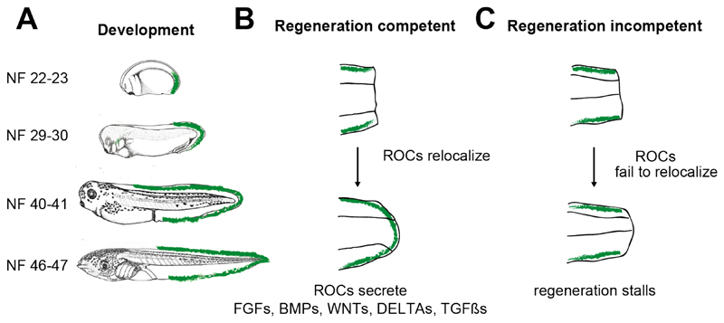
Unlike mammals, Xenopus laevis tadpoles have a high regenerative potential. To characterize this regenerative response, we performed single-cell RNA sequencing after tail amputation. By comparing naturally occurring regeneration-competent and -incompetent tadpoles, we identified a previously unrecognized cell type, which we term the regeneration-organizing cell (ROC). ROCs are present in the epidermis during normal tail development and specifically relocalize to the amputation plane of regeneration-competent tadpoles, forming the wound epidermis. Genetic ablation or manual removal of ROCs blocks regeneration, whereas transplantation of ROC-containing grafts induces ectopic outgrowths in early embryos. Transcriptional profiling revealed that ROCs secrete ligands associated with key regenerative pathways, signaling to progenitors to reconstitute lost tissue. These findings reveal the cellular mechanism through which ROCs form the wound epidermis and ensure successful regeneration.


Conflict of interest statement

Competing interests

The authors declare no competing interests.

Figures

Fig. 1
Fig. 1. Pooled transcriptional cell state atlas of the Xenopus laevis tail before and after amputation.
(A) Samples were prepared for single-cell RNA-seq analysis from regeneration-competent and incompetent tadpoles, collecting either intact tails, or tails at various stages following amputation: 1-3 days post amputation (dpa) for regeneration-competent, and 1 dpa for regeneration-incompetent tadpoles. Developmental timing is indicated for each sample (days post-fertilisation, dpf). Samples were processed separately for sequencing and then pooled for UMAP visualisation (Methods). Each dot represents a single cell; colour indicates main tissue group (n≥2 for each sample). (B) Cluster identities based on established cell type markers. For details of cluster annotations, see main text, fig. S1 and Methods.
Fig. 2
Fig. 2. Comparison of scRNA-seq samples discriminate gene expression and cell state changes that take place during development from those associated with the response to amputation or regeneration.
Examples of cell-specific gene expression changes that take place (A) during development, (B) in response to amputation, and (C) in response to regeneration. Grey dots: cells from samples at all respective time points; red dots: cells from selected time point and condition. Black and white filled arrows indicate presence and absence of populations, respectively, when comparing intact tail to 1 dpa samples. Panel A shows a continuous change in the gene expression profile of Goblet cells that takes place during development both in regeneration-competent and incompetent tail; panel B shows gene expression changes that take place in motor neurons in response to amputation, both in regeneration-competent and incompetent tail; and panel C shows differential gene expression changes that take place in epidermis between regeneration-competent and incompetent tail, identifying a cell state change specific to regeneration.
Fig. 3
Fig. 3. Regeneration-organizing cells (ROCs) characterize the specialized wound epidermis in regeneration competent tadpole.
(A) ROCs express high Lef1 mRNA level, and reappear after amputation specifically in regeneration-competent tadpoles. Grey dots: TP63 positive epidermal clusters; circled dots: selected sample. Relative Lef1 expression visualized for each cell. (B) ROCs (TP63+/LEF1+ cells, denoted by asterisks) are localized along the midline edge of the epidermis in intact tails. Green, pbin7Lef; Red, TP63. Scale bar: 500 μm. (C) ROCs (LEF1+) remain along the posterior trunk following amputation (asterisks), but are removed from the amputation plane (empty arrowheads). ROCs specifically reappear in the amputation plane of 1 dpa regeneration-competent tadpoles (filled arrowhead). hpa: hours post-amputation. Green, pbin7LEF:GFP. Scale bars: 250 μm; a total of ≥3 tadpoles per conditions were imaged from 2 biological replicates. (D) Quantification of TP63+/LEF1+ cells at the amputation plane (mean ± standard deviation) shows a significant reduction in regeneration-incompetent tadpoles at 1 dpa (n=12 and n=11 for competent and incompetent samples, respectively, both from 2 biological replicates). *: p < 0.001.
Fig. 4
Fig. 4. Regeneration-organizing cells relocation to the amputation area mediates tail regeneration.
(A) Nitroreductase (NTR)/Metronidazole-mediated ablation of ROCs during regeneration. pbin7LEF:GFP/Krt.L:NTR F0 transgenic tadpoles (bottom) show successful cell ablation at 3 dpa: GFP positive ROCs are present in control (plain arrowhead) but lost in MTZ treated animal (empty arrowhead). Scale, 1 mm. (B) ROC-ablated tadpoles cannot regenerate (n=11 from 2 biological replicates). (C) Schematic of ROCs-containing region manual removal protocol. Green colored area indicate ROCs localization. (D) Manual removal of posterior trunk ROCs at the same time as tail amputation reduces regeneration (n= 45 from 5 biological replicates), but manual removal of posterior trunk ROCs 12-16 hours post amputation does not negatively affect regeneration (n= 95 from 3 biological replicates). (E) Time-lapse images of ROCs relocating to the amputation plane, as assessed by pbin7LEF:GFP. Asterisks denote cells with brighter GFP that can be tracked (n=8 from 3 biological replicates). Scale bar, 500 μm.
Fig. 5
Fig. 5. ROCs act as a signalling centre coordinating progenitor outgrowth during tail regeneration.
(A) Expression of FGF ligands (Top) and receptor (Bottom) shown for selected cell types as a boxplot (outliers not shown). (B) Bar plot indicating the change in the fraction of cells in G2/M and S phases between regeneration-competent 2 dpa and incompetent intact tail samples, all taken at 5 dpf. (C) (Left) Removal of large or small ROC-containing tissues causes tail development defects in donors (n= 20), (Right) grafting these regions to the trunk enables tail-enriched or fin-enriched distal growth in hosts, respectively. Matching donor-acceptor pairs are shown 2 days post-grafting (n= 20 from 3 biological replicates). (D) Non-labelled grafts to CMV:GFP positive embryos induce outgrowth containing GFP positive cells; donor tissues are at the tip of the ectopic structure (n=12 from 3 biological replicates). Green, CMV:GFP. Scale bars: full tadpoles, 1 mm; zoomed grafts and merged graft images, 500 μm.
Fig. 6
Fig. 6. ROC-based model of tail regeneration.
Transcriptional signature of ROCs first appears in NF stage 22-23 embryos at the tip of the tail-bud, then expand towards the edges of the epidermis midline from the tail tip to the posterior trunk during development (table S2). Relocalization of ROCs to the wound area forms the specialized wound epidermis and is a hallmark of successful tail regeneration.

Comment in

References

    1. Tanaka EM. The Molecular and Cellular Choreography of Appendage Regeneration. Cell. 2016;165:1598–1608. - PubMed
    1. Stoick-Cooper CL, Moon RT, Weidinger G. Advances in signaling in vertebrate regeneration as a prelude to regenerative medicine. Genes & Development. 2007;21:1292–1315. - PubMed
    1. Chen C-H, Poss KD. Regeneration Genetics. Annu Rev Genet. 2017;51:63–82. - PMC - PubMed
    1. Li J, Zhang S, Amaya E. The cellular and molecular mechanisms of tissue repair and regeneration as revealed by studies in Xenopus. Regeneration. 2016;3:198–208. - PMC - PubMed
    1. Beck CW, Christen B, Slack JMW. Molecular Pathways Needed for Regeneration of Spinal Cord and Muscle in a Vertebrate. Developmental Cell. 2003;5:429–439. - PubMed

Publication types

Substances