

Proteome-wide detection of S-nitrosylation targets and motifs using bioorthogonal cleavable-linker-based enrichment and switch technique
- PMID: 31097712
- PMCID: PMC6522481
- DOI: 10.1038/s41467-019-10182-4
Proteome-wide detection of S-nitrosylation targets and motifs using bioorthogonal cleavable-linker-based enrichment and switch technique
Abstract
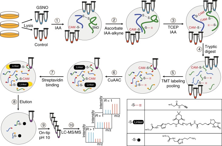
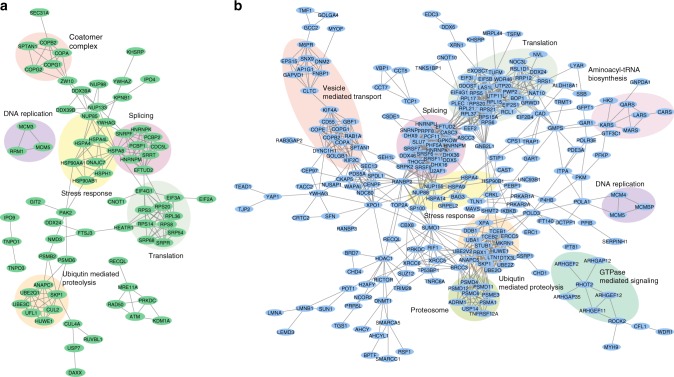
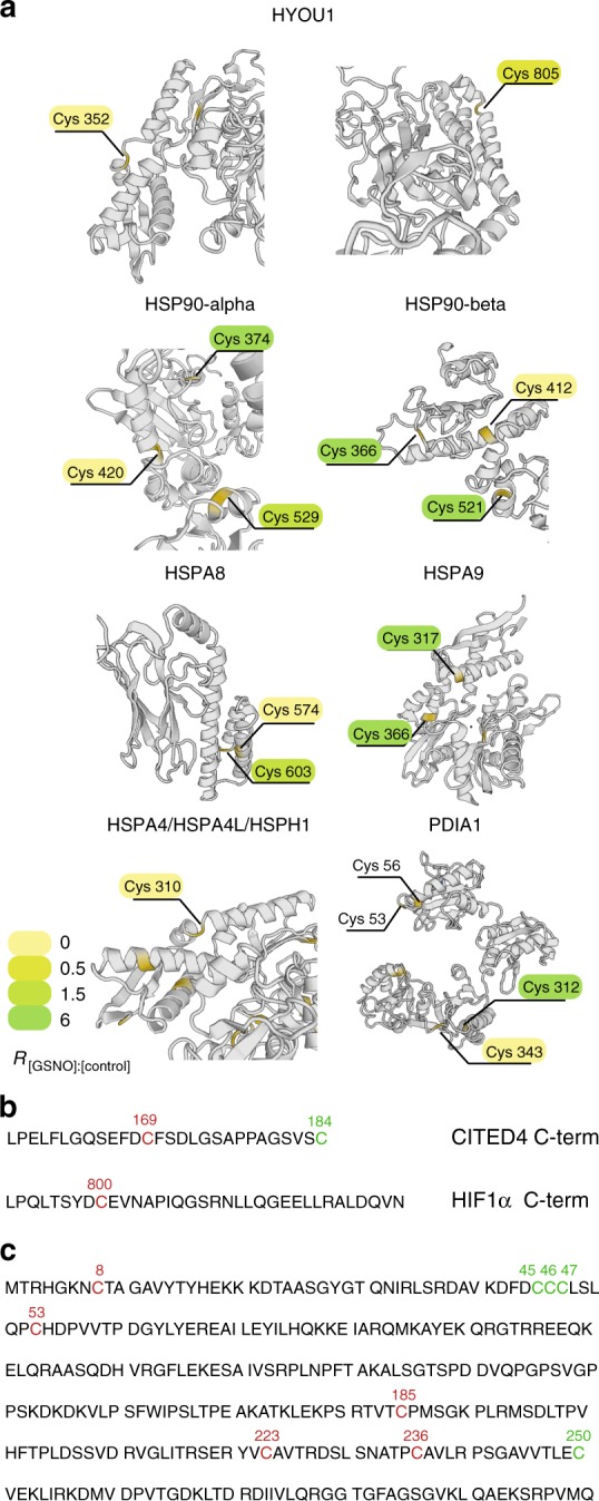
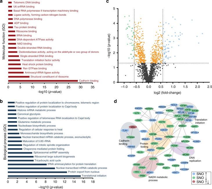
Cysteine modifications emerge as important players in cellular signaling and homeostasis. Here, we present a chemical proteomics strategy for quantitative analysis of reversibly modified Cysteines using bioorthogonal cleavable-linker and switch technique (Cys-BOOST). Compared to iodoTMT for total Cysteine analysis, Cys-BOOST shows a threefold higher sensitivity and considerably higher specificity and precision. Analyzing S-nitrosylation (SNO) in S-nitrosoglutathione (GSNO)-treated and non-treated HeLa extracts Cys-BOOST identifies 8,304 SNO sites on 3,632 proteins covering a wide dynamic range of the proteome. Consensus motifs of SNO sites with differential GSNO reactivity confirm the relevance of both acid-base catalysis and local hydrophobicity for NO targeting to particular Cysteines. Applying Cys-BOOST to SH-SY5Y cells, we identify 2,151 SNO sites under basal conditions and reveal significantly changed SNO levels as response to early nitrosative stress, involving neuro(axono)genesis, glutamatergic synaptic transmission, protein folding/translation, and DNA replication. Our work suggests SNO as a global regulator of protein function akin to phosphorylation and ubiquitination.
Conflict of interest statement
The authors declare no competing interests.
Figures


Similar articles
-
Quantitative Proteomics Analysis of VEGF-Responsive Endothelial Protein S-Nitrosylation Using Stable Isotope Labeling by Amino Acids in Cell Culture (SILAC) and LC-MS/MS.Biol Reprod. 2016 May;94(5):114. doi: 10.1095/biolreprod.116.139337. Epub 2016 Apr 13. Biol Reprod. 2016. PMID: 27075618 Free PMC article.
-
Dual Labeling Biotin Switch Assay to Reduce Bias Derived From Different Cysteine Subpopulations: A Method to Maximize S-Nitrosylation Detection.Circ Res. 2015 Oct 23;117(10):846-57. doi: 10.1161/CIRCRESAHA.115.307336. Epub 2015 Sep 3. Circ Res. 2015. PMID: 26338901 Free PMC article.
-
Identification and quantification of S-nitrosylation by cysteine reactive tandem mass tag switch assay.Mol Cell Proteomics. 2012 Feb;11(2):M111.013441. doi: 10.1074/mcp.M111.013441. Epub 2011 Nov 29. Mol Cell Proteomics. 2012. PMID: 22126794 Free PMC article.
-
Solid-phase capture for the detection and relative quantification of S-nitrosoproteins by mass spectrometry.Methods. 2013 Aug 1;62(2):130-7. doi: 10.1016/j.ymeth.2012.10.001. Epub 2012 Oct 11. Methods. 2013. PMID: 23064468 Free PMC article. Review.
-
Stabilising cysteinyl thiol oxidation and nitrosation for proteomic analysis.J Proteomics. 2013 Oct 30;92:160-70. doi: 10.1016/j.jprot.2013.06.019. Epub 2013 Jun 21. J Proteomics. 2013. PMID: 23796488 Review.
Cited by
-
Oxidative Stress in Age-Related Neurodegenerative Diseases: An Overview of Recent Tools and Findings.Antioxidants (Basel). 2023 Jan 5;12(1):131. doi: 10.3390/antiox12010131. Antioxidants (Basel). 2023. PMID: 36670993 Free PMC article. Review.
-
Nitric oxide inhibits endothelial cell apoptosis by inhibiting cysteine-dependent SOD1 monomerization.FEBS Open Bio. 2022 Feb;12(2):538-548. doi: 10.1002/2211-5463.13362. Epub 2022 Jan 11. FEBS Open Bio. 2022. PMID: 34986524 Free PMC article.
-
Adenylyl cyclase isoforms 5 and 6 in the cardiovascular system: complex regulation and divergent roles.Front Pharmacol. 2024 Apr 3;15:1370506. doi: 10.3389/fphar.2024.1370506. eCollection 2024. Front Pharmacol. 2024. PMID: 38633617 Free PMC article. Review.
-
Enrichable cross-linkers for mapping direct protein interactions.Genome Biol. 2025 Jul 15;26(1):205. doi: 10.1186/s13059-025-03669-5. Genome Biol. 2025. PMID: 40665372 Free PMC article.
-
Intracellular Accumulation of α-Synuclein Aggregates Promotes S-Nitrosylation of MAP1A Leading to Decreased NMDAR-Evoked Calcium Influx and Loss of Mature Synaptic Spines.J Neurosci. 2022 Dec 14;42(50):9473-9487. doi: 10.1523/JNEUROSCI.0074-22.2022. Epub 2022 Nov 22. J Neurosci. 2022. PMID: 36414406 Free PMC article.
References
-
- Lane P, Hao G, Gross SS. S-nitrosylation is emerging as a specific and fundamental posttranslational protein modification: head-to-head comparison with O-phosphorylation. Sci. STKE. 2001;2001:re1. - PubMed
Publication types
MeSH terms
Substances
Grants and funding
LinkOut - more resources
Full Text Sources
Molecular Biology Databases
