. 2019 May 16;10(1):2178.
doi: 10.1038/s41467-019-09963-8.

Innate lymphocyte-induced CXCR3B-mediated melanocyte apoptosis is a potential initiator of T-cell autoreactivity in vitiligo

Affiliations

Innate lymphocyte-induced CXCR3B-mediated melanocyte apoptosis is a potential initiator of T-cell autoreactivity in vitiligo

Meri K Tulic et al. Nat Commun. .

Abstract

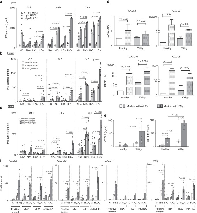
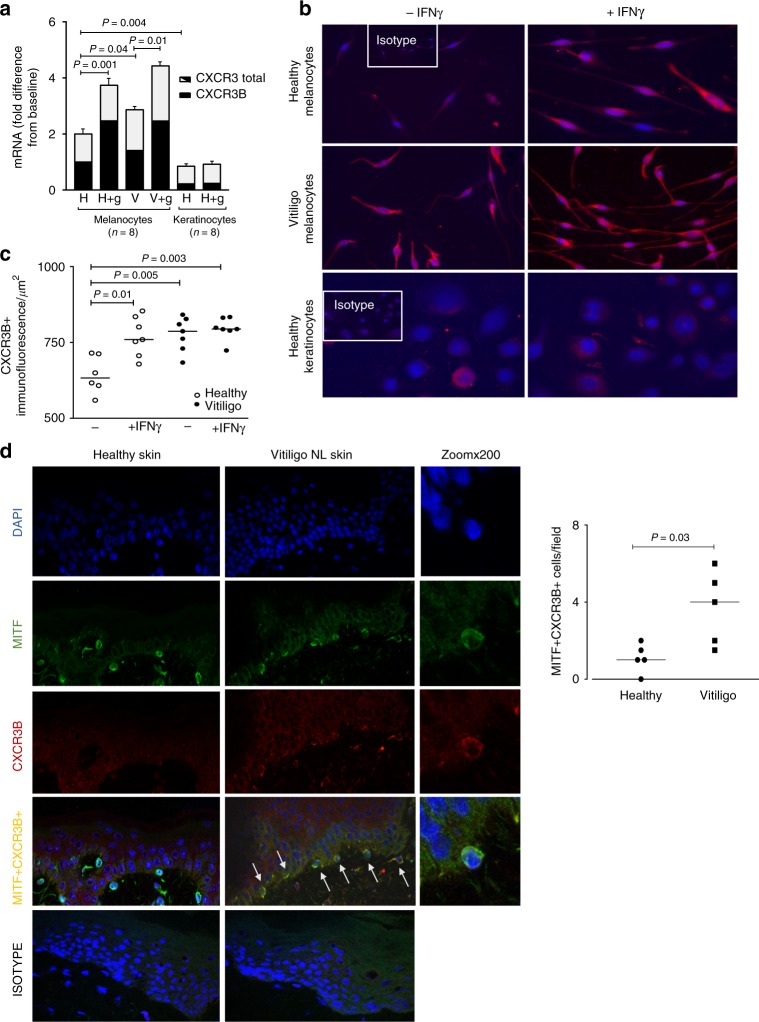
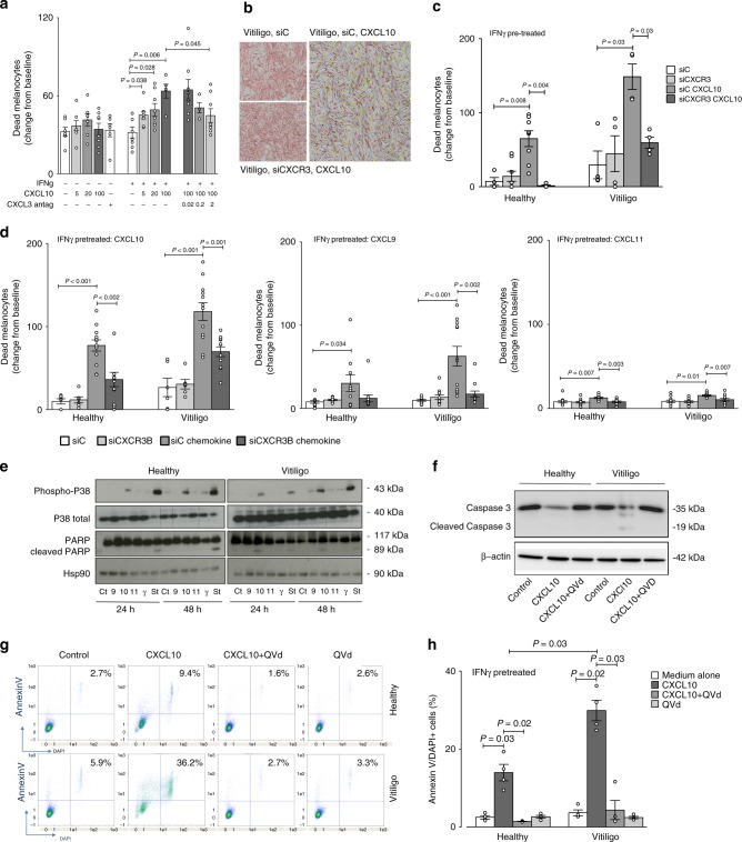
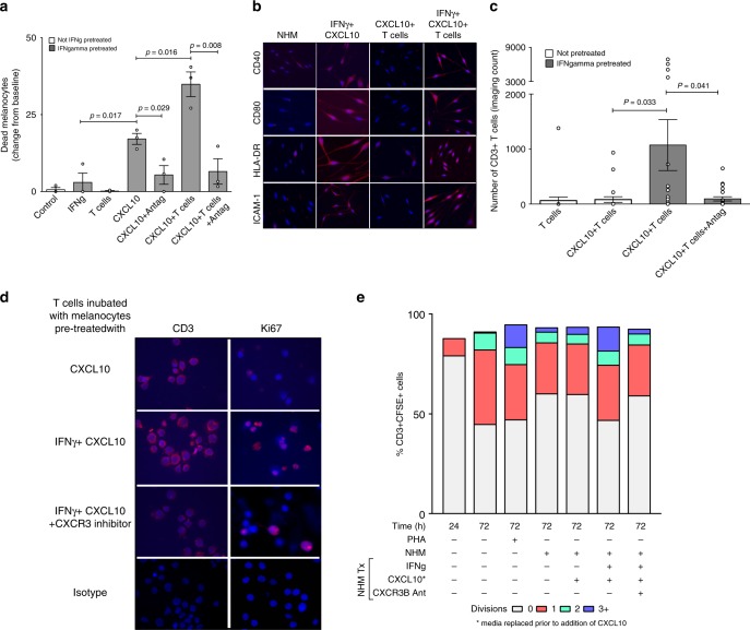
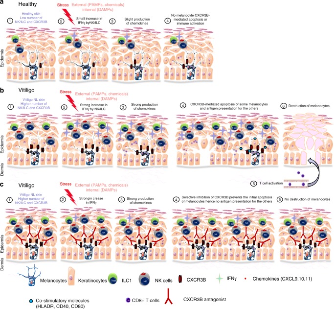
T-cells play a crucial role in progression of autoimmunity, including vitiligo, yet the initial steps triggering their activation and tissue damage remain unknown. Here we demonstrate increased presence of type-1 innate lymphoid cells (NK and ILC1)-producing interferon gamma (IFNγ) in the blood and in non-lesional skin of vitiligo patients. Melanocytes of vitiligo patients have strong basal expression of chemokine-receptor-3 (CXCR3) isoform B which is directly regulated by IFNγ. CXCR3B activation by CXCL10 at the surface of cultured human melanocytes induces their apoptosis. The remaining melanocytes, activated by the IFNγ production, express co-stimulatory markers which trigger T-cell proliferation and subsequent anti-melanocytic immunity. Inhibiting the CXCR3B activation prevents this apoptosis and the further activation of T cells. Our results emphasize the key role of CXCR3B in apoptosis of melanocytes and identify CXCR3B as a potential target to prevent and to treat vitiligo by acting at the early stages of melanocyte destruction.


Conflict of interest statement

The authors declare no competing interests.

Figures

Fig. 1
Fig. 1
Increased detection of innate immune cells in non-lesional skin of vitiligo patients. Immunofluorescence depicting NK cells (a, b, c) and ILC1 (d, e) in the skin and in the peripheral blood (f, g) of vitiligo patients (n = 6) and healthy controls (n = 5). Number of yellow-immunoreactive CD56 + GranzymeB+ NK cells and their semi-quantification in non-lesional (NL) and lesional (L) skin of vitiligo patient compared to healthy skin are shown in a and the number of CD56+IFNγ+NK in b. The presence of NK cells (CD3-CD56+) was confirmed by immunohistochemistry showing cells to be located just under the epidermis of non-lesional (NL) skin (c bottom left and magnified in 1–4) but less in lesional (L) (c top) or healthy skin (c top right). d Depicts Tbet+CD161+ILCs in the skin and their quantification. In the peripheral blood total NK and separately, cytokine producing NKs (CD56bright) and cytotoxic NKs (CD56dim) were examined in 6 healthy and 11 vitiligo patients and results represented as % of total PBMC (e). The percentage of ILC1, ILC2 and ILC3 in the blood of healthy (n = 8) and vitiligo (n = 10) patients is shown in f. Results are shown as individual dot plots with a line at median (ad) or as means ± SEM (e, f)
Fig. 2
Fig. 2
Effect of stressed innate immune cells on melanocyte function. IFNγ production by sorted NK and ILCs from blood of healthy control (c, n = 6–10) and vitiligo (v, n = 6–10) subjects following in vitro stimulation with H2O2 (0.1–10 μM) (a), HMGB1 (250–1000 ng/ml) (b) or HSP70 (TKD, 250–1500 ng/ml) (c) for 24, 48 or 72 h. The effect of exogenous IFNγ (50 ng/ml) on CXCL4, CXCL9, CXCL10, CXCL11 mRNA (d) and CXCL9, CXCL10 protein (e) production by healthy (n = 4–7) and vitiligo melanocytes (n = 7–9). In f chemokine and IFNγ production was measured 24 h post stimulation of healthy (white bars, n = 4) or vitiligo (grey bars, n = 4) melanocytes with patient’s own autologous NK or ILCs (alone or in combination) which were pre-stressed with H2O2 for 48 h before addition of innate cells to patients own melanocytes. Positive control condition represents melanocytes directly pre-stimulated with IFNγ (50 ng/ml) for the same duration of time. PCR results are normalized to house-keeping gene SB and expressed as fold change in expression relative to the pool of healthy skin samples. Results are shown as individual dot plots with a line either at median (ac) or at mean ± SEM (e, f)
Fig. 3
Fig. 3
Expression of CXCR3B in human melanocytes and their regulation by IFNγ. a Total CXCR3 mRNA (black + white bars) and CXCR3B mRNA (black bars) in healthy and vitiligo primary melanocytes (n = 8) and healthy keratinocytes (n = 5) before and after exposure to IFNγ 50 ng/ml for 24 h. Results are normalized to unstimulated melanocytes from healthy subjects and expressed as mean ± SEM. b Immuno-detection of CXCR3B protein and c its semi-quantification in melanocytes extracted from healthy skin (open circles, n = 6–7) and skin from vitiligo patients (closed circles, n = 7) before and after IFNγ stimulation. d In situ detection of CXCR3B+ melanocytes (MITF+CXCR3B+) in vitiligo non-lesional (NL) skin and their quantification (n = 5) compared to healthy skin (n = 5). Results are shown as individual dot plots with a line at median
Fig. 4
Fig. 4
CXCL10 activates melanocytic CXCR3B to induce apoptosis. a Effect of CXCL10 (5, 20, 100 pg/ml) on cell viability in unstimulated or IFNγ stimulated (50 ng/ml for 48 h) melanocytes from vitiligo patients, in presence or absence of CXCR3 antagonist AS612568 (0.02 μM, 0.2 μM or 2 μM) (n = 5–8). Cell viability was monitored using lncuCyte® live cell fluorescence imaging system. b Illustrates live IncuCyte images of vitiligo melanocytes 24 h after exposure to CXCL10 in cells transfected with siC or siCXCR3. Melanocytes were tracked with CellTrackerTM Red CMPTX dye and dead cells tracked with IncuCyte® Cytotox Green reagent. Co-localised yellow cells represent dead melanocytes. The effect of siCXCR3 (or its siC) on CXCL10 (100 pg/ml)-induced death of healthy (n = 4–8) and vitiligo (n = 4) melanocytes are shown in c. In d healthy and vitiligo melanocytes (n = 6–12) were transfected with siCXCR3B (or its siC) and melanocyte death shown at 24 h following CXCL10, CXCL9 or CXCL11 (100 pg/ml) stimulation. In separate experiments, cell lysates was used to study the signalling pathway induced by chemokines, IFNγ (50 ng/ml) or Staurosporine (positive control, 1 μg/ml) at 24 or 48 h post stimulation, measuring the expression levels of phosphorylated and total p38 and total and cleaved poly(ADP-ribose) polymerase (PARP) by Western Blot analysis (e). HSP90 was used as an internal loading control. Representative blot of 3 separate experiments is shown. Total and cleaved caspase-3 activity (f) and proportion of apoptotic cells (counted as % Annexin V+DAPI+cells in FACS analysis) (g) from healthy (n = 4) and vitiligo (n = 4) melanocytes stimulated with 100 pg/ml CXCL10 for 24 h in presence or absence of QVd OPh (10 μM, caspase inhibitor) (h). Results are shown as individual dot plots with a line at mean ± SEM
Fig. 5
Fig. 5
T cells enhance CXCL10-induced melanocyte death by induction of adaptive immunity. a CXCL10-induced death of vitiligo melanocytes in presence or absence of patients own autologous T cells. As above, IFNγ-pretreated melanocytes were exposed to CXCL10 in presence or absence of CXCR3 antagonist AS612568 (2 μM). The next day, media was replaced and 3 days later patient’s own CD3+ T cell were sorted and added to the melanocytes (n = 3) prior to initiation of IncuCyte. b At the end of the experiment (~48 h), supernatant was collected, remaining cells trypsinised and cytospin sections prepared for immunofluorescence detection of co-stimulatory (CD40, CD80, HLA-DR) and adhesion (ICAM-1) molecules on melanocytes. c T cell-induced potentiation of melanocyte death in IFNγ-pretreated melanocytes (compared to untreated melanocytes) was associated with a parallel increase in the number of CD3+ T cells which was supported by increased expression of Ki67+cells in the same cytospin sections d. Results are shown as individual dot plots with a line at mean ± SEM. e T cell proliferation was quantified by flow analysis measuring the percentage of CD3+CSFE+ cells undergoing 0, 1, 2 or 3+divisions (n = 3). Labelled cells at time zero was used as a negative reference, unstimulated cells left in culture for 72 h before labelling as a control and cells stimulated with PHA for 72 h (Phytohemagglutinin, 5 μg/ml) as a positive control
Fig. 6
Fig. 6
Initial steps in the anti-melanocytic immune activation in vitiligo. a Schematic representation of normal skin. A low number of NK and ILC1 cells are present in the skin. External stress on the skin induces only a small increase in IFNγ by NK/ILC which is not sufficient to initiate their chemokines production, hence not initiating melanocyte apoptosis or immune activation. b Schematic representation of vitiligo skin. The basal number of NK and ILC1 cells is increased. Damage-associated molecular pattern (DAMP) molecules of genetically predisposed melanocytes can activate ILC1 and NK (1). Alternatively, ILC1 and NK can be also activated by external stress and pathogen-associated molecular pattern molecules (PAMP) (2). These NK and ILC cells are more sensitive to stress compared to healthy individuals and in response, they produce large amount of IFNγ (2) that induces the secretion of chemokines by keratinocytes and to a lesser extent by melanocytes (3). These chemokines (mostly CXCL10), bind chemokine receptor CXCR3B at the surface of melanocytes and induces their initial apoptosis and release of melanocytic antigens. The remaining melanocytes stimulated by IFNγ express co-stimulatory (CD40, CD80, HLA-DR) and adhesion (ICAM-1) molecules and start presenting their own antigens to the naive T cells attracted by the chemokines (5), thus initiating the initial steps involved in anti-melanocytic immunity in the skin (6). c Schematic representation of vitiligo skin treated with a selective inhibition of the B isoform of CXCR3. DAMPs and PAMPs induce a strong production of IFNγ by NK and ILC1 cells followed by a potent production of chemokines. However, the selective inhibition of CXCR3B prevents the initial melanocytic apoptosis and subsequent presentation of melanocytic antigens without impairing T cell function

References

    1. Spritz RA. Six decades of vitiligo genetics: genome-wide studies provide insights into autoimmune pathogenesis. J. Invest. Dermatol. 2012;132:268–273. doi: 10.1038/jid.2011.321. - DOI - PMC - PubMed
    1. Jin Y, et al. Genome-wide association analyses identify 13 new susceptibility loci for generalized vitiligo. Nat. Genet. 2012;44:676–680. doi: 10.1038/ng.2272. - DOI - PMC - PubMed
    1. Jin Y, et al. Genome-wide association studies of autoimmune vitiligo identify 23 new risk loci and highlight key pathways and regulatory variants. Nat. Genet. 2016;48:1418–1424. doi: 10.1038/ng.3680. - DOI - PMC - PubMed
    1. Chatterjee S, et al. A quantitative increase in regulatory T cells controls development of vitiligo. J. Invest. Dermatol. 2014;134:1285–1294. doi: 10.1038/jid.2013.540. - DOI - PMC - PubMed
    1. Mosenson JA, et al. Mutant HSP70 reverses autoimmune depigmentation in vitiligo. Sci. Transl. Med. 2013;5:174ra128. doi: 10.1126/scitranslmed.3005127. - DOI - PMC - PubMed

Publication types

MeSH terms