. 2020 Jan;27(1):146-160.
doi: 10.1038/s41418-019-0345-2. Epub 2019 May 16.

Sam50-Mic19-Mic60 axis determines mitochondrial cristae architecture by mediating mitochondrial outer and inner membrane contact

Affiliations

Sam50-Mic19-Mic60 axis determines mitochondrial cristae architecture by mediating mitochondrial outer and inner membrane contact

Junhui Tang et al. Cell Death Differ. 2020 Jan.

Abstract

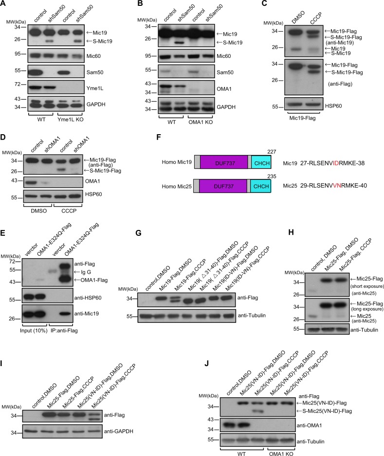
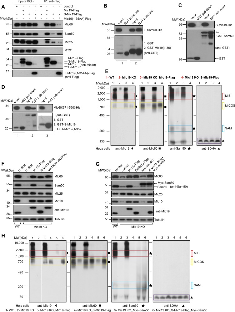
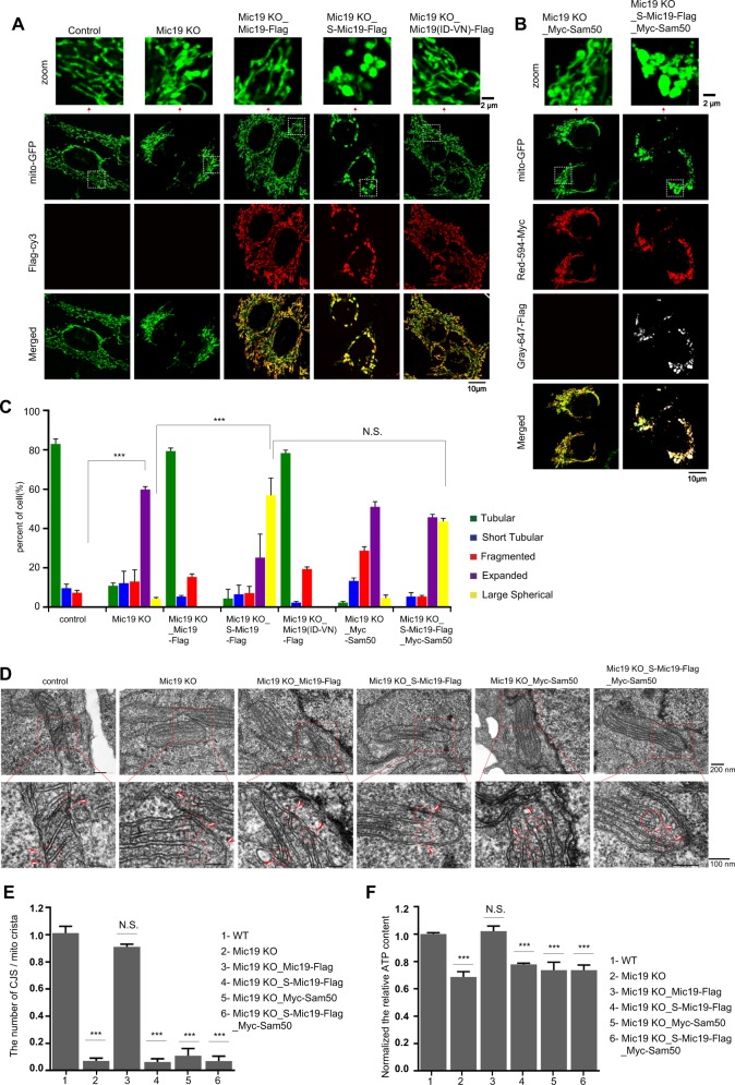
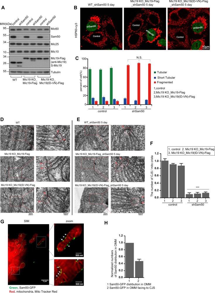
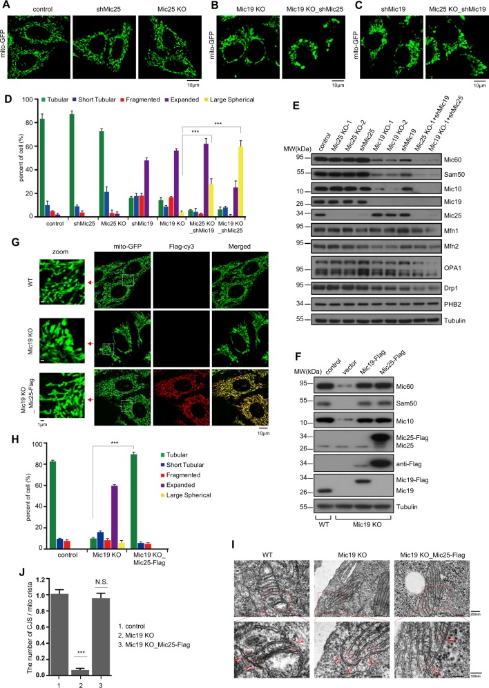
Mitochondrial cristae are critical for efficient oxidative phosphorylation, however, how cristae architecture is precisely organized remains largely unknown. Here, we discovered that Mic19, a core component of MICOS (mitochondrial contact site and cristae organizing system) complex, can be cleaved at N-terminal by mitochondrial protease OMA1 under certain physiological stresses. Mic19 directly interacts with mitochondrial outer-membrane protein Sam50 (the key subunit of SAM complex) and inner-membrane protein Mic60 (the key component of MICOS complex) to form Sam50-Mic19-Mic60 axis, which dominantly connects SAM and MICOS complexes to assemble MIB (mitochondrial intermembrane space bridging) supercomplex for mediating mitochondrial outer- and inner-membrane contact. OMA1-mediated Mic19 cleavage causes Sam50-Mic19-Mic60 axis disruption, which separates SAM and MICOS and leads to MIB disassembly. Disrupted Sam50-Mic19-Mic60 axis, even in the presence of SAM and MICOS complexes, causes the abnormal mitochondrial morphology, loss of mitochondrial cristae junctions, abnormal cristae distribution and reduced ATP production. Importantly, Sam50 displays punctate distribution at mitochondrial outer membrane, and acts as an anchoring point to guide the formation of mitochondrial cristae junctions. Therefore, we propose that Sam50-Mic19-Mic60 axis-mediated SAM-MICOS complexes integration determines mitochondrial cristae architecture.


Conflict of interest statement

The authors declare that they have no conflict of interest.

Figures

Fig. 1
Fig. 1
Mic19 can be cleaved by mitochondrial protease OMA1. a-b Wild-type (WT), Yme1L KO (a) or OMA1 KO (b) HCT116 cells were infected by retrovirus particles containing control or shSam50, and further cultured for 7 days. Cell lysates were analyzed by Western blotting using the indicated antibodies. c HeLa cells stably expressing Human Mic19-Flag were treated with DMSO or CCCP (20 μM, 4 h). Cell lysates were analyzed by Western blotting with anti-Flag, anti-Mic19, or anti-HSP60 antibodies. The black arrowhead indicates the specific protein. d HeLa cells stably expressing Mic19-Flag were infected by lentivirus particles containing control or shOMA1, and cultured for an additional 5 days. Cells were then treated with DMSO or CCCP (20 μM) for 4 h. Mic19-Flag cleavage was examined by Western blotting using anti-Flag antibodies. e 293 T cells were transiently transfected with control (empty vector) or plasmid coding for OMA1E324Q-Flag (which glutamic acid (E) at residue 324 was substituted by glutamine (Q) that leads to OMA1 catalytically dead). After 48 h transfection, the cells were harvested and solubilized with IP lysis buffer, and cell lysates were then immunoprecipitated with anti-Flag M2 affinity gel, followed by Western blotting using the indicated antibodies. f The schematic presentation of human Mic19 and Mic25 domain. ‘DUF737’ indicates the domain of unknown function; ‘CHCH’ indicates the coiled-coil helix-coiled-coil helix domain. The comparison of N-terminal 27–38 amino acids (AA) of Mic19 and N-terminal 29–40AA of Mic25 were showed on the right. g HeLa cells stably expressing Mic19-Flag and the indicated Mic19 mutants (Flag-tagged) were treated with DMSO or CCCP (20 μM, 4 h), and cell lysates were then analyzed by Western blotting using anti-Flag or anti-Tubulin antibodies. “Δ” indicates deletion of residues. h-j HeLa cells expressing Mic25-Flag (h) or the indicated Mic25 mutants (i), and WT or OMA1 KO HCT116 cells expressing Mic25 mutants (j) were treated with DMSO or CCCP (20 μM, 4 h). Cell lysates were then analyzed by Western blotting using the indicated antibodies
Fig. 2
Fig. 2
Mic19 cleavage disrupts Sam50-Mic19-Mic60 axis and disassembles the MIB supercomplex. a Cell lysates of 293 T cells expressing control (empty vector), Mic19-Flag, S-Mic19-Flag, and Mic19 (1-35aa)-Flag were used for co-immunoprecipitation (co-IP) assay with anti-Flag M2 resin, and the protein samples were subjected to immunoblot using the indicated antibodies. bd GST or His fused proteins were expressed in E. coli, cell extracts were used for GST pull-down assay by using the Pierce Glutathione Agarose, and the protein samples were analyzed by Western blotting using antibodies against GST or His. The examination of the direct interaction between Mic19 (1-35AA) and Sam50 (b). The demonstration of the direct interaction between S-Mic19 and Sam50 (c). The detection of direct interaction between Mic19 (1-35AA) or S-Mic19 with Mic60 (371-590AA) (d). e Mitochondria were extracted from WT, Mic19 KO, or Mic19ΔgRNA-Flag or S-Mic19ΔgRNA-Flag expressed Mic19 KO HeLa cells, then non-denatured protein samples from mitochondria were analyzed for MIB, MICOS and SAM complexes by BN-PAGE and Western blotting using indicated antibodies. The bands of the complexes are labeled with corresponding boxes. The MIB complex is approximately 2000kDa, and the MICOS is ~700 kDa. SAM complex is approximately 250 kDa, ~ 125 kDa unidentified complex containing Sam50 is showed. Mitochondrial supercomplex II is detected by anti-SDHA, served as loading control. “Mic19∆gRNA” is a Mic19 mutation (2 nucleotides of Mic19 cDNA were mutated, but the amino acid sequences of Mic19 are not changed) in the sequence of guide RNA (gRNA) against Mic19 gene. Mic19∆gRNA could not be recognized by the Mic19 KO Cas9/CRISPR system. f Control (empty vector), Mic19ΔgRNA-Flag, S-Mic19ΔgRNA-Flag, and Mic19(ID33-34VN)ΔgRNA-Flag were stably expressed in Mic19 KO HeLa cells respectively. Cell lysates were analyzed by Western blot using indicated antibodies. g Cell lysates of WT and Mic19 KO HeLa cells expressing the indicated protein were analyzed for expression of MICOS subunits by Western blotting using the indicated antibodies. h Mitochondria were extracted from indicated cells, BN-PAGE and Western blotting analysis for the related cell lines, the non-denatured protein samples from mitochondria were analyzed for MIB, MICOS and SAM complexes by BN-PAGE and Western blotting. The bands of the complexes are labeled with corresponding boxes
Fig. 3
Fig. 3
Disruption of Sam50-Mic19-Mic60 axis leads to abnormal mitochondrial morphology and loss of crista junctions. a Mic19 KO HeLa cells stably expressing mito-GFP (a mitochondrial marker) were infected with retrovirus particles containing empty vector, Mic19ΔgRNA-Flag, S-Mic19ΔgRNA-Flag, and Mic19(ID33-34VN)ΔgRNA-Flag respectively. Cells were then immunostained with anti-Flag antibody, and mitochondrial morphology was visualized by confocal microscope. Local magnifications of all morphological graphs were shown on the top panel. b Myc-Sam50, or S-Mic19ΔgRNA-Flag plus Myc-Sam50 were expressed in Mic19 KO HeLa cells stably expressing mito-GFP, respectively. Cells were then immunostained with anti-Flag or anti-Myc antibodies, Mitochondrial morphology was visualized by confocal microscope. c Mitochondrial morphology described in “A and B” was counted according to the criteria detailed in“Materials and Methods”. All data represent the means ± SD of three independent experiments (100 cells per independent experiment). Statistical significance was assessed by student’s t-test, ***P < 0.001 and N.S indicates none significance. d Mitochondrial cristae junctions structure in WT, Mic19 KO, and Mic19 KO HeLa cells expressing Mic19-Flag, S-Mic19-Flag, Myc-Sam50 or S-Mic19-Flag plus Myc-Sam50 were analyzed by transmission electron microscope (TEM). The ultrathin section sample was observed using an electron microscope (JEM-1400 plus, Tokyo, Japan) under the condition of an accelerating voltage of 100 kV. e Statistics of the mitochondrial ultrastructure in the cells described in (d). Error bars represent means ± SD of three independent experiments in which 100 mitochondrial cristae and the number of the corresponding CJs were counted. ***P < 0.001 vs WT. f The relative ATP level of WT, Mic19 KO, and Mic19 KO HeLa cells expressing Mic19-Flag, S-Mic19-Flag, Myc-Sam50 or S-Mic19-Flag plus Myc-Sam50 were measured using an ATP assay kit. Error bars represent means ± SD of three independent experiments, ***P < 0.001 vs WT
Fig. 4
Fig. 4
Sam50 may act as an anchoring point for Sam50-Mic19-Mic60 axis to guide the formation of mitochondrial crista junctions. a Knockdown of Sam50 (shSam50, 5 days) in WT, Mic19 KO, and Mic19ΔgRNA-Flag or Mic19 (ID33-34VN)ΔgRNA-Flag expressed Mic19 KO HeLa cells were performed by infecting cells with control or shSam50 lentiviral particles, cell lysates were then analyzed by Western blotting using indicated antibodies. Tubulin was used as loading control. b WT, Mic19ΔgRNA-Flag, or Mic19 (ID33-34VN)ΔgRNA-Flag expressed Mic19 KO HeLa cells were infected with control or shSam50 lentiviral particles. 5 days after infection, shSam50 and control cells were then mixed and grown on the same glass slide, and were fixed and immunostained with anti-HSP60 antibody, mitochondrial morphology was visualized by confocal microscope. shSam50 (shRNAi against Sam50) plasmid, which also encodes a nuclear localization signal fused GFP (nu-GFP), was used to deplete Sam50, and empty vector without nu-GFP was acted as control. Nu-GFP positive cells indicate “shSam50 cells”, and cells without nu-GFP expression indicate“control cells”. c Mitochondrial morphology of indicated cell lines was classified into three types (tubular, short tubular, and fragmented) and counted. Bars represent means ± S.D. of three independent experiments. d-e The mitochondrial cristae junction structures of WT, Mic19ΔgRNA-Flag or Mic19 (ID33-34VN)ΔgRNA-Flag expressed Mic19 KO HeLa cells with or without Sam50 knockdown were analyzed by transmission electron microscope (TEM). f Statistics of the mitochondrial ultrastructure in the indicated cell lines. The mean value and standard deviations (S.D.) were calculated from 3 independent experiments in which 100 mitochondrial cristae and the number of corresponding CJs were counted, ***P < 0.001. g Sam50-GFP transiently expressed COS7 cells were stained with MitoTracker® Red CMXRos dye for labeling mitochondria, and the living cells were visualized and imaged with Hessian-SIM operating at 2D-SIM configuration. A stack of SIM images was processed by Image J software. Boxed regions in the left image are magnified and shown on the right panel. The white arrowhead indicated the co-localization of Sam50-GFP with crista junctions. h Quantification of Sam50-GFP signals in OMM over against to the CJs structure. 300 Sam50-GFP signals were counted, and the relative number of the co-localization of Sam50-GFP with crista junctions were shown
Fig. 5
Fig. 5
Restoration of Sam50-X-Mic60 axis by overexpression of Mic25 in Mic19 KO cells can reconstruct mitochondrial morphology and crista junctions. ac WT (a), Mic19 KO (b), or Mic25 KO (c) HeLa cells expressing mito-GFP were knocked down for Mic19 (shMic19) or Mic25 (shMic25) respectively, and mitochondrial morphology was analyzed by confocal microscope. d Mitochondrial morphology in cell lines described in “a-c” was classified and counted according to the criteria detailed in “Materials and Methods”, and error bars represent means ± S.D. of three independent experiments, ***P < 0.001. e Cell lysates of the indicated cells described in “a-c” were analyzed by Western blotting using the indicated antibodies. Tubulin protein was used as loading control. f Cell lysates of WT, Mic19 KO, and Mic19-Flag or Mic25-Flag overexpressed Mic19 KO HeLa cells were analyzed for protein levels by Western blotting using the indicated antibodies. g Mic25-Flag was stably overexpressed in Mic19 KO cells expressing mito-GFP, and immunostained with anti-Flag antibody. Mitochondrial morphology in WT, Mic19 KO, and Mic25-Flag overexpressed Mic19 KO HeLa cells was assessed by confocal microscope. h Statistical analysis of mitochondrial morphology in the cells described in “g” was performed according to the criteria detailed in “Materials and Methods”, the mean value and standard deviations (S.D.) were calculated from 3 independent experiments in which 100 cells were scored, ***P < 0.001. i Mitochondrial crista junction structure of WT, Mic19 KO, or Mic25 overexpressed Mic19 KO HeLa cells were analyzed by transmission electron microscope (TEM). j Statistics of the mitochondrial ultrastructure in the indicated cells described in “h”. The mean value and standard deviations (S.D.) were calculated from 3 independent experiments in which 100 mitochondrial cristae and the number of corresponding CJs were counted, ***P < 0.001

Similar articles

Cited by

References

    1. Mannella CA. Structure and dynamics of the mitochondrial inner membrane cristae. Biochim Biophys Acta. 2006;1763:542–8. - PubMed
    1. Demongeot J, Glade N, Hansen O, Moreira A. An open issue: the inner mitochondrial membrane (IMM) as a free boundary problem. Biochimie. 2007;89:1049–57. - PubMed
    1. Huynen MA, Muhlmeister M, Gotthardt K, Guerrero-Castillo S, Brandt U. Evolution and structural organization of the mitochondrial contact site (MICOS) complex and the mitochondrial intermembrane space bridging (MIB) complex. Biochim Biophys Acta. 2016;1863:91–101. - PubMed
    1. van der Laan M, Horvath SE, Pfanner N. Mitochondrial contact site and cristae organizing system. Curr Opin Cell Biol. 2016;41:33–42. - PubMed
    1. Rabl R, Soubannier V, Scholz R, Vogel F, Mendl N, Vasiljev-Neumeyer A, et al. Formation of cristae and crista junctions in mitochondria depends on antagonism between Fcj1 and Su e/g. J Cell Biol. 2009;185:1047–63. - PMC - PubMed

Publication types

Substances