. 2019 Jul;24(4):749-761.
doi: 10.1007/s12192-019-01001-2. Epub 2019 May 16.

Expression and localization of heat-shock proteins during skeletal muscle cell proliferation and differentiation and the impact of heat stress

Affiliations

Expression and localization of heat-shock proteins during skeletal muscle cell proliferation and differentiation and the impact of heat stress

Savant S Thakur et al. Cell Stress Chaperones. 2019 Jul.

Abstract

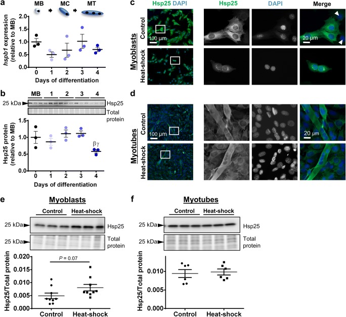
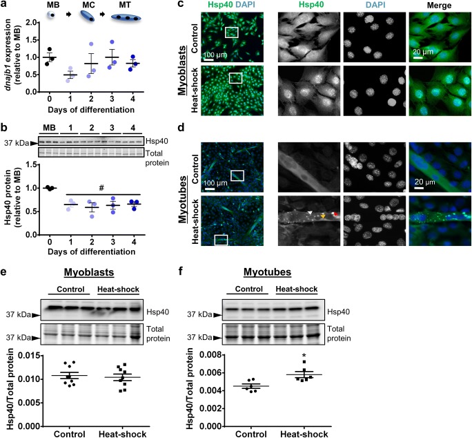
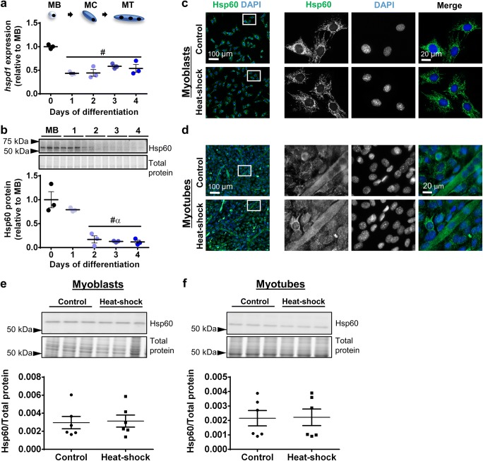
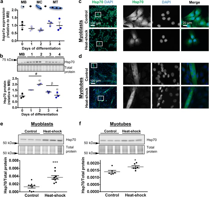
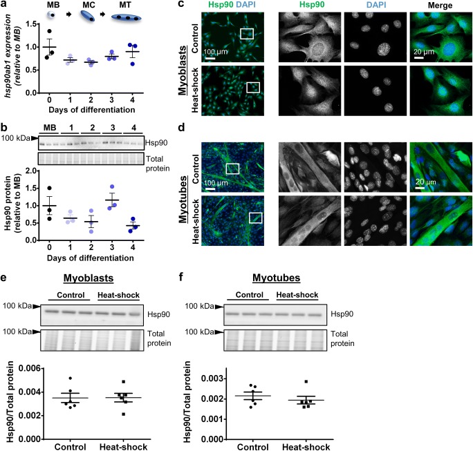
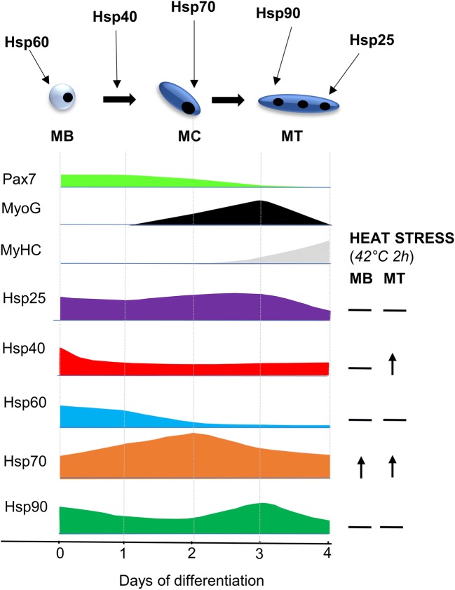
Skeletal myogenesis is a coordinated sequence of events associated with dramatic changes in cell morphology, motility, and metabolism, which causes cellular stress and alters proteostasis. Chaperones, such as heat-shock proteins (HSPs), play important roles in limiting cellular stresses and maintaining proteostasis, but whether HSPs are specifically involved in myogenesis is not well understood. Here, we characterized gene and protein expression and subcellular localization of various HSPs in proliferating C2C12 myoblasts and differentiating myotubes under control conditions and in response to heat stress. Hsp25, Hsp40, and Hsp60 protein expression declined by 48, 35, and 83%, respectively, during differentiation. In contrast, Hsp70 protein levels doubled during early differentiation. Hsp25 was predominantly localized to the cytoplasm of myoblasts and myotubes but formed distinct aggregates in perinuclear spaces of myoblasts after heat-shock. Hsp40 was distributed diffusely throughout the cytoplasm and nucleus and, after heat-shock, translocated to the nucleus of myoblasts but formed aggregates in myotubes. Hsp60 localized to the perinuclear space in myoblasts but was distributed more diffusely across the cytoplasm in myotubes. Hsp70 was expressed diffusely throughout the cytoplasm and nucleus and translocated to the nucleus after heat-shock in myoblasts, but not in myotubes. Hsp90 was expressed diffusely across the cytoplasm in both myoblasts and myotubes under control conditions and did not change in response to heat-shock. These findings reveal distinct and different roles for HSPs in the regulation of myogenic cell proliferation and differentiation.

Keywords: C2C12; Heat-shock proteins; Molecular chaperones; Muscle development; Myogenesis; Skeletal muscle.


Figures

Fig. 1
Fig. 1
Hsp25 gene expression, protein expression, and subcellular localization in proliferating and differentiating C2C12 cells. a The mRNA expression of gene encoding Hsp25 (hspb1) was examined by semi-quantitative real-time PCR (qPCR) in proliferating myoblasts (MB or day 0) and C2C12 cells differentiated for 1–4 days. Gene transcript levels were normalized to cDNA content and expressed relative to MB levels. b Representative Western blot showing protein expression of Hsp25 in MB and at days 1–4. Densitometry values were normalized to total protein and expressed relative to MB levels. Individual data points and the mean (horizontal line) ± SEM shown; n = 3 replicates/timepoint. βP < 0.05 vs. day 2; γP < 0.05 vs. day 3. cd Representative immunofluorescence images of C2C12 myoblasts (c) and myotubes (d) stained with DAPI and endogenous Hsp25 in the absence of (control; top row) and after heat-shock treatment at 42 °C for 2 h (bottom row). Arrow heads indicate a subpopulation of cells with low Hsp25 expression. Scale bars as indicated. Boxed regions shown at higher magnification. ef Western blot analysis showing protein expression of Hsp25 in lysates prepared from control and heat-shock-treated myoblasts (e) and myotubes (f). Densitometry values were normalized to total protein content assessed from the stain-free gel image. Individual data points and the mean (horizontal line) ± SEM are shown; n = 6–9 replicates/timepoint
Fig. 2
Fig. 2
Hsp40 gene expression, protein expression, and subcellular localization in proliferating and differentiating C2C12 cells. a The mRNA expression of gene encoding Hsp40 (dnajb1) was examined by qPCR in MB and at days 1–4. Gene transcript levels were normalized to cDNA content and expressed relative to MB levels. b Representative Western blot showing protein expression of Hsp40 in MB and at days 1–4. Densitometry values were normalized to total protein and expressed relative to MB levels. Individual data points and the mean (horizontal line) ± SEM shown; n = 3 replicates/timepoint. #P < 0.05 vs. MB. cd Representative immunofluorescence images of C2C12 myoblasts (c) and myotubes (d) stained with DAPI and endogenous Hsp40 in the absence of (control; top row) and after heat-shock treatment at 42 °C for 2 h (bottom row). In heat-shocked myotubes, Hsp40 formed large punctate aggregates (red arrowhead), submyonuclear aggregates (orange arrowhead), and smaller cytoplasmic aggregates (white arrowhead). Scale bars as indicated. Boxed regions shown at higher magnification. ef Western blot analysis showing protein expression of Hsp40 in lysates prepared from control and heat-shock-treated myoblasts (e) and myotubes (f). Densitometry values were normalized to total protein content assessed from the stain-free gel image. Individual data points and the mean (horizontal line) ± SEM are shown; n = 6–9 replicates/timepoint. *P < 0.05 vs. control
Fig. 3
Fig. 3
Hsp60 gene expression, protein expression, and subcellular localization in proliferating and differentiating C2C12 cells. a The mRNA expression of gene encoding Hsp60 (hspd1) was examined qPCR in MB and at days 1–4. Gene transcript levels were normalized to cDNA content and expressed relative to MB levels. b Representative Western blot showing protein expression of Hsp60 in MB and at days 1–4. Densitometry values were normalized to total protein and expressed relative to MB levels. Individual data points and the mean (horizontal line) ± SEM shown; n = 3 replicates/timepoint. #P < 0.05 vs. MB; αP < 0.05 vs. day 1. cd Representative immunofluorescence images of C2C12 myoblasts (c) and myotubes (d) stained with DAPI and endogenous Hsp60 in the absence of (control; top row) and after heat-shock treatment at 42 °C for 2 h (bottom row). Scale bars as indicated. Boxed regions shown at higher magnification. ef Western blot analysis showing protein expression of Hsp60 in lysates prepared from control and heat-shock-treated myoblasts (e) and myotubes (f). Densitometry values were normalized to total protein content assessed from the stain free gel image. Individual data points and the mean (horizontal line) ± SEM are shown; n = 6 replicates/timepoint
Fig. 4
Fig. 4
Hsp70 gene expression, protein expression, and subcellular localization in proliferating and differentiating C2C12 cells. a The mRNA expression of gene encoding Hsp70 (hspa1a) was examined by qPCR in MB and at days 1–4. Gene transcript levels were normalized to cDNA content and expressed relative to MB levels. b Representative Western blot showing protein expression of Hsp70 in MB and at days 1–4. Densitometry values were normalized to total protein and expressed relative to MB levels. Individual data points and the mean (horizontal line) ± SEM shown; n = 3 replicates/timepoint. #P < 0.05 vs. MB; αP < 0.05 vs. day 1; βP < 0.05 vs. day 2. cd Representative immunofluorescence images of C2C12 myoblasts (c) and myotubes (d) stained with DAPI and endogenous Hsp70 in the absence of (control; top row) and after heat-shock treatment at 42 °C for 2 h (bottom row). Scale bars as indicated. Boxed regions shown at higher magnification. ef Western blot analysis showing protein expression of Hsp70 in lysates prepared from control and heat-shock-treated myoblasts (e) and myotubes (f). Densitometry values were normalized to total protein content assessed from the stain free gel image. Individual data points and the mean (horizontal line) ± SEM are shown; n = 6–9 replicates/timepoint. *P < 0.05, ***P < 0.001 vs. control
Fig. 5
Fig. 5
Hsp90 gene expression, protein expression, and subcellular localization in proliferating and differentiating C2C12 cells. a The mRNA expression of gene encoding Hsp90 (hsp90ab1) was examined by qPCR in MB and at days 1–4. Gene transcript levels were normalized to cDNA content and expressed relative to MB levels. b Representative Western blot showing protein expression of Hsp90 in MB and at days 1–4. Densitometry values were normalized to total protein and expressed relative to MB levels. Individual data points and the mean (horizontal line) ± SEM shown; n = 3 replicates/timepoint. cd Representative immunofluorescence images of C2C12 myoblasts (c) and myotubes (d) stained with DAPI and endogenous Hsp90 in the absence of (control; top row) and after heat-shock treatment at 42 °C for 2 h (bottom row). Scale bars as indicated. Boxed regions shown at higher magnification. ef Western blot analysis showing protein expression of Hsp90 in lysates prepared from control and heat-shock-treated myoblasts (e) and myotubes (f). Densitometry values were normalized to total protein content assessed from the stain-free gel image. Individual data points and the mean (horizontal line) ± SEM are shown; n = 6 replicates/timepoint
Fig. 6
Fig. 6
Coordinated expression of HSPs during myogenesis under control and heat-shock conditions. Protein expression profile of Hsp25, Hsp40, Hspo60, Hsp70, and Hsp90 in relation to myogenic markers and each other in myoblasts (MB) and during days 1–4 of cell differentiation. Figure depicts phases of myogenesis with proposed Hsp involvement and changes in Hsps with heat stress

References

    1. Bentzinger CF, Wang YX, Rudnicki MA. Building muscle: molecular regulation of myogenesis. Cold Spring Harb Perspect Biol. 2012;4:a008342. doi: 10.1101/cshperspect.a008342. - DOI - PMC - PubMed
    1. Botos J, Xian W, Smith DF, Smith CL. Progesterone receptor deficient in chromatin binding has an altered cellular state. J Biol Chem. 2004;279:15231–15239. doi: 10.1074/jbc.M309718200. - DOI - PubMed
    1. Bruey JM, Ducasse C, Bonniaud P, Ravagnan L, Susin SA, Diaz-Latoud C, Gurbuxani S, Arrigo AP, Kroemer G, Solary E, Garrido C. Hsp27 negatively regulates cell death by interacting with cytochrome c. Nat Cell Biol. 2000;2:645–652. doi: 10.1038/35023595. - DOI - PubMed
    1. Cheng H, Cenciarelli C, Nelkin G, Tsan R, Fan D, Cheng-Mayer C, Fidler IJ. Molecular mechanism of hTid-1, the human homolog of Drosophila tumor suppressor l(2)Tid, in the regulation of NF-kappaB activity and suppression of tumor growth. Mol Cell Biol. 2005;25:44–59. doi: 10.1128/MCB.25.1.44-59.2005. - DOI - PMC - PubMed
    1. Cloutier P, Coulombe B. Regulation of molecular chaperones through post-translational modifications: decrypting the chaperone code. Biochim Biophys Acta. 2013;1829:443–454. doi: 10.1016/j.bbagrm.2013.02.010. - DOI - PMC - PubMed

Publication types

MeSH terms

Substances