. 2020 Jan;30(1):36-45.
doi: 10.1111/bpa.12734. Epub 2019 Jun 5.

Aβ oligomers trigger and accelerate Aβ seeding

Affiliations

Aβ oligomers trigger and accelerate Aβ seeding

Natalie Katzmarski et al. Brain Pathol. 2020 Jan.

Abstract

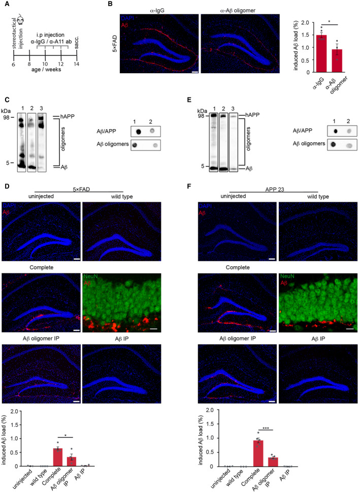
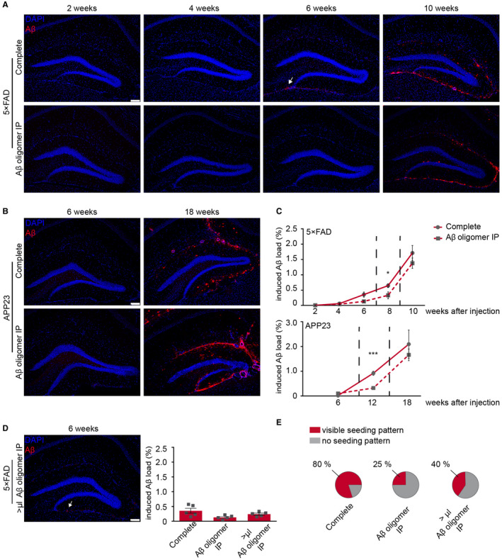
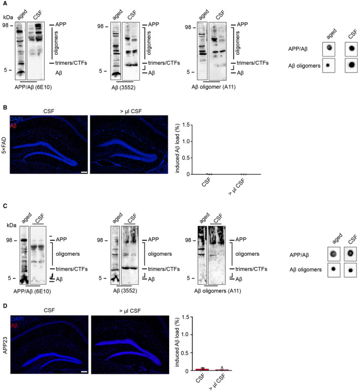
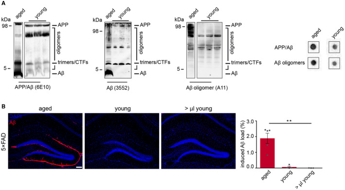
Aggregation of amyloid-β (Aβ) that leads to the formation of plaques in Alzheimer's disease (AD) occurs through the stepwise formation of oligomers and fibrils. An earlier onset of aggregation is obtained upon intracerebral injection of Aβ-containing brain homogenate into human APP transgenic mice that follows a prion-like seeding mechanism. Immunoprecipitation of these brain extracts with anti-Aβ oligomer antibodies or passive immunization of the recipient animals abrogated the observed seeding activity, although induced Aβ deposition was still evident. Here, we establish that, together with Aβ monomers, Aβ oligomers trigger the initial phase of Aβ seeding and that the depletion of oligomeric Aβ delays the aggregation process, leading to a transient reduction of seed-induced Aβ deposits. This work extends the current knowledge about the role of Aβ oligomers beyond its cytotoxic nature by pointing to a role in the initiation of Aβ aggregation in vivo. We conclude that Aβ oligomers are important for the early initiation phase of the seeding process.

Keywords: Alzheimer's disease; Aβ oligomers; Aβ seeding; amyloid-β plaques.


Figures

Figure 1
Figure 1
Reduction of Aβ oligomers diminishes exogenously induced Aβ deposition. A. Schematic representation of the passive immunization paradigm. 6‐week‐old 5×FAD transgenic mice were intracerebrally injected with Aβ‐containing brain homogenate (10% w/v) from aged mice, followed by weekly intraperitoneal administration of either A11 antibody or IgG for 5 weeks. B. Immunofluorescence staining against Aβ with antibody 3552 (red) and quantification of hippocampal‐induced Aβ load revealed significantly reduced Aβ load in mice passively immunized with A11 antibody compared to control immunized mice with IgG (n = 3–5 mice per group) Mann–Whitney test: *P = 0.036. C. Immunoblot analysis with Aβ‐specific antibody 6E10 of brain homogenate from 5×FAD that was used for injections. D. Reduction of Aβ oligomers in the injected brain homogenate significantly reduced seed‐induced Aβ deposition in 5×FAD tg mice. E. Immunoblot analysis with Aβ‐specific antibody 6E10 of brain homogenate from APP23 transgenic mice that was used for injections. Note that Aβ oligomers were significantly reduced in the aged brain homogenate after several rounds of immunoprecipitation with A11 antibody (lane 2) and total Aβ after immunoprecipitation with antibody 3552 (lane 3). Representative dot blots confirmed the low abundance of total Aβ/APP and Aβ oligomers in the A11‐depleted brain homogenate. F. Reduction of Aβ oligomers in the injected brain homogenate significantly reduced seed‐induced Aβ deposition in APP23 mice. Injection of wt brain homogenate or Aβ‐depleted brain homogenate failed to induce seeding in 5×FAD and APP23 transgenic mice. Confocal images of NeuN‐positive neurons (green) and Aβ (red) confirm that the seed‐induced Aβ deposits are located extracellularly. Unpaired t‐test for 5×FAD mice (n = 4–5 mice per group): *P = 0.03 and for APP23: ***P = 0.0002. Scale bar in (B, D and F) indicates 100 µm in the overview and 10 µm in the higher magnification images in (D and F).
Figure 2
Figure 2
Seed‐induced Aβ load aligns with longer incubation time. A. 6‐week‐old 5×FAD transgenic mice were injected with complete brain homogenate or Aβ oligomer depleted brain homogenate and incubated for 2, 4, 6 or 10 weeks. First seed‐induced Aβ deposits appeared at 6 weeks post‐injection in mice injected with complete brain homogenate (white arrow). B. 24‐week‐old APP23 transgenic mice were injected with complete brain homogenate or Aβ oligomer‐depleted brain homogenate and incubated for 6 or 18 weeks. C. The induced Aβ load was nearly similar 10 weeks after injection in 5×FAD mice (upper graph) and was not significantly different 18 weeks after injection in APP23 mice (lower graph). The cohort of mice for the 8‐week time point is reused from Figure 1D,F as indicated by the dashed lines. D. The injection of a higher volume (>μL) of A11‐depleted brain homogenate shows an earlier onset of induced Aβ deposition (white arrow) and a slight but not significant increase at 6 weeks post‐injection (n = 5 mice per group; One‐way ANOVA: F(2,12) = 2.495 P = 0.1241; n.s.; Tukey's multiple comparisons test n.s.). E. In 80% of 5×FAD injected with complete brain homogenate a typical seeding pattern was visible compared to only 25% when A11‐depleted homogenate was injected and 40% when a higher volume (>μL) of the latter brain homogenate was intracerebrally injected. Scale bar in (A, B andD) represents 100 μm. Indicated is the mean ± S.E.M.
Figure 3
Figure 3
Seeding capacity of Aβ oligomers from murine CSF. A. CSF from young, 9‐week‐old 5×FAD mice revealed the presence of Aβ oligomers but no monomeric Aβ that was confirmed by dot blot analysis. B. Despite these high levels of oligomeric Aβ in the CSF of young mice, this preparation failed to induce Aβ deposition. C. Immunoblot and dot blot analyses confirmed the abundance of oligomeric Aβ in the CSF of 21‐week‐old APP23 transgenic mice as well (each lane represents a different CSF probe) when compared to aged brain homogenate. D. Undiluted murine CSF from APP23 mice was injected into young, predepositing APP23 transgenic mice and analyzed after a 3‐month incubation period. Neither CSF from APP23 mice nor higher volumes of the CSF revealed any seed‐induced Aβ depositions. Scale bar in (B, D) indicates 100 µm. (B) n(2.5 µL CSF) = 3; n(5 µL CSF) = 3. (D) n(2.5 µL CSF) = 3; n(5 µL CSF) = 4. Indicated is the mean ± S.E.M.
Figure 4
Figure 4
Seeding capacity of Aβ oligomers from young 5×FAD mice. A. Immunoblot analysis using SDS‐PAGE and dot blot assay (right panel) revealed the presence of Aβ oligomers but no monomeric Aβ in brain homogenates of young, 16‐week‐old 5×FAD transgenic mice. Membranes were probed with hAβ/APP‐specific antibody 6E10, 3552 or Aβ oligomer‐specific antibody A11. The presence of Aβ oligomers in young brain homogenate was also confirmed by dot blot analysis. B. Immunofluorescence staining against Aβ with antibody 3552 (red) showed robust induced Aβ deposition when 10% w/v of aged brain homogenate was injected, whereas no amyloid deposition was observed with young brain homogenate even with higher volumes. Scale bar in (B) represents 100 μm. One‐way ANOVA F(2,9) = 21.09; P = 0.0004; Bonferroni's multiple comparisons test **P< 0.0021). n(aged brain homogenate) = 5; n(2.5 μL young brain homogenate) = 4; n(5.0 μL young brain homogenate) = 3. Indicated is the mean ± S.E.M.

References

    1. Bachhuber T, Katzmarski N, McCarter JF, Loreth D, Tahirovic S, Kamp F et al (2015) Inhibition of amyloid‐beta plaque formation by alpha‐synuclein. Nat Med 21:802–807. - PubMed
    1. Cohen SI, Linse S, Luheshi LM, Hellstrand E, White DA, Rajah L et al (2013) Proliferation of amyloid‐beta42 aggregates occurs through a secondary nucleation mechanism. Proc Natl Acad Sci U S A 110:9758–9763. - PMC - PubMed
    1. Collins SR, Douglass A, Vale RD, Weissman JS (2004) Mechanism of prion propagation: amyloid growth occurs by monomer addition. PLoS Biol 2:e321. - PMC - PubMed
    1. DeMattos RB, Bales KR, Parsadanian M, O'Dell MA, Foss EM, Paul SM, Holtzman DM (2002) Plaque‐associated disruption of CSF and plasma amyloid‐beta (Abeta) equilibrium in a mouse model of Alzheimer's disease. J Neurochem 81:229–236. - PubMed
    1. Duran‐Aniotz C, Morales R, Moreno‐Gonzalez I, Hu PP, Fedynyshyn J, Soto C (2014) Aggregate‐depleted brain fails to induce Abeta deposition in a mouse model of Alzheimer's disease. PLoS One 9:e89014. - PMC - PubMed

Publication types

LinkOut - more resources