. 2019 Jun 1;142(6):1660-1674.
doi: 10.1093/brain/awz112.

Network localization of cervical dystonia based on causal brain lesions

Affiliations

Network localization of cervical dystonia based on causal brain lesions

Daniel T Corp et al. Brain. .

Abstract

Cervical dystonia is a neurological disorder characterized by sustained, involuntary movements of the head and neck. Most cases of cervical dystonia are idiopathic, with no obvious cause, yet some cases are acquired, secondary to focal brain lesions. These latter cases are valuable as they establish a causal link between neuroanatomy and resultant symptoms, lending insight into the brain regions causing cervical dystonia and possible treatment targets. However, lesions causing cervical dystonia can occur in multiple different brain locations, leaving localization unclear. Here, we use a technique termed 'lesion network mapping', which uses connectome data from a large cohort of healthy subjects (resting state functional MRI, n = 1000) to test whether lesion locations causing cervical dystonia map to a common brain network. We then test whether this network, derived from brain lesions, is abnormal in patients with idiopathic cervical dystonia (n = 39) versus matched controls (n = 37). A systematic literature search identified 25 cases of lesion-induced cervical dystonia. Lesion locations were heterogeneous, with lesions scattered throughout the cerebellum, brainstem, and basal ganglia. However, these heterogeneous lesion locations were all part of a single functionally connected brain network. Positive connectivity to the cerebellum and negative connectivity to the somatosensory cortex were specific markers for cervical dystonia compared to lesions causing other neurological symptoms. Connectivity with these two regions defined a single brain network that encompassed the heterogeneous lesion locations causing cervical dystonia. These cerebellar and somatosensory regions also showed abnormal connectivity in patients with idiopathic cervical dystonia. Finally, the most effective deep brain stimulation sites for treating dystonia were connected to these same cerebellar and somatosensory regions identified using lesion network mapping. These results lend insight into the causal neuroanatomical substrate of cervical dystonia, demonstrate convergence across idiopathic and acquired dystonia, and identify a network target for dystonia treatment.

Keywords: cerebellum; cervical dystonia; functional connectivity; lesions; somatosensory cortex.


Figures

Figure 1
Figure 1
Lesion locations causing cervical dystonia. A systematic literature search identified 25 cases of cervical dystonia with an identifiable lesion location that could be traced onto a standard brain atlas. Case numbers correspond to those in Table 1 and Supplementary Table 1, which provides additional clinical details of cases listed in Table 1.
Figure 2
Figure 2
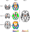
Lesion network mapping technique. In step one, lesions causing cervical dystonia were traced onto a standard atlas. In step two, connectivity between each lesion location and the rest of the brain was computed using a normative dataset of resting state functional connectivity scans from 1000 healthy individuals, and a standard seed-based approach. In step three, functional connectivity maps were thresholded, binarized (functionally connected or not), and overlapped to identify voxels connected to the greatest number of lesion locations.
Figure 3
Figure 3
Lesion network mapping of cervical dystonia. (A) Regions positively correlated (orange/yellow) or negatively correlated (blue/green) to lesion locations causing cervical dystonia (thresholded at 90% or 23/25 cases). From left to right: thalamus (z = 10); globus pallidus (z = −2); midbrain (z = −13); cerebellum (z = −32), and somatosensory cortex (projected onto the brain surface). (B) Regions that are both sensitive and specific to lesions causing cervical dystonia, with significantly greater (positive or negative) functional connectivity to lesion locations causing cervical dystonia compared to control lesion locations (conjunction across four separate specificity analyses).
Figure 4
Figure 4
Lesions causing cervical dystonia are part of a commonly connected brain network. The combination of positive connectivity to our cerebellum region of interest and negative connectivity to our somatosensory region of interest defines a network of regions (blue) that encompasses 24 of 25 lesion locations causing cervical dystonia (red). Case 8 lesion location falls immediately adjacent to this network.
Figure 5
Figure 5
Relevance to idiopathic cervical dystonia. Connectivity with our lesion-derived cerebellar region of interest (A, red) and somatosensory region of interest (B, blue) is abnormal in patients with idiopathic cervical dystonia. Patients with idiopathic cervical dystonia had a loss of negative functional connectivity from our cerebellar region of interest to the right sensorimotor cortex (z = 38) (orange/yellow) (A). Patients with idiopathic cervical dystonia also had loss of negative connectivity from our somatosensory region of interest to regions in the thalamus/basal ganglia and anterior cingulate (z = 12) (orange/yellow), and loss of positive connectivity to the sensorimotor and occipital cortex (z = 27) (blue/green) (B). All images were corrected with threshold-free cluster enhancement PFWE < 0.05. Corresponding average (SEM) Fischer z transformed correlation coefficients (Fz) are shown in box and whisker plots. Middle line within plot represents median, and cross shows mean. CD = idiopathic cervical dystonia patients; HV = healthy volunteers.
Figure 6
Figure 6
Relevance to deep brain stimulation. Lesions causing cervical dystonia are negatively connected to the somatosensory cortex and positively to the cerebellum (A). Globus pallidus interna DBS locations associated with good clinical response in cervical dystonia (B) and all dystonia patients (C) also show negative functional connectivity to the somatosensory cortex and positive functional connectivity to the cerebellum. Voxels associated with good response also show similar connectivity profile when controlling for voxels associated with poor response (D).

References

    1. Adolphs R. Human lesion studies in the 21st century. Neuron 2016; 90: 1151–53. - PMC - PubMed
    1. Albanese A, Bhatia K, Bressman SB, DeLong MR, Fahn S, Fung VS, et al.Phenomenology and classification of dystonia: a consensus update. Mov Disord 2013; 28: 863–73. - PMC - PubMed
    1. Antelmi E, Di Stasio F, Rocchi L, Erro R, Liguori R, Ganos C, et al.Impaired eye blink classical conditioning distinguishes dystonic patients with and without tremor. Parkinsonism Relat Disord 2016; 31: 23–7. - PubMed
    1. Ashburner J. SPM: a history. Neuroimage 2012; 62: 791–800. - PMC - PubMed
    1. Avanzino L, Ravaschio A, Lagravinese G, Bonassi G, Abbruzzese G, Pelosin E. Adaptation of feedforward movement control is abnormal in patients with cervical dystonia and tremor. Clin Neurophysiol 2018; 129: 319–26. - PubMed

Publication types