Review
. 2019 Jul;112(1):1-15.
doi: 10.1111/mmi.14267. Epub 2019 Jun 17.

The pathogenic actinobacterium Rhodococcus equi: what's in a name?

Affiliations
Review

The pathogenic actinobacterium Rhodococcus equi: what's in a name?

José A Vázquez-Boland et al. Mol Microbiol. 2019 Jul.

Abstract

Rhodococcus equi is the only recognized animal pathogenic species within an extended genus of metabolically versatile Actinobacteria of considerable biotechnological interest. Best known as a horse pathogen, R. equi is commonly isolated from other animal species, particularly pigs and ruminants, and causes severe opportunistic infections in people. As typical in the rhodococci, R. equi niche specialization is extrachromosomally determined, via a conjugative virulence plasmid that promotes intramacrophage survival. Progress in the molecular understanding of R. equi and its recent rise as a novel paradigm of multihost adaptation has been accompanied by an unusual nomenclatural instability, with a confusing succession of names: "Prescottia equi", "Prescotella equi", Corynebacterium hoagii and Rhodococcus hoagii. This article reviews current advances in the genomics, biology and virulence of this pathogenic actinobacterium with a unique mechanism of plasmid-transferable animal host tropism. It also discusses the taxonomic and nomenclatural issues around R. equi in the light of recent phylogenomic evidence that confirms its membership as a bona fide Rhodococcus.


Figures

Figure 1
Figure 1
R. equi lung infection and colony morphology. A. Purulent bronchopneumonia in foal with multifocal abscesses. Courtesy of Dr. U. Fogarty, Irish Equine Centre. B. Typical mucous appearance of R. equi colonies (LB agar incubated at 30°C for 48 h).
Figure 2
Figure 2
Genomic relatedness of R. equi isolates. Modified from Anastasi et al. (2016). A. Circular diagram of R. equi 103S (= NCTC 13926, DSM 104936) chromosome (5.02‐Mpb, outer ring with forward and reverse strands) compared to draft genomes of representative isolates from different sources and genetic lineages (inner rings). BLASTn alignments, red colour indicates > 98% sequence identity. HGT regions in 103S (arrow heads) coincide with gaps in the DNA alignments, indicating they are strain‐specific or less conserved. B. Core‐genome maximum‐likelihood phylogeny of R. equi isolates in A. Top, unrooted tree; reference genome isolate 103S and type strain of the species are indicated. Bottom, tree rooted with the closely related species, R defluvii Ca11T. The star‐like topology in the early branchings of the R. equi lineage suggests that the species’ diversification occurred through rapid clonal radiation from the common progenitor. See also Fig. 5.
Figure 3
Figure 3
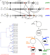
The three host‐specific R. equi virulence plasmids. Comparison of pVAPA (equine type) and pVAPB (porcine type) circular virulence plasmids and the recently characterized linear pVAPN plasmid (ruminant type) with closest homologs from environmental biodegrader Rhodococcus spp. Regions of significant similarity are connected with grey stripes. The vap PAIs are shaded in light blue. Gene colour code: Hypothetical proteins (gray), conjugation or DNA replication/recombination/metabolism (red), DNA mobility genes (magenta), transcriptional regulators (blue), secreted proteins (dark green), membrane proteins (pale green), metabolic functions (yellow), vap family genes (black) and pseudogenes (brown).Green and pale red bars below the genes indicate conjugation and replication/partitioning functional modules respectively; dashed underline indicates HGT region. Modified from Valero‐Rello et al. (2015)
Figure 4
Figure 4
Structure and evolution of the host‐specific vap PAIs. Modified from Valero‐Rello et al. (2015). A. Genetic structure of the vap PAIs from pVAPA (15.1 kb), pVAPB (21.5 kb) and pVAPN (15.9 kb). PAI genes in grey (non‐vap genes, in darker shade the vir operon) or black (vap genes). Genes outside the PAIs in white. PAI boundaries indicated by yellow arrowheads. The figure schematizes the evolutionary relationships of the vap genes as inferred from phylogenetic analysis, gene duplication/loss analysis (panel B) and genetic structure comparison. Straight lines connect allelic variants of same vap gene ancestor; those of vapA have red surround, curved lines indicate vap gene duplications. Crosses denote vap genes that were lost. Asterisks indicate pseudogenes. B. Gene duplication and loss in R. equi vap multigene family. Constructed with notung v2.6 from a vap gene ML tree. The analysis indicates that the common ancestor of the three host‐specific PAIs contained seven vap genes which evolved by gene duplication from a single ancestor vap gene. C. Fate of the vap PAI during host‐driven R. equi virulence plasmid evolution.
Figure 5
Figure 5
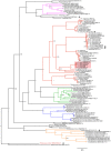
Whole‐genome Corynebacteriales ML tree. Nodes indicate bootstrap values. Tree constructed with five R.equi genomes, 47 non‐equi Rhodococus genomes including representatives from the major 16S rRNA gene clades (Goodfellow et al., 1998; McMinn et al., 2000; Jones and Goodfellow, 2012; Ludwig et al., 2012), and 57 genomes from 11 Corynebacteriales genera. Rooted with Streptomyces albus NBRC 1304T (outgroup). (T) indicates type strain. Genome used for R. equi type strain DSM 20307T = ATCC 6939T is assembly acc. no. LWTX00000000 (Anastasi et al., 2016). Major genera are highlighted in different colour. Black arrowheads indicate misclassifications revealed by the phylogenomic analysis. One of them is R. rhodnii NRRL B‐16535T (GenBank assembly acc. no. GCA_000720375.1); this probably represents a sequence mislabelling or strain mixup. Modified from Anastasi et al. (2016).

References

    1. Anastasi, E. , MacArthur, I. , Scortti, M. , Alvarez, S. , Giguere, S. and Vazquez‐Boland, J.A. (2016) Pangenome and phylogenomic analysis of the pathogenic actinobacterium Rhodococcus equi . Genome Biology and Evolution, 8, 3140–3148. - PMC - PubMed
    1. Barabote, R.D. and Saier, M.H. Jr . (2005) Comparative genomic analyses of the bacterial phosphotransferase system. Microbiology and Molecular Biology Reviews, 69, 608–634. - PMC - PubMed
    1. von Bargen, K. and Haas, A. (2009) Molecular and infection biology of the horse pathogen Rhodococcus equi . FEMS Microbiology Reviews, 33, 870–891. - PubMed
    1. Bryan, L.K. , Clark, S.D. , Diaz‐Delgado, J. , Lawhon, S.D. and Edwards, J.F. (2017) Rhodococcus equi infections in dogs. Veterinary Pathology, 54, 159–163. - PubMed
    1. Byrne, G.A. , Russell, D.A. , Chen, X. and Meijer, W.G. (2007) Transcriptional regulation of the virR operon of the intracellular pathogen Rhodococcus equi . Journal of Bacteriology, 189, 5082–5089. - PMC - PubMed

Publication types

LinkOut - more resources