

A three-wave longitudinal study of subcortical-cortical resting-state connectivity in adolescence: Testing age- and puberty-related changes
- PMID: 31099959
- PMCID: PMC6767490
- DOI: 10.1002/hbm.24630
A three-wave longitudinal study of subcortical-cortical resting-state connectivity in adolescence: Testing age- and puberty-related changes
Abstract
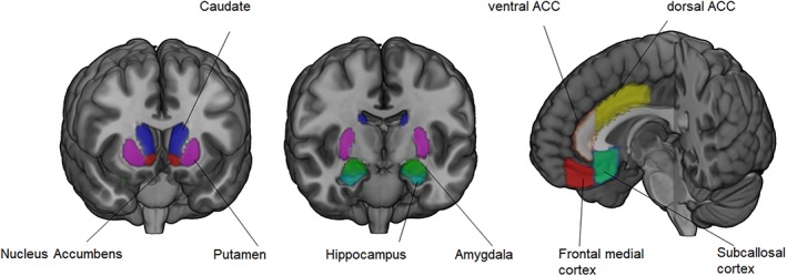
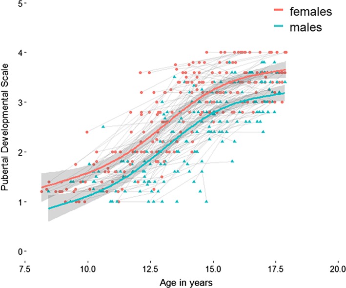
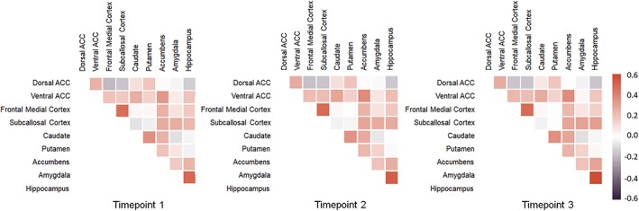
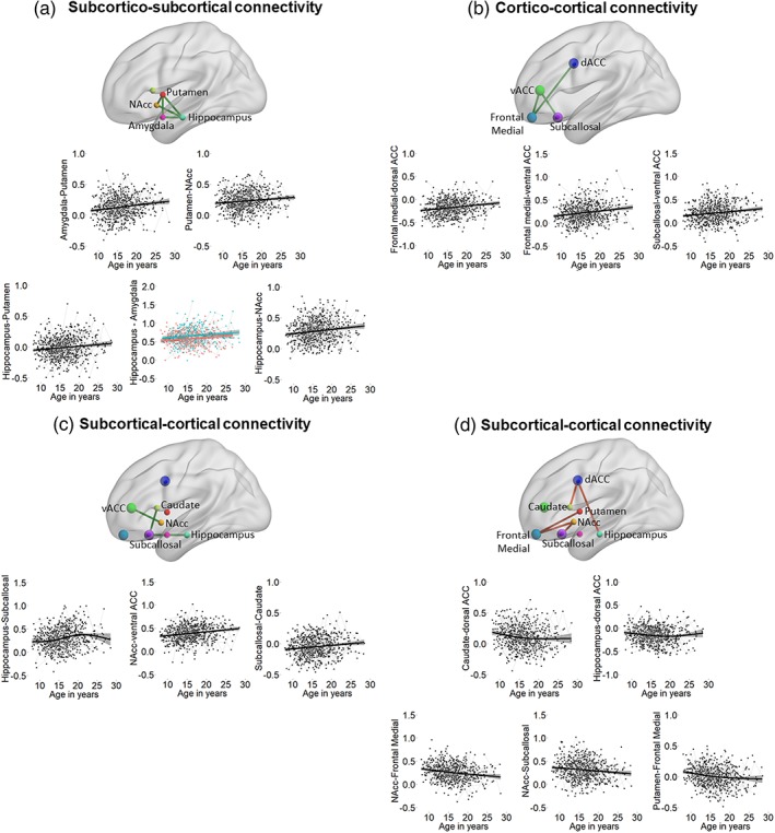
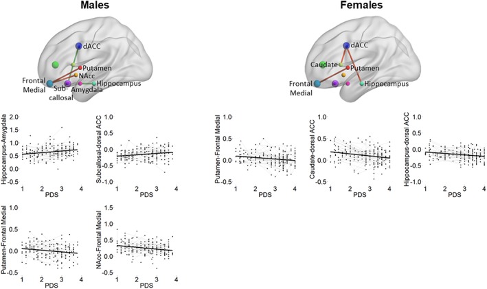
Adolescence is the transitional period between childhood and adulthood, characterized by substantial changes in reward-driven behavior. Although reward-driven behavior is supported by subcortical-medial prefrontal cortex (PFC) connectivity, the development of these circuits is not well understood. Particularly, while puberty has been hypothesized to accelerate organization and activation of functional neural circuits, the relationship between age, sex, pubertal change, and functional connectivity has hardly been studied. Here, we present an analysis of resting-state functional connectivity between subcortical structures and the medial PFC, in 661 scans of 273 participants between 8 and 29 years, using a three-wave longitudinal design. Generalized additive mixed model procedures were used to assess the effects of age, sex, and self-reported pubertal status on connectivity between subcortical structures (nucleus accumbens, caudate, putamen, hippocampus, and amygdala) and cortical medial structures (dorsal anterior cingulate, ventral anterior cingulate, subcallosal cortex, frontal medial cortex). We observed an age-related strengthening of subcortico-subcortical and cortico-cortical connectivity. Subcortical-cortical connectivity, such as, between the nucleus accumbens-frontal medial cortex, and the caudate-dorsal anterior cingulate cortex, however, weakened across age. Model-based comparisons revealed that for specific connections pubertal development described developmental change better than chronological age. This was particularly the case for changes in subcortical-cortical connectivity and distinctively for boys and girls. Together, these findings indicate changes in functional network strengthening with pubertal development. These changes in functional connectivity may maximize the neural efficiency of interregional communication and set the stage for further inquiry of biological factors driving adolescent functional connectivity changes.
Keywords: adolescence; functional connectivity; longitudinal; pubertal development; resting-state.
© 2019 The Authors. Human Brain Mapping published by Wiley Periodicals, Inc.
Conflict of interest statement
The authors declare no conflict of interest.
Figures
References
-
- Achterberg, M. , Bakermans‐Kranenburg, M. J. , van IJzendoorn, M. H. , van der Meulen, M. , Tottenham, N. , & Crone, E. A. (2018). Distinctive heritability patterns of subcortical‐prefrontal cortex resting state connectivity in childhood: A twin study. NeuroImage, 175, 138–149. - PubMed
-
- Alexander, G. E. , Crutcher, M. D. , & DeLong, M. R. (1990). Basal ganglia‐thalamocortical circuits: Parallel substrates for motor, oculomotor, “prefrontal” and “limbic” functions. Progress in Brain Research, 85, 119–146. - PubMed
Publication types
MeSH terms
Grants and funding
LinkOut - more resources
Full Text Sources
Medical
Miscellaneous
