. 2019 May 17;14(5):e0216603.
doi: 10.1371/journal.pone.0216603. eCollection 2019.

Lipophilic statins inhibit YAP nuclear localization, co-activator activity and colony formation in pancreatic cancer cells and prevent the initial stages of pancreatic ductal adenocarcinoma in KrasG12D mice

Affiliations

Lipophilic statins inhibit YAP nuclear localization, co-activator activity and colony formation in pancreatic cancer cells and prevent the initial stages of pancreatic ductal adenocarcinoma in KrasG12D mice

Fang Hao et al. PLoS One. .

Abstract

We examined the impact of statins on Yes-associated Protein (YAP) localization, phosphorylation and transcriptional activity in human and mouse pancreatic ductal adenocarcinoma (PDAC) cells. Exposure of sparse cultures of PANC-1 and MiaPaCa-2 cells to cerivastatin or simvastatin induced a striking re-localization of YAP from the nucleus to the cytoplasm and inhibited the expression of the YAP/TEAD-regulated genes Connective Tissue Growth Factor (CTGF) and Cysteine-rich angiogenic inducer 61 (CYR61). Statins also prevented YAP nuclear import and expression of CTGF and CYR61 stimulated by the mitogenic combination of insulin and neurotensin in dense culture of these PDAC cells. Cerivastatin, simvastatin, atorvastatin and fluvastatin also inhibited colony formation by PANC-1 and MiaPaCa-2 cells in a dose-dependent manner. In contrast, the hydrophilic statin pravastatin did not exert any inhibitory effect even at a high concentration (10 μM). Mechanistically, cerivastatin did not alter the phosphorylation of YAP at Ser127 in either PANC-1 or MiaPaCa-2 cells incubated without or with neurotensin and insulin but blunted the assembly of actin stress fiber in these cells. We extended these findings with human PDAC cells using primary KC and KPC cells, (expressing KrasG12D or both KrasG12D and mutant p53, respectively) isolated from KC or KPC mice. Using cultures of these murine cells, we show that lipophilic statins induced striking YAP translocation from the nucleus to the cytoplasm, inhibited the expression of Ctgf, Cyr61 and Birc5 and profoundly inhibited colony formation of these cells. Administration of simvastatin to KC mice subjected to diet-induced obesity prevented early pancreatic acini depletion and PanIN formation. Collectively, our results show that lipophilic statins restrain YAP activity and proliferation in pancreatic cancer cell models in vitro and attenuates early lesions leading to PDAC in vivo.


Conflict of interest statement

The authors have declared that no competing interests exist.

Figures
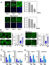
Fig 1
Fig 1. Statins induce re-distribution of YAP to the cytoplasm and inhibit YAP/TEAD-regulated genes in PDAC cells.
A, PANC-1 and Mia PaCa-2 cells were fixed with 4% paraformaldehyde 1, 3 and 7 days after plating, as indicated. B, PANC-1 and Mia PaCa-2 cells were incubated in either absence or presence of 0.3 μM cerivastatin (Cer) added 1 day after plating and for 24 h. either with or without 250 μM mevalonic acid Then, the cells were fixed with 4% paraformaldehyde. In A and B the cultures were stained with an antibody that detects total YAP. Bars represent the ratio of nuclear/cytoplasm (50 to 75 cells) mean ± S.E. with similar results obtained in three independent experiments (T-test p values comparing the indicated groups to control were **p<0.01). C and D, PANC-1 and MiaPaCa-2 cells were incubated in either absence or presence of cerivastatin (Cer) at 0.1 and 1 μM or simvastatin (sim) at 0.3 and 3 μM, as indicated. Statins were added 1 day after plating and the incubation continued for 24 h. RNA was then isolated and the relative levels (n = 3) of CTGF (C) or CYR61 (D) mRNA compared with 18s mRNA was measured by RT-qPCR. Data are presented as mean ± SEM of 5 independent experiments for CTGF and 3 independent experiments for CYR61. Cerivastatin and simvastatin groups vs control *p<0.05, **p<0.01.
Fig 2
Fig 2. Cerivastatin inhibits YAP nuclear localization induced by stimulation with neurotensin and insulin in confluent PDAC cells.
Confluent cultures of PANC-1 (A) or Mia PaCa-2 (B) cells were incubated either in the absence or presence of 0.3μM cerivastatin (Cer) for 24 h. The cultures were then stimulated without (-) or with a combination of 5 nM neurotensin and 10 ng/ml insulin (NT+Ins) for 60 min. The cultures were then washed, fixed with 4% paraformaldehyde and stained with an antibody that detects total YAP and with Hoechst 33342 to visualize the cell nuclei. Bars represent the ratio of nuclear/cytoplasm (100 to 125 cells). Cerivastatin significantly decreased NT+Ins nuclear localization of YAP as compared with untreated controls (**P < 0.01) as shown by t-test.
Fig 3
Fig 3. Exposure to lipophilic statins inhibits the expression of CTGF and CYR61 induced by stimulation with neurotensin and insulin in PDAC cells.
Confluent cultures of PANC-1 cells (upper panel) or MiaPaca-2 cells (lower panel) were incubated for 24 h either without or with cerivastatin (Cer) at 0.1 and 0.3 μM or simvastatin (sim) at 3 and 10 μM, as indicated. The cultures were then stimulated either without (-) or with a combination of 5 nM neurotensin and 10 ng/ml insulin (NT+Ins) for 60 min. RNA was isolated and the relative levels (n = 3) of CTGF or CYR61 mRNA compared with 18s mRNA was measured by RT-qPCR. Data are presented as mean ± SEM. Similar results were obtained in 3 independent experiments. Exposure to cerivastatin and simvastatin significantly decreased relative mRNA levels of CTGF and CYR61 as compared with controls (**P < 0.01) as shown by paired t-test.
Fig 4
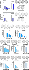
Fig 4. Lipophilic statins inhibit colony-formation by PDAC cells.
Colony formation assays were performed as described in the Materials and Methods section. PANC-1 or MiaPaCa-2 cells were incubated for 10 days (PANC-1 cells) or 8 days (Mia PaCa-2 cells) with various concentrations of lipophilic statins, such as cerivastatin, simvastatin, atorvastatin, fluvastatin or the hydrophilic statin pravastatin, as indicated. Typical pictures are shown for selected conditions The bars represent the number of colonies (mean ± SEM; n = 4 dishes per condition). Similar results were obtained in 6 independent experiments for cerivastatin, 4 for simvastatin and 3 for atorvastatin, fluvastatin and pravastatin.
Fig 5
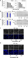
Fig 5. Mechanism of action of statins in PDAC cells.
A, The geranylgeranyl transferase inhibitor GGTI 298 inhibits colony formation by PDAC cells. Colony formation assays were performed as described in the Materials and Methods section. PANC-1 or MiaPaCa-2 cells were incubated for 10 days (PANC-1 cells) or 8 days (Mia PaCa-2 cells) with or without GGTI 298 at the indicated concentrations. The bars represent the number of colonies (mean ± SEM; n = 4 dishes per condition). B and C, Treatment with cerivastatin does not alter YAP phosphorylation at Ser127. PANC-1 (B) and Mia PaCa-2 (C) cells were treated for 24 h either in the absence or presence of cerivastatin (Cer, 0.3 μM) for 24h. The cultures were then stimulated with 5 nM neurotensin and 10 ng/ml insulin (NT+Ins) for 30 min as indicated, and lysed with SDS–PAGE sample buffer. The samples were analyzed by SDS-PAGE and immunoblotting with YAP Ser127 and YAP antibodies. Quantification of phosphorylated YAP Ser127 was performed using Multi Gauge V3.0. from 9 independent experiments, T-test p values comparing the indicated groups to control were * p< 0.05; **p < .0.01. D and E, Exposure to either cerivastatin or simvastatin does not alter YAP phosphorylation at Ser127. PANC-1 and Mia PaCa-2 cells were treated for 24 h either in the absence or presence of cerivastatin (Cer) or simvastatin (Sim) at the indicated concentrations (in μM) for 24h. The cultures were then stimulated with 5 nM neurotensin and 10 ng/ml insulin (NT+Ins) for 30 min as indicated, and lysed with SDS–PAGE sample buffer. The samples were analyzed by SDS-PAGE and immunoblotting with antibodies that detect phospho YAP Ser127, YAP, phospho LATS Thr1079 and LATS2 antibodies. Equal loading was verified by immunoblotting with actin antibody. Similar results were obtained in 4 independent experiments. F and G, Cerivastatin inhibits the assembly of actin stress fibers induced by stimulation with insulin and neurotensin. PANC-1 (F) and Mia PaCa-2 cells (G) were treated for 24 h either in the absence or presence of cerivastatin at the indicated concentrations for 24h prior to stimulation with 5 nM neurotensin and 10 ng/ml insulin (NT+Ins) for 30 min. The cultures were then washed, fixed with 4% paraformaldehyde, and stained with TRITC-conjugated phalloidin and Hoechst 33342. Similar results were obtained in 3 independent experiments.
Fig 6
Fig 6. Statins inhibit YAP nuclear localization, colony formation and the expression of Ctgf, Cyr61 and Birc5 in KC cells.
A, KC cells 1 day after plating were incubated either in absence or presence of 0.3 μM cerivastatin (Cer) or 3 μM simvastatin (Ser) for 24 h. Then, the cells were then fixed with 4% paraformaldehyde and stained with an antibody that detects total YAP and with Hoechst 33342 to visualize the cell nuclei. Bars represent the ratio of nuclear/cytoplasm (50 to 75 cells). **P 0.01 as shown by paired t-test. B, KC cells were incubated either in absence or presence of cerivastatin (Cer) or simvastatin (Sim) at the indicated concentrations. Statins were added 1 day after plating and the incubation continued for 24 h. RNA was then isolated and the relative levels (n = 3) of Ctgf, Cyr61 and Birc5 mRNA compared with 18s mRNA were measured by RT-qPCR. Data are presented as mean ± SEM. Similar results were obtained in 3 independent experiments. C, KC cells were incubated for 6 days with various concentrations of cerivastatin or simvastatin, as indicated. The bars represent the number of colonies (mean ± SEM; n = 4 cultures per condition).
Fig 7
Fig 7. Simvastatin prevents the disruption in histologic pancreatic architecture induced by DIO in KC mice.
A and B, Representative H&E staining of the pancreas of a KC mouse fed the HFCD (A) or HFCD plus simvastatin (B). C, Percentage (%) of intact acini in KC mice fed the CD, HFCD or HFCD plus simvastatin at 3 months. D, Pancreatitis score in KC mice fed CD, HFCD or HFCD plus simvastatin at 3 months. E, Percentage (%) of PanIN-3 lesions in KC mice fed CD, HFCD or HFCD plus simvastatin at 3 months. F, Simvastatin has no effect on weight gain in KC mice fed with either CD, HFCD, or HFCD plus simvastatin at 3 months. The number of mice used were: CD = 10; HFCD = 14; HFCD+simvastatin = 14. Values are means ± s.d. *p < 0.05 vs. CD, #p < 0.05 vs. HFCD.

References

    1. Siegel RL, Miller KD, Jemal A. Cancer statistics, 2018. CA Cancer J Clin. 2018;68(1):7–30. 10.3322/caac.21442 - DOI - PubMed
    1. Rahib L, Smith BD, Aizenberg R, Rosenzweig AB, Fleshman JM, Matrisian LM. Projecting Cancer Incidence and Deaths to 2030: The Unexpected Burden of Thyroid, Liver, and Pancreas Cancers in the United States. Cancer Res. 2014;74(11):2913–21. 10.1158/0008-5472.CAN-14-0155 - DOI - PubMed
    1. Hingorani SR, Petricoin EF, Maitra A, Rajapakse V, King C, Jacobetz MA, et al. Preinvasive and invasive ductal pancreatic cancer and its early detection in the mouse. Cancer Cell. 2003;4(6):437–50. . - PubMed
    1. Maitra A, Fukushima N, Takaori K, Hruban RH. Precursors to invasive pancreatic cancer. Adv Anat Pathol. 2005;12(2):81–91. . - PubMed
    1. Jones S, Zhang X, Parsons DW, Lin JC, Leary RJ, Angenendt P, et al. Core signaling pathways in human pancreatic cancers revealed by global genomic analyses. Science. 2008;321(5897):1801–6. 10.1126/science.1164368 . - DOI - PMC - PubMed

Publication types

MeSH terms

Substances