. 2019 May 17;14(5):e0217145.
doi: 10.1371/journal.pone.0217145. eCollection 2019.

Improvement in diagnostic quality of structural and angiographic MRI of the brain using motion correction with interleaved, volumetric navigators

Affiliations

Improvement in diagnostic quality of structural and angiographic MRI of the brain using motion correction with interleaved, volumetric navigators

Mads Andersen et al. PLoS One. .

Abstract

Introduction: Subject movements lead to severe artifacts in magnetic resonance (MR) brain imaging. In this study we evaluate the diagnostic image quality in T1-weighted, T2-weighted, and time-of-flight angiographic MR sequences when using a flexible, navigator-based prospective motion correction system (iMOCO).

Methods: Five healthy volunteers were scanned during different movement scenarios with and without (+/-) iMOCO activated. An experienced neuroradiologist graded images for image quality criteria (grey-white-matter discrimination, basal ganglia, and small structure and vessel delineation), and general image quality on a four-grade scale.

Results: In scans with deliberate motion, there was a significant improvement in the image quality with iMOCO compared to the scans without iMOCO in both general image impression (T1 p<0.01, T2 p<0.01, TOF p = 0.03) and in anatomical grading (T1 p<0.01, T2 p<0.01, TOF p = 0.01). Subjective image quality was considered non-diagnostic in 91% of the scans with motion -iMOCO, but only in 4% of the scans with motion +iMOCO. iMOCO performed best in the T1-weighted sequence and least well in the angiography sequence. iMOCO was not shown to have any negative effect on diagnostic image quality, as no significant difference in diagnostic quality was seen between scans -iMOCO and +iMOCO with no deliberate movement.

Conclusion: The evaluation showed that iMOCO enables substantial improvements in image quality in scans affected by subject movement, recovering important diagnostic information in an otherwise unusable scan.


Conflict of interest statement

MA is an employee of Philips Danmark A/S.

Figures
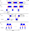
Fig 1
Fig 1. Overview of the switching between host sequences and navigators with associated timings.
a. The 3D T1-weighted sequence without an interleaved navigator. bd. Sequence layouts after incorporation of navigators. In all cases, the host sequence and navigator were defined as two different sequences in the standard user interface. The red arrows represent the switch between sequences.
Fig 2
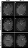
Fig 2. Examples of T1-weighted scans with (+) and without (-) iMOCO for different types of movement.
Panels ad are from subject 1, while panels e and f are from subject 5. The scans in a, b, d, and f were given a grading of 3 in all quality criteria. For the scans with motion -iMOCO, panel c was given a grading of 1 in all criteria, while panel e was given a grading of 2 in all regions except for the frontal horns, the septum pellucidum, and the anterior commissure and the columns of the fornix (not illustrated), where grade 3 was given.
Fig 3
Fig 3. Examples of T2-weighted scans with (+) and without (-) iMOCO for different types of movement.
Panels a‒d are from subject 1, whereas panels e and f are from subject 5. The scans in a and b were given a grading of 3 in all quality criteria except for two scored structures that are not illustrated (insula, extreme capsule, and claustrum; and putamen), which received a grading of 2. The scan in panel c was graded 1 in all criteria, whereas panel d was given a grading of 2 in all criteria except for frontal horn and septum pellucidum (both not illustrated), which were graded 3. With jerk motion, the artefacts were less problematic, and the scan represented in e was given a grading of 2 in all criteria while f was graded 3.
Fig 4
Fig 4. Examples of TOF scans with (+) and without (-) iMOCO for different types of movement.
Panels ad are from subject 1, while e and f are from subject 7. The scans in panels a and b were given a grading of 3 in all quality criteria. The scans in c and e were performed with motion but -iMOCO, and were graded 1 in all criteria. The corresponding cases +iMOCO were given higher gradings, i.e. panel d was grade 3 in all criteria except general image impression (grade 2), and panel f was grade 2 in all criteria.
Fig 5
Fig 5. Summary of the grading of image quality.
Panels ad show the pooled grades of all anatomical structures, given for the three types of sequence (T1, T2, and TOF), while panels eh show the general image impression (GII) grades for the three sequences.
Fig 6
Fig 6. Examples of position parameters obtained from the navigators in the T1 sequence, for the different motion scenarios.
The translations and rotations shown are the accumulated motion updates performed, and thus reflect the patient position relative to the starting position. The motion score reflects the change in position between every navigator and leads to reacquisition of the previously acquired k-space segment in the T1 sequence when above 1 mm.

References

    1. Andre JB, Bresnahan BW, Mossa-Basha M, Hoff MN, Smith CP, Anzai Y, et al. Towards Quantifying the Prevalence, Severity, and Cost Associated With Patient Motion During Clinical MR Examinations. J Am Coll Radiol. Elsevier Inc; 2015;12(7):689–95. - PubMed
    1. Afacan O, Erem B, Roby DP, Roth N, Roth A, Prabhu SP, et al. Evaluation of motion and its effect on brain magnetic resonance image quality in children. Pediatr Radiol. 2016. November 3;46(12):1728–35. 10.1007/s00247-016-3677-9 - DOI - PMC - PubMed
    1. Tornqvist E, Mansson A, Larsson EM, Hallstrom I. Impact of extended written information on patient anxiety and image motion artifacts during magnetic resonance imaging. Acta Radiol. England; 2006. June;47(5):474–80. - PubMed
    1. Maclaren J, Herbst M, Speck O, Zaitsev M. Prospective motion correction in brain imaging: A review. Magn Reson Med. 2013. May 8;69:621–36. 10.1002/mrm.24314 - DOI - PubMed
    1. Maclaren J, Armstrong BSR, Barrows RT, Danishad K a, Ernst T, Foster CL, et al. Measurement and Correction of Microscopic Head Motion during Magnetic Resonance Imaging of the Brain. Hess CP, editor. PLoS One. 2012. November 7;7(11):e48088 10.1371/journal.pone.0048088 - DOI - PMC - PubMed

Publication types