. 2019 May 16;177(5):1136-1152.e18.
doi: 10.1016/j.cell.2019.04.011.

A Site of Vulnerability on the Influenza Virus Hemagglutinin Head Domain Trimer Interface

Affiliations

A Site of Vulnerability on the Influenza Virus Hemagglutinin Head Domain Trimer Interface

Sandhya Bangaru et al. Cell. .

Abstract

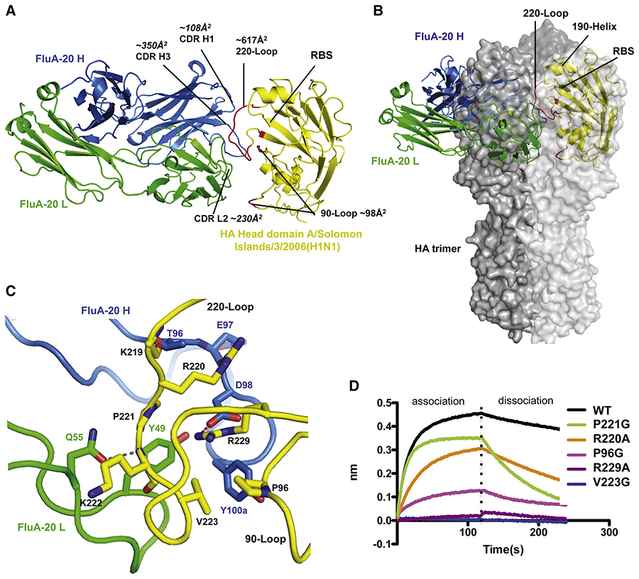
Here, we describe the discovery of a naturally occurring human antibody (Ab), FluA-20, that recognizes a new site of vulnerability on the hemagglutinin (HA) head domain and reacts with most influenza A viruses. Structural characterization of FluA-20 with H1 and H3 head domains revealed a novel epitope in the HA trimer interface, suggesting previously unrecognized dynamic features of the trimeric HA protein. The critical HA residues recognized by FluA-20 remain conserved across most subtypes of influenza A viruses, which explains the Ab's extraordinary breadth. The Ab rapidly disrupted the integrity of HA protein trimers, inhibited cell-to-cell spread of virus in culture, and protected mice against challenge with viruses of H1N1, H3N2, H5N1, or H7N9 subtypes when used as prophylaxis or therapy. The FluA-20 Ab has uncovered an exceedingly conserved protective determinant in the influenza HA head domain trimer interface that is an unexpected new target for anti-influenza therapeutics and vaccines.

Keywords: B-lymphocytes; antibodies; antibody-dependent cell cytotoxicity; antigen-antibody reactions; hemagglutinin glycoproteins; influenza A virus; influenza virus; monoclonal; viral.


Conflict of interest statement

DECLARATION OF INTERESTS. J.E.C. has served as a consultant for Takeda Vaccines, Sanofi Pasteur, Pfizer, and Novavax, is on the Scientific Advisory Boards of CompuVax and Meissa Vaccines, and is Founder of IDBiologics, Inc. AG-S is inventor of patents owned by the Icahn School of Medicine at Mount Sinai in the field of influenza virus vaccines and Abs. All other authors declare no conflict of interest. Vanderbilt University has applied for a patent related to the FluA-20 Ab.

Figures

Figure 1.
Figure 1.. Network analysis of sequences clonally related to FluA-20 and FluA-20 reactivity to diverse HAs.
(A) Timeline showing the vaccination history of FluA-20 donor and the time points from which FluA-20 (triangle) and its clonally related siblings (circles) were identified. (B) Nodes represent unique sequences observed, with the size of the node correlating to the count of replicate sequences observed. The color of each node denotes the time point at which it was found; white day 5, yellow day 6, orange day 11 and pink day 14. The black node represents the VH4–61/JH4 germline sequence, and the gray node represents an inferred common ancestor. The maroon, triangle-shaped node represents FluA-20. Edges drawn between nodes show that those sequences are more closely related to each other than to any other sequence. Edge distances are arbitrary and used only to visually clarify the graph. The somatic variants of FluA-20 that were expressed and tested are indicted. (C) ELISA binding EC50 (ng/mL) values for FluA-20, recombinant FluA-20 (rFluA-20) and unmutated common ancestor of FluA-20 (FluA-20-UCA) to HAs derived from different strains representing group 1 (green) and group 2 (blue) IAVs. The table is displayed in purple-white color scale corresponding to strong-weak binding, respectively. The > symbol indicates that binding was not observed at concentrations ≤10 μg/mL.
Figure 2.
Figure 2.. MAb FluA-20 exhibits protection in vivo against diverse IAV subtypes
(A) Body weight change in mice that received FluA-20 prophylactically prior to sub-lethal challenge with IAV strains from H1N1, H3N2, H5N1 or H7N9. Mice were treated with 10 mg/kg of either FluA-20 or a similarly prepared control Ab to an unrelated target and challenged 24 h later with either H1N1 A/Netherlands/602/2009 or H3N2 A/X-31 (6:2 PR8 backbone) or H5N1 A/barn swallow/Hong Kong/D10–1161/2010 (7:1 PR8 backbone) or H7N9 A/Shanghai/1/2013 (6:2 PR8 backbone). The weight loss of mice (n=5) was measured daily for 14 days after inoculation (day 0). The experiments were performed twice with similar results. (B) Survival and weight change in mice (n=10) prophylactically treated with FluA-20 (1 or 3 or 10 mg/kg) or 10 mg/kg of control IgG or PBS prior to lethal challenge with mouse adapted H1N1 A/California/04/2009. One experimental group was treated with 30 mg/kg/day of oseltamivir for 5 days post-challenge as a positive control. *** P < 0.001, compared to placebo-treated group; +++ P < 0.001, ++ P < 0.01, compared to DENV 2D22-treated group. (C) Percentage survival in mice prophylactically treated with 10 mg/kg of either FluA-20 or a recombinant form of CR6261 or control IgG (MRSA-147) prior to lethal challenge with H1N1 A/California/04/2009 virus. (D) Weight change in mice that were sub-lethally challenged with H1N1 A/California/04/2009 virus prior to therapeutic treatment with 10 mg/kg of either mAbs FluA-20 or a recombinant form of CR6261 or control IgG (MRSA-147) on day 1 post-inoculation. (E) Survival and weight change in mice lethally challenged with H3N2 and H5N1 viruses (same strains as panel (A)) prior to therapeutic treatment with 10 mg/kg of either mAbs FluA-20 or control IgG on days 1, 2 and 4 post-inoculation. Each group was compared to the mock-treated group in A to E. Body weight change data in B, E are shown only for the animals that survived at each indicated time point. The weights in A, B, D and E are shown as the group mean and the SEM.
Figure 3.
Figure 3.. FluA-20 targets the 220-loop and the 90-loop at the trimer interface of the H1 head domain.
(A) Structural overview of rFluA-20 Fab in complex with the head domain of H1 HA (A/Solomon Islands/3/2006). FluA-20 Fab is shown as a backbone trace in blue heavy chain (H) and green light chain (L). The backbone of the HA head domain is shown as a yellow trace and residues contacted by FluA-20 are red. (B) The H1 head domain is superimposed with one protomer colored in light grey surface from an HA trimer structure (PDB 4M4Y). The adjacent HA protomers are shown with dark grey solid surface. The variable domain of FluA-20 would clash with a large area of the head domain from an adjacent protomer in the HA trimer model. (C) FluA-20 interaction with H1. The salt bridge interaction between Asp98 (H) to Arg229 is shown as a red dashed line. A hydrogen bond between Asn55 (H) to Lys222 is presented with a grey line. Two additional hydrogen bonds are between the side chain of Thr96 (H) to main-chain carbonyl of Lys219 and Arg220 side chain to the main-chain carbonyl of Glu97 (H). Other hydrophobic residues that contribute to the interaction are shown with side chains. (D) The binding traces of HA head domain, or its mutants (at the concentration of 0.5 μM), to immobilized rFluA-20 Fab in BLI assay are presented.
Figure 4.
Figure 4.. FluA-20 interacts with H3 head domain.
(A) The structure of rFluA-20 in complex with a H3 head domain (A/Hong Kong/3/1968) is presented similarly to Figure 3, with the H3 head domain colored in wheat. The H3 residues interacting with FluA-20 are red and the Ab footprint size on HA is analyzed. The H3 head domain is superimposed with one protomer of an H3 trimer structure (PDB 4FNK, shown as surface with different shade of grey for each protomer). (B) Interaction of FluA-20 with H3 HA. A salt bridge between R229 from HA and Asp98 (H) of FluA-20 is shown with a red line. Hydrogen bonds between Gln55 (L) to main chain amide of Trp222 and Asn53 (L) to Arg224 are presented with grey lines. Several hydrophobic residues that contribute to the interaction are shown with their side chains.
Figure 5.
Figure 5.. Critical residues involved in FluA-20 binding to different HAs.
(A, B) Principal residues that FluA-20 recognizes in HA head domains are highly conserved across various HA subtypes. The binding core of FluA-20 in complex with H1 (A) or H3 (B) HA is highlighted by a salt bridge between Asp98 (H) and Arg229, which is enclosed by a circle of hydrophobic residues, including Pro96, Pro221, and Val223 of HA, Tyr49 (L) and Tyr100a of FluA-20. The conservation of the core residues in each HA subtypes is indicated by identity percentages. (C, D, E and F) Simulation of FluA-20 binding to HAs from other subtypes. The head domains of H5 (C), H13 (D) or H7 HA (E and F) are superimposed with H3 HA (colored in wheat) in complex with FluA-20. (C) H5 (VN/1203) has Ser221 (red circle), instead of Pro221 in H1 and H3 subtypes. The KD values of FluA-20 Fab binding to either wt H5 or H5_S221P mutant were determined by BLI assay. (D) Instead of the salt bridge interaction between Asp98 (H) of FluA-20 and Arg229 in other HAs, H13 HA possesses two aromatic residues, Tyr223 and Trp229, that contribute to the binding by aromatic stacking with Y100a. (E) H7 HA of A/Shanghai/2/2013, in orange, aligns well to H3_FluA20 complex structure. (F) H7 HA of A/New York/107/2003 (grey) was aligned to the H3_FluA-20 complex structure. This H7 strain has a truncated 220-loop and is missing residues 221–228. (G, H) FluA-20 accommodates variability in the HA 220-loop of H1 (G) or H3 (H) HA. Residues 219, 222, and 224 in the FluA-20 epitope exhibit considerable variation in various subtypes. However, FluA-20 forms hydrogen bond interactions with the main chains of these variable residues (in grey lines), and the approach angle of FluA-20 successfully avoids contacts or collisions with bulky and variable side chains.
Figure 6.
Figure 6.. FluA-20 binding is inhibited by HA cleavage potentially due to dynamic changes in the trimer.
(A) The association and disassociation traces of HA0 or cleaved HAs from H1 (A/California/04/2009) or H7 (A/Shanghai/02/2003) to immobilized rFluA-20. The HA was tested at 1μM concentration (B). HEK293F cells were either untransfected (UT) or transiently transfected with full-length H3 (A/Hong Kong/1/1968) HA cDNA for HA surface expression. The cells were either left untreated or treated with TPCK trypsin and then incubated with 10 μg/mL of mAb CR9114 or mAb FluA-20 followed by incubation with secondary Ab. Ab binding to cleaved and uncleaved HA on the cell surface was determined by flow cytometric analysis. The error bars represent mean ± SD of technical replicates. Statistical significance was calculated using the unpaired two-tailed t-test. Data are representative of two independent experiments. (C) Deuterium exchange comparison of cleaved HA trimer to HA0 trimer from A/Netherlands/219/2003(H7N7) by HDX-MS. One HA protomer in the model (PDB 4DJ6) is shown in colored backbone trace. Peptides with slower deuterium exchange in cleaved HA are blue, and peptides with faster exchange are red. Peptides in grey represent no difference in deuterium exchange rate and peptides in black indicate peptides that were not covered in the MS data.
Figure 7.
Figure 7.. FluA-20 inhibits cell-cell spread, disrupts the uncleaved HA trimer protein, and does not require Fc-effector function for in vivo protection.
(A, B, C) demonstrate that FluA-20 diminishes cell-to-cell spread of influenza virus. (A) Representative images of 6-well plate wells with influenza virus A/Hong Kong/1/1968 foci developed on MDCK monolayers after 48 hours of incubation at presence of 10 μg/mL of irrelevant control mAb MRSA-147, FluA-20, CR9114, or equimolar concentration of zanamivir; foci were immunostained and images were captured by CTL (Cellular Technology Ltd.). Images are representative of 3 replicates of 2 independent experiments (B) Quantitative determination of foci area reduction. Foci area calculated by ImageJ software and represented as percentage of total well area. Each value represents mean focus area ± SD. (C) Concentration-dependent effect of focus area reduction. Each value represents the mean focus area ± SD. Figures (D) and (E) corresponds to the in vivo protective efficacy of engineered Fc mutant variants of mAb FluA-20. BALB/c mice were inoculated i.p. with 10 mg/kg of mAb on the day before challenge by the i.n. route with 1.24 × 10⁴ FFU of A/California/04/2009 virus and monitored for 14 days. The control group was treated with mAb specific to an unrelated target. The protective efficacy of mAbs was assessed by weight change (D) and clinical score (E). The dotted line indicates the IACUC-stipulated endpoint for humane euthanasia. Data are cumulative of 2 independent experiments and show the mean value ± SEM, using 5–10 mice per group. Multiple group comparisons were performed using two-way ANOVA with Tukey’s post-test for panel A. The results of comparison between rFluA-20 IgG1-N297A-treated (grey) and rFluA-20 IgG1-treated (blue) groups demonstrate a significant difference in weight change between these two groups (denoted with * symbol), although the N297A Fc region mutation that abrogates FcR binding had a negligible impact of on overall protection. (F) Selected 2D class averages of H1 HA trimer (A/California/04/2009) after a 20-second incubation with FluA-20 Fab. All of the Fabs complexed HA were in monomeric form, while a few apo HA trimers were observed. All 2D class averages are shown in Figure S7B. FluA-20 Fab is blue and HA is white. (G) Illustration showing that FluA-20 Fab (heavy chain in blue and light chain in green) dissociates native HA trimer (grey), as assessed by negative-stain EM data shown in (F) and Figure S7B.

Comment in

References

    1. Adams PD, Afonine PV, Bunkoczi G, Chen VB, Davis IW, Echols N, Headd JJ, Hung LW, Kapral GJ, Grosse-Kunstleve RW, et al. (2010). PHENIX: a comprehensive Python-based system for macromolecular structure solution. Acta Crystallogr. D Biol. Crystallogr 66, 213–221. - PMC - PubMed
    1. Aiyegbo MS, Eli IM, Spiller BW, Williams DR, Kim R, Lee DE, Liu T, Li S, Stewart PL, and Crowe JE Jr. (2014). Differential accessibility of a rotavirus VP6 epitope in trimers comprising type I, II, or III channels as revealed by binding of a human rotavirus VP6-specific antibody. J. Virol 88, 469–476. - PMC - PubMed
    1. Al-Hubeshy ZB, Coleman A, Nelson M, and Goodier MR (2011). A rapid method for assessment of natural killer cell function after multiple receptor crosslinking. J. Immunol. Methods 366, 52–59. - PubMed
    1. Alter G, Malenfant JM, and Altfeld M (2004). CD107a as a functional marker for the identification of natural killer cell activity. J. Immunol. Methods 294, 15–22. - PubMed
    1. Altman MO, Bennink JR, Yewdell JW, and Herrin BR (2015). Lamprey VLRB response to influenza virus supports universal rules of immunogenicity and antigenicity. eLife 4, 07467. - PMC - PubMed

Publication types

MeSH terms

Substances