. 2019 May 16;24(10):1882.
doi: 10.3390/molecules24101882.

Antifungal Activities of 4″,6″-Disubstituted Amphiphilic Kanamycins

Affiliations

Antifungal Activities of 4″,6″-Disubstituted Amphiphilic Kanamycins

Madher N Alfindee et al. Molecules. .

Abstract

Amphiphilic kanamycins derived from the classic antibiotic kanamycin have attracted interest due to their novel bioactivities beyond inhibition of bacteria. In this study, the recently described 4″,6″-diaryl amphiphilic kanamycins reported as inhibitors of connexin were examined for their antifungal activities. Nearly all 4″,6″-diaryl amphiphilic kanamycins tested had antifungal activities comparable to those of 4″,6″-dialkyl amphiphilic kanamycins, reported previously against several fungal strains. The minimal growth inhibitory concentrations (MICs) correlated with the degree of amphiphilicity (cLogD) of the di-substituted amphiphilic kanamycins. Using the fluorogenic dyes, SYTOXTM Green and propidium iodide, the most active compounds at the corresponding MICs or at 2×MICs caused biphasic dye fluorescence increases over time with intact cells. Further lowering the concentrations to half MICs caused first-order dye fluorescence increases. Interestingly, 4×MIC or 8×MIC levels resulted in fluorescence suppression that did not correlate with the MIC and plasma membrane permeabilization. The results show that 4″,6″-diaryl amphiphilic kanamycins are antifungal and that amphiphilicity parameter cLogD is useful for the design of the most membrane-active versions. A cautionary limitation of fluorescence suppression was revealed when using fluorogenic dyes to measure cell-permeation mechanisms with these antifungals at high concentrations. Finally, 4″,6″-diaryl amphiphilic kanamycins elevate the production of cellular reactive oxygen species as other reported amphiphilic kanamycins.

Keywords: Cryptococcus neoformans; SYTOXTM green; amphiphilic kanamycin; antifungal; kinetic membrane permeabilization; propidium iodide.


Conflict of interest statement

No conflicts of interest declared.

Figures

Figure 1
Figure 1
Structure of aminoglycosides and selected kanamycins (AKs).
Figure 2
Figure 2
Structures of SYTOXTM green and propidium iodide (PI).
Figure 3
Figure 3
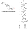
Structure of selected AKs.
Figure 4
Figure 4
Relationship of the water/1-octanol distribution coefficient (cLogD) vs. minimal growth inhibitory concentration (MIC).
Figure 5
Figure 5
Phase contrast (left panels ) and fluorescent (right panels) images of C. neoformans H99. (A) and (B): Images of cells treated with SYTOXTM green alone; (C) and (D): Images of cells treated with SYTOXTM green and 1% Triton X-100; (E) and (F): Images of cells treated with SYTOXTM green and compound 7 (1×MIC); (G) and (H): Images of cells treated with SYTOXTM green and compound 11 (1xMIC); (I) and (J): Images of cells treated with SYTOXTM green and compound 13 (256 μg/mL).
Figure 6
Figure 6
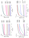
Kinetic membrane permeabilization of C. neoformans H99. (A) Cells treated with 4″,6″-diaryl AKs using SYTOXTM green; (B) cells treated with 4″,6″-diaryl AKs using PI; (C) cells treated with 4″,6″-dialkyl AKs using SYTOXTM green; (D) cells treated with 4″,6″-dialkyl AKs using PI. The figure with standard deviation is available in Supplementary Materials.
Figure 7
Figure 7
Kinetic membrane permeabilization of C. neoformans H99 using varied concentrations of AKs. (A) Cells treated with compound 7 using SYTOXTM green; (B) cells treated with compound 7 using PI; (C cells treated with compound 10 using SYTOXTM green; (D): cells treated with compound 10 using PI; (E cells treated with compound 11 using SYTOXTM green; (F): cells treated with compound 11 using PI. The figure with standard deviation is available in Supplementary Materials.
Figure 8
Figure 8
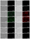
Images of C. neoformans treated with varied concentrations of compound 11. (A1,A2) Cells treated with only SYTOXTM green; (B1,B2) cells treated with Triton X-100 and SYTOXTM green; (C1,C2) cells treated with 0.5×MIC of 11 and SYTOXTM green; (D1,D2) cells treated with 1×MIC of 11 and SYTOXTM green; (E1,E2) cells treated with 2×MIC of 11 and SYTOXTM green; (F1,F2) cells treated with 4×MIC of 11 and SYTOXTM green; (G1,G2) cells treated with 8×MIC of 11 and SYTOXTM green; (H1,H2) cells treated with only PI; (I1,I2) cells treated with Triton X-100 and PI; (J1,J2) cells treated with 0.5×MIC of 11 and PI; (K1,K2) cells treated with 1×MIC of 11 and PI; (L1,L2) cells treated with 2×MIC of 11 and PI; (M1,M2) cells treated with 4×MIC of 11 and PI; (N1,N2) cells treated with 8×MIC of 11 and PI.
Figure 9
Figure 9
Reactive oxygen species (ROS) study for 4″,6″-disubstituted AKs and compound 14 structure.

References

    1. Brown G.D., Denning D.W., Levitz S.M. Tackling human fungal infections. Science. 2012;336:647. doi: 10.1126/science.1222236. - DOI - PubMed
    1. Armstrong-James D., Meintjes G., Brown G.D. A neglected epidemic: Fungal infections in HIV/AIDS. Trends Microbiol. 2014;22:120–127. doi: 10.1016/j.tim.2014.01.001. - DOI - PubMed
    1. Dutcher J.D. The discovery and development of amphotericin b. Chest. 1968;54:296–298. doi: 10.1378/chest.54.Supplement_1.296. - DOI - PubMed
    1. Donovick R., Gold W., Pagano J.F., Stout H.A. Amphotericins a and b, antifungal antibiotics produced by a streptomycete. I. In vitro studies. Antibiot. Annu. 1955;3:579–586. - PubMed
    1. Lyman C.A., Walsh T.J. Systemically administered antifungal agents. Drugs. 1992;44:9–35. doi: 10.2165/00003495-199244010-00002. - DOI - PubMed

MeSH terms