Review
. 2019 May 16;11(5):448.
doi: 10.3390/v11050448.

Antiviral RNAi in Insects and Mammals: Parallels and Differences

Affiliations
Review

Antiviral RNAi in Insects and Mammals: Parallels and Differences

Susan Schuster et al. Viruses. .

Abstract

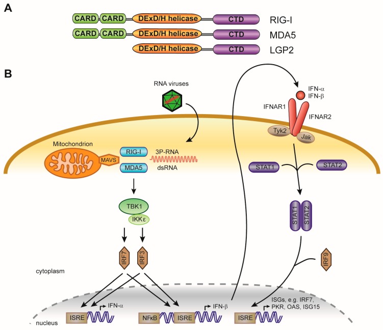
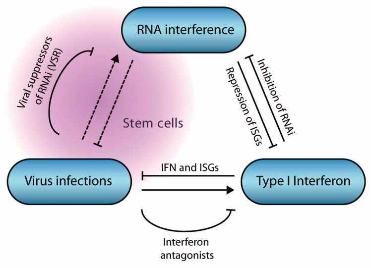
The RNA interference (RNAi) pathway is a potent antiviral defense mechanism in plants and invertebrates, in response to which viruses evolved suppressors of RNAi. In mammals, the first line of defense is mediated by the type I interferon system (IFN); however, the degree to which RNAi contributes to antiviral defense is still not completely understood. Recent work suggests that antiviral RNAi is active in undifferentiated stem cells and that antiviral RNAi can be uncovered in differentiated cells in which the IFN system is inactive or in infections with viruses lacking putative viral suppressors of RNAi. In this review, we describe the mechanism of RNAi and its antiviral functions in insects and mammals. We draw parallels and highlight differences between (antiviral) RNAi in these classes of animals and discuss open questions for future research.

Keywords: RNA interference; RNA virus; antiviral defense; innate immunity; interferon; small interfering RNA; stem cells.


Conflict of interest statement

The authors declare no conflict of interest.

Figures

Figure 1
Figure 1
The small interfering RNA (siRNA) pathway in Drosophila melanogaster. Double-stranded RNA precursors of different sources are processed by Dicer-2 into short interfering RNAs of ~21 nt in size. The siRNA duplex is loaded into an Argonaute2 containing RISC complex, where one strand (passenger) is degraded, and the guide strand is retained. The guide strand mediates target RNA recognition through Watson-Crick base pairing, followed by target cleavage (slicing) by Argonaute. Loqs-PD is required for endo-siRNA biogenesis, but dispensable for viral siRNA (vsiRNA) biogenesis.
Figure 2
Figure 2
Dicer proteins process double-stranded RNA (dsRNA) into small interfering RNA (siRNA). (A) Schematic representation of the domain organization of human Dicer protein [40]. RIIIa, RNase-IIIa; RIIIb, RNase-IIIb (B) Cryo-EM structure of human Dicer. Protein domains are colored in accordance to the scheme in A. The structure was determined by Liu et al. [41], and the published PDB file (5ZAM) was edited in Yasara View [42]. Drosophila Dicer-2 has a similar domain structure and L-shaped Cryo-EM structure as human Dicer [40]. (C) Schematic representation of the recognition and cleavage of dsRNA with a 3’ overhang and dsRNA with blunt termini by Drosophila Dicer-2, proposed by Sinha and colleagues [40]. Substrates with a 3’ overhang were proposed to bind the PAZ-Platform domains (referred to as PAZ in panel A) via the 3’ terminal overhang. Blunt-ended termini bind to the helicase domain and the dsRNA threads through this domain, after which cleavage occurs by the two RNaseIII domains. The latter mode results in processive, ATP-dependent cleavage of dsRNA and may contribute to efficient production of vsiRNAs for antiviral defense.
Figure 3
Figure 3
Argonaute proteins are at the core of small RNA silencing pathways. (A) Schematic representation of the domain organization of mammalian Argonaute and the conserved residues required for slicer activity. (B) Crystal structure of human AGO2 in association with a guide RNA and a target RNA base pairing from nucleotide 2 to 8. Protein domains are colored in accordance to the scheme in A. The structure was determined by Schirle and colleagues [49] and the published PDB file (4W5Q) was edited in Yasara View. (C) Schematic representation of target slicing by Argonaute proteins.
Figure 4
Figure 4
The RNA interference (RNAi) pathway in mammals. A single Dicer protein processes long dsRNA into siRNAs and pre-miRNAs into miRNA duplexes. These small RNAs are loaded into an Argonaute containing RISC complex, from which one of the strands is eliminated and degraded. The other strand, referred to as guide strand (for siRNAs) or the mature miRNA (for miRNAs), is retained and used to guide Argonaute onto target RNAs, resulting in cleavage (siRNA) or translational inhibition or target RNA destabilization (miRNA). The scheme shows the cytoplasmic stage of the miRNA pathway; the nuclear stage (pri-miRNA transcription, processing, and pre-miRNA nuclear export) is not shown.
Figure 5
Figure 5
Cytosolic recognition of foreign nucleic acids and activation of interferon stimulated genes (ISGs). (A) Domain structure of the cytosolic RNA sensors RIG-I and MDA5, showing the CARD signaling domain, the DExD/H-box helicase domain, and the C terminal domain (CTD). LGP2 lacks the CARD signaling domain. (B) Schematic representation of the interferon response in mammals. RIG-I and MDA5 recognize non-self viral RNA signatures, including the presence of a 5’ triphosphate moiety on RNA (3P-RNA) or long dsRNA. Upon detection of foreign nucleic acids, the CARD domains transduce the signal to mitochondrial antiviral-signaling protein (MAVS) located at mitochondrial membranes, leading to the phosphorylation and activation interferon response factors (IRF) 3 and 7. Upon activation, IRF3 and IRF7 form homodimers and translocate to the nucleus, where they bind Interferon-Stimulated Response Elements (ISRE) to activate transcription of type I interferons (IFN-α and IFN-β). Type I IFNs translocate across the cell membrane, after which they signal in a paracrine or autocrine manner via the interferon-α/β receptor (consisting of two subunits, IFNAR1 and IFNAR2). This activates the JAK-STAT pathway, leading to phosphorylation of STAT transcription factors (signal transducer and activator of transcription). Phosphorylated STAT1 and STAT2 heterodimerize, and translocate to the nucleus to activate expression of broad range of ISGs.
Figure 6
Figure 6
Interactions between viruses, RNA interference (RNAi), and the interferon pathway in mammals. Virus infection induces the expression of type I interferons, leading to the expression of Interferon stimulated genes (ISGs) that collectively restrict virus infection. The interferon pathway inhibits RNAi via multiple mechanisms, whereas miRNAs inhibit expression of ISGs. Virus infection induces an antiviral RNAi response under specific conditions, in stem cells or in absence of viral suppressors of RNAi.

Similar articles

Cited by

References

    1. Fire A., Xu S., Montgomery M.K., Kostas S.A., Driver S.E., Mello C.C. Potent and specific genetic interference by double-stranded RNA in Caenorhabditis elegans. Nature. 1998;391:806–811. doi: 10.1038/35888. - DOI - PubMed
    1. Ghildiyal M., Zamore P.D. Small silencing RNAs: An expanding universe. Nat. Rev. Genet. 2009;10:94–108. doi: 10.1038/nrg2504. - DOI - PMC - PubMed
    1. Schirle N.T., Kinberger G.A., Murray H.F., Lima W.F., Prakash T.P., MacRae I.J. Structural analysis of human Argonaute-2 bound to a modified siRNA guide. J. Am. Chem. Soc. 2016;138:8694–8697. doi: 10.1021/jacs.6b04454. - DOI - PMC - PubMed
    1. Elkayam E., Kuhn C.D., Tocilj A., Haase A.D., Greene E.M., Hannon G.J., Joshua-Tor L. The structure of human argonaute-2 in complex with miR-20a. Cell. 2012;150:100–110. doi: 10.1016/j.cell.2012.05.017. - DOI - PMC - PubMed
    1. Ketting R.F. The many faces of RNAi. Dev. Cell. 2011;20:148–161. doi: 10.1016/j.devcel.2011.01.012. - DOI - PubMed

Publication types

LinkOut - more resources