Review
. 2019 Sep 8:73:43-67.
doi: 10.1146/annurev-micro-020518-115907. Epub 2019 May 17.

Posttranscription Initiation Control of Gene Expression Mediated by Bacterial RNA-Binding Proteins

Affiliations
Review

Posttranscription Initiation Control of Gene Expression Mediated by Bacterial RNA-Binding Proteins

Paul Babitzke et al. Annu Rev Microbiol. .

Abstract

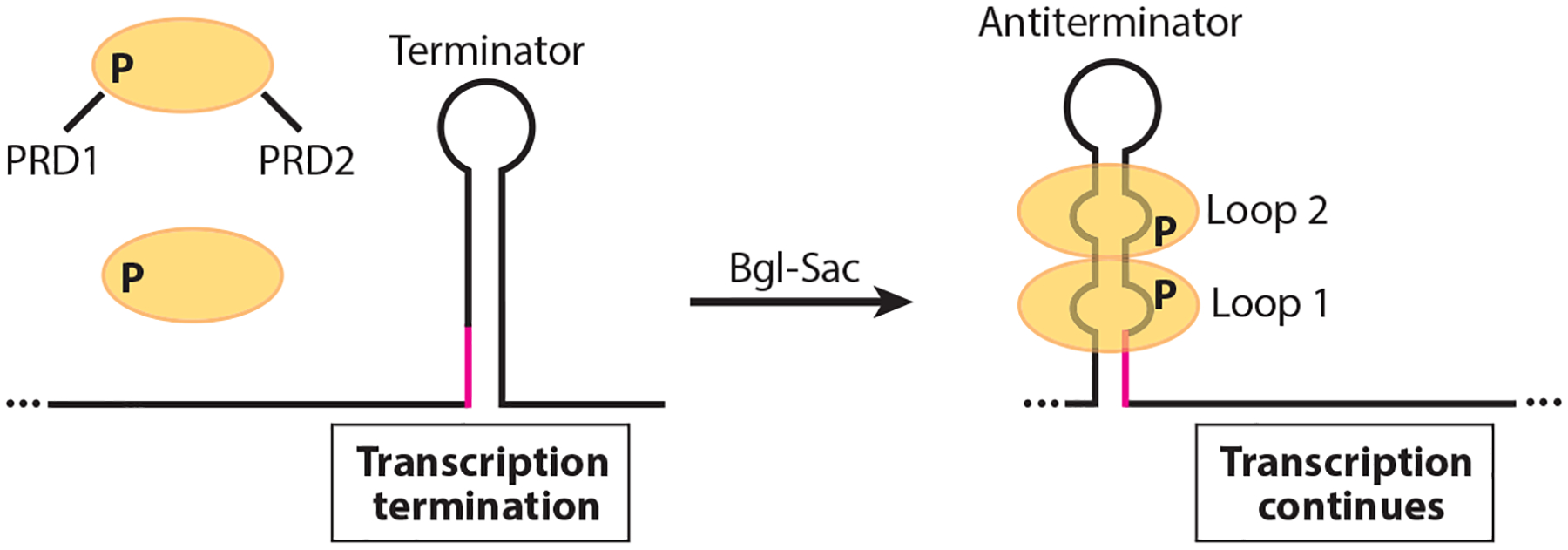
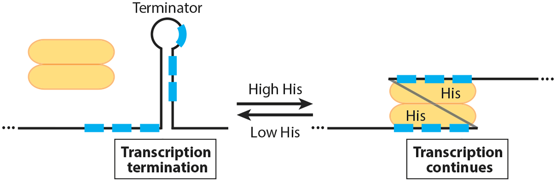
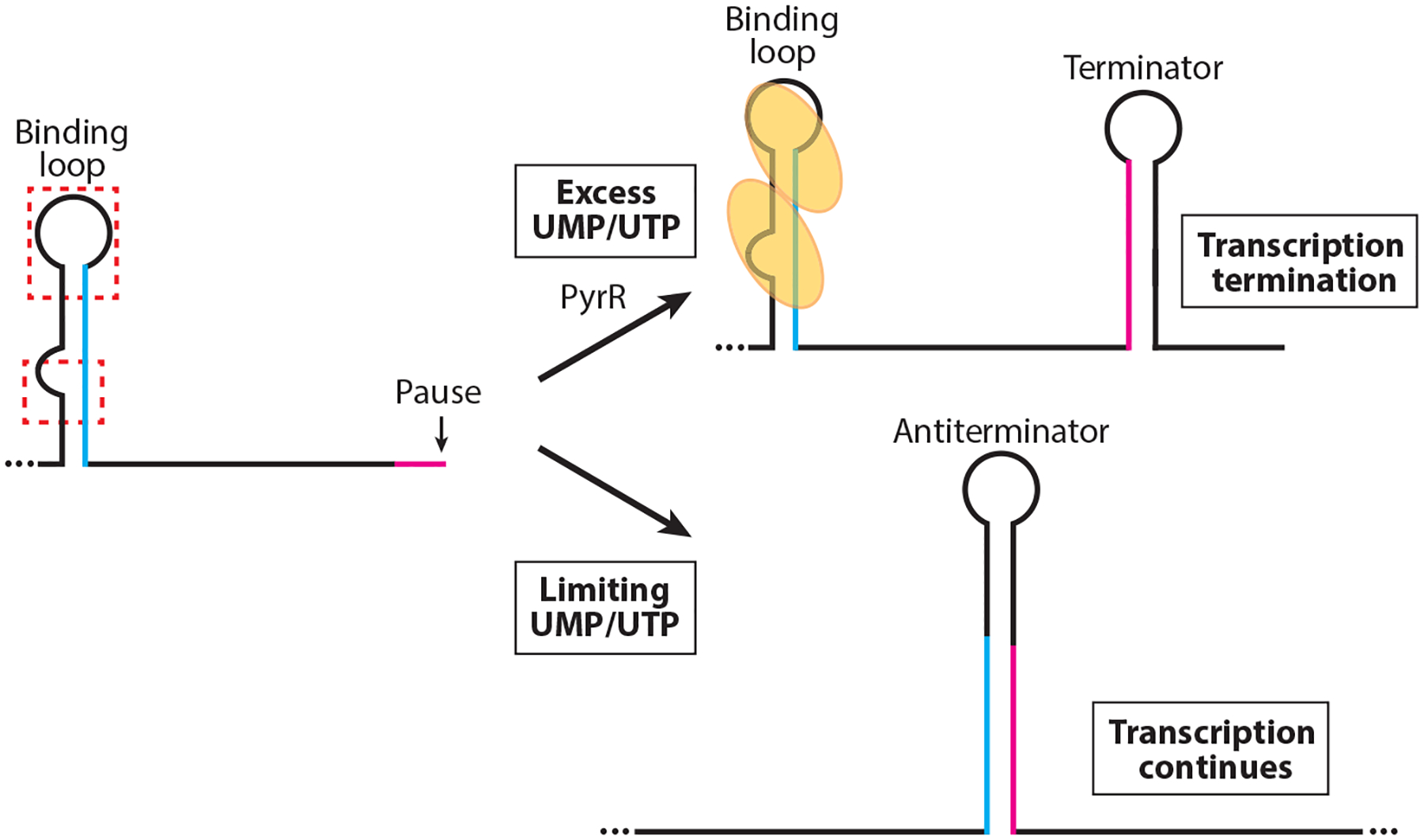
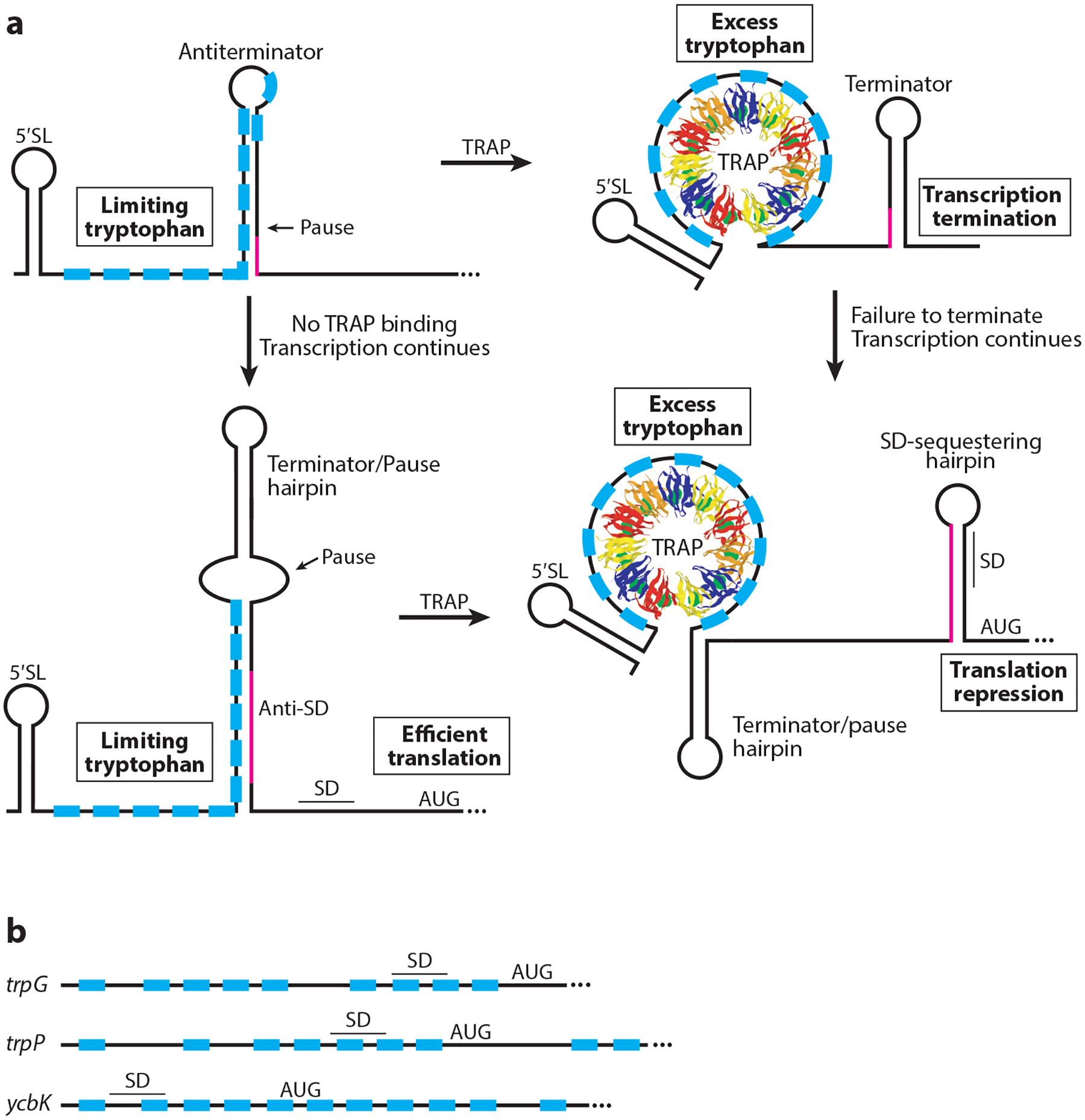
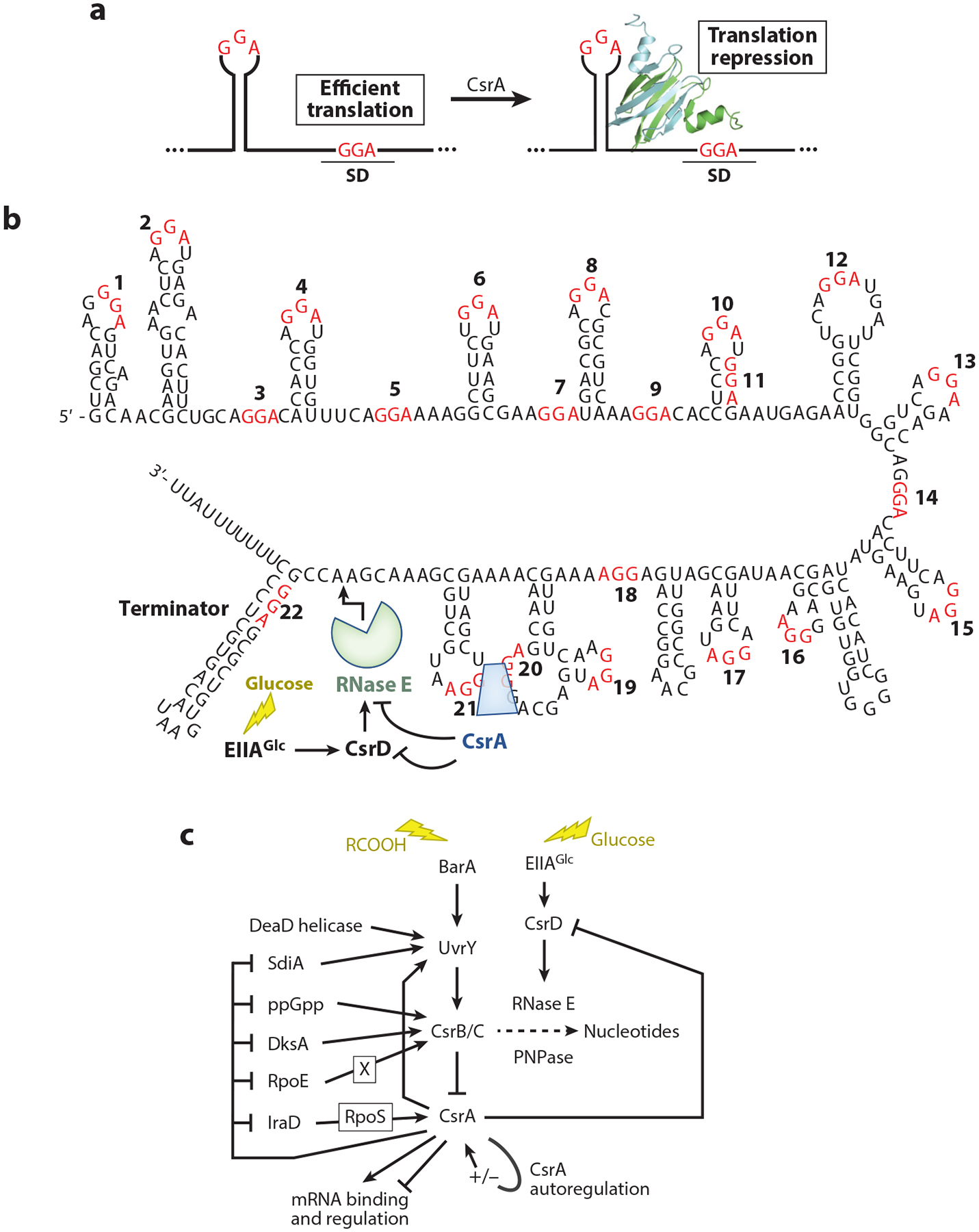
RNA-binding proteins play vital roles in regulating gene expression and cellular physiology in all organisms. Bacterial RNA-binding proteins can regulate transcription termination via attenuation or antitermination mechanisms, while others can repress or activate translation initiation by affecting ribosome binding. The RNA targets for these proteins include short repeated sequences, longer single-stranded sequences, RNA secondary or tertiary structure, and a combination of these features. The activity of these proteins can be influenced by binding of metabolites, small RNAs, or other proteins, as well as by phosphorylation events. Some of these proteins regulate specific genes, while others function as global regulators. As the regulatory mechanisms, components, targets, and signaling circuitry surrounding RNA-binding proteins have become better understood, in part through rapid advances provided by systems approaches, a sense of the true nature of biological complexity is becoming apparent, which we attempt to capture for the reader of this review.

Keywords: RNA-binding protein; antitermination; attenuation; gene regulation; sRNA; translation.


Figures

Figure 1
Figure 1
Model of the Bgl-Sac family antitermination mechanism. In the absence of the cognate carbon source the antitermination protein (yellow ovals) is rendered inactive by phosphorylation of PRD1. In the presence of the carbon source, PRD1 is dephosphorylated and PRD2 is phosphorylated, resulting in protein dimerization. The active dimer then binds to its cognate RNA target, thereby stabilizing an otherwise weak antiterminator structure. As a consequence, an overlapping intrinsic terminator is unable to form (overlap in magenta), leading to transcription of the operon involved in catabolism of the cognate carbon source. In the absence of bound protein, the terminator hairpin forms and transcription halts upstream of the coding sequence.
Figure 2
Figure 2
Model of the HutP antitermination mechanism. Under low histidine conditions, the HutP dimer (yellow) is inactive and an intrinsic terminator causes transcription to terminate in the hutP-hutH intercistronic region. In the presence of excess histidine, histidine-activated HutP binds to six NAG triplet repeats (blue boxes) in the nascent hut transcript, thereby preventing formation of the intrinsic terminator such that transcription continues into the downstream histidine utilization genes.
Figure 3
Figure 3
Model of the PyrR-mediated transcription attenuation mechanism. During transcription RNA polymerase pauses downstream of the PyrR-binding loop and within the overlap between the antiterminator and terminator structures (magenta). PyrR (yellow ovals) is not activated in limiting UMP/UTP conditions and does not bind to the binding loops in the 5′ UTR, or to the pyrR-pyrP and pyrP-pyrB intergenic regions. Once RNA polymerase resumes transcription, the antiterminator forms, which prevents formation of the mutually exclusive terminator hairpin (overlap in blue). In excess UMP/UTP conditions, UMP/UTP-activated PyrR binds to two regions of the binding loop (red dashed boxes). Pausing allows additional time for PyrR binding. Bound PyrR promotes transcription termination by preventing formation of the antiterminator structure.
Figure 4
Figure 4
Models of TRAP-mediated transcription attenuation and translation repression mechanisms. The 11 TRAP subunits are shown in different colors, and tryptophan is shown as green spheres. (a, Top) Transcription attenuation mechanism. TRAP is not activated in limiting tryptophan conditions and does not bind to the RNA. Thus formation of the antiterminator results in transcription of the downstream genes. In excess tryptophan conditions, tryptophan-activated TRAP binds to the triplet repeats (blue boxes). Pausing may provide additional time for TRAP binding. Bound TRAP prevents formation of the antiterminator, resulting in transcription termination upstream of the coding sequences. Overlap between the antiterminator and terminator structures is shown in magenta. (a, Bottom) trpE translation control model. In tryptophan-limiting conditions the RNA adopts a structure such that the trpE SD sequence is single stranded and available for ribosome binding. When tryptophan is in excess, tryptophan-activated TRAP binds to its target sequence such that the trpE SD–sequestering hairpin forms, causing translational repression. RNA polymerase pausing provides additional time for TRAP binding. Overlap between the two alternative structures is shown in magenta. (b) TRAP-binding sites overlapping the trpG, trpP, and ycbK SD sequence and translation initiation regions. Bound TRAP represses translation of each gene. Abbreviations: 5′ SL, 5′ stem-loop; SD, Shine-Dalgarno; TRAP, trp RNA–binding attenuation protein. Adapted with permission from Reference .
Figure 5
Figure 5
Csr-mediated regulatory pathways. (a) Common CsrA-mediated translation repression mechanism. CsrA dimers, depicted as green and blue ribbons, bind to two sites, one of which overlaps the Shine-Dalgarno (SD) sequence such that bound CsrA blocks ribosome binding. GGA motifs of CsrA-binding sites are in red. (b) CsrB sequence, secondary structure, and RNA-decay pathway. GGA motifs are numbered and shown in red. (c) Circuitry influencing the Csr system. The Csr system of Escherichia coli includes CsrA autoregulation, negative-feedback loops among the Csr components, and reciprocal interactions with other global regulatory systems. Autoregulation and feedback loops fine-tune CsrA activity and support robust regulatory responses, while interactions with other global regulatory systems integrate the Csr system into stress responses. The CsrA ribbon diagram in panel a is adapted with permission from Reference .
Figure 6
Figure 6
Regulation by the RNA-binding protein RapZ and its two related small RNA targets, GlmZ and GlmY. (a) Regulatory circuitry and (b) RapZ:GlmZ recognition involving stem-loop 2 (SL2) promote RNase E recognition and sequence-independent cleavage at the single-stranded base-pairing site of GlmZ. GlmY competes with GlmZ for RapZ binding without undergoing RapZ-mediated RNase E cleavage.
Figure 7
Figure 7
Target small RNAs (sRNAs) of FinO/ProQ domain proteins. Putative ProQ-binding sites in yellow. Regions involved in base-pairing with target mRNAs are circled in red. Guanine-cytosine pairs are depicted by double bars, adenine-uracil pairs are depicted by single bars, and bars with circles represent guanine-uracil pairs. (a) cis-Acting FinP sRNA from Escherichia coli F plasmid, which is bound by FinO. (b) trans-Acting RocR sRNA from Legionella pneumophila, which is bound by RocC. (c) trans-Acting RaiZ sRNA from Salmonella enterica, which is dependent on ProQ.

References

    1. Adamson DN, Lim HN. 2013. Rapid and robust signaling in the CsrA cascade via RNA-protein interactions and feedback regulation. PNAS 110:13120–25 - PMC - PubMed
    1. Altegoer F, Rensing SA, Bange G. 2016. Structural basis for the CsrA-dependent modulation of translation initiation by an ancient regulatory protein. PNAS 113:10168–73 - PMC - PubMed
    1. Amster-Choder O 2005. The bgl sensory system: a transmembrane signaling pathway controlling transcriptional antitermination. Curr. Opin. Microbiol 8:127–34 - PubMed
    1. Amster-Choder O, Houman F, Wright A. 1989. Protein phosphorylation regulates transcription of the β-glucoside utilization operon in E. coli. Cell 58:847–55 - PubMed
    1. Amster-Choder O, Wright A. 1992. Modulation of the dimerization of a transcriptional antiterminator protein by phosphorylation. Science 257:1395–98 - PubMed

Publication types

MeSH terms

LinkOut - more resources