. 2019 May 17;20(1):386.
doi: 10.1186/s12864-019-5773-3.

A deficiency in SUMOylation activity disrupts multiple pathways leading to neural tube and heart defects in Xenopus embryos

Affiliations

A deficiency in SUMOylation activity disrupts multiple pathways leading to neural tube and heart defects in Xenopus embryos

Michelle M Bertke et al. BMC Genomics. .

Abstract

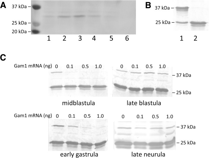
Background: Adenovirus protein, Gam1, triggers the proteolytic destruction of the E1 SUMO-activating enzyme. Microinjection of an empirically determined amount of Gam1 mRNA into one-cell Xenopus embryos can reduce SUMOylation activity to undetectable, but nonlethal, levels, enabling an examination of the role of this post-translational modification during early vertebrate development.

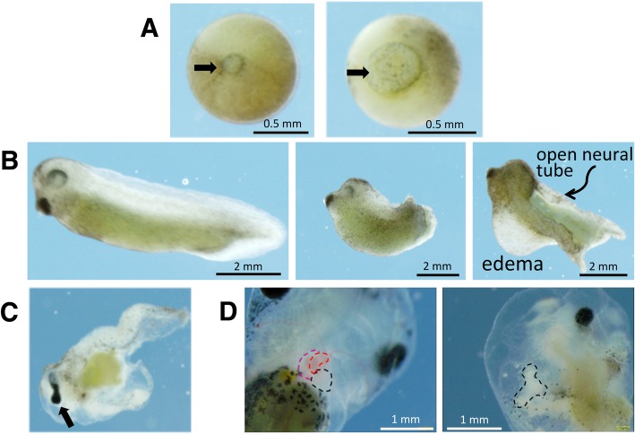
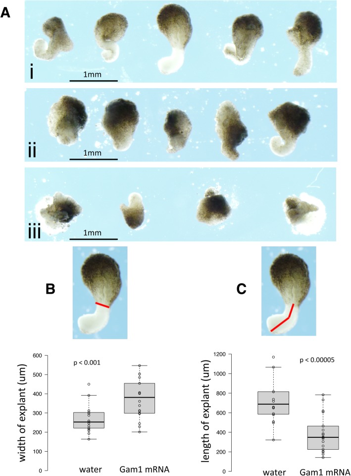
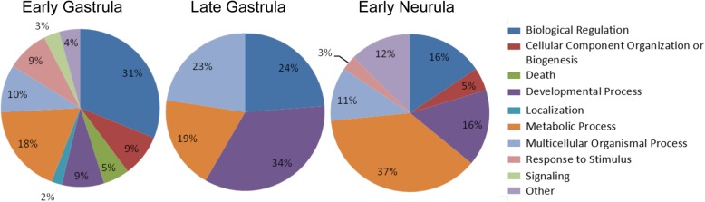
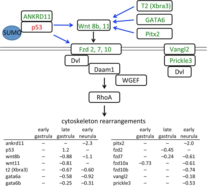
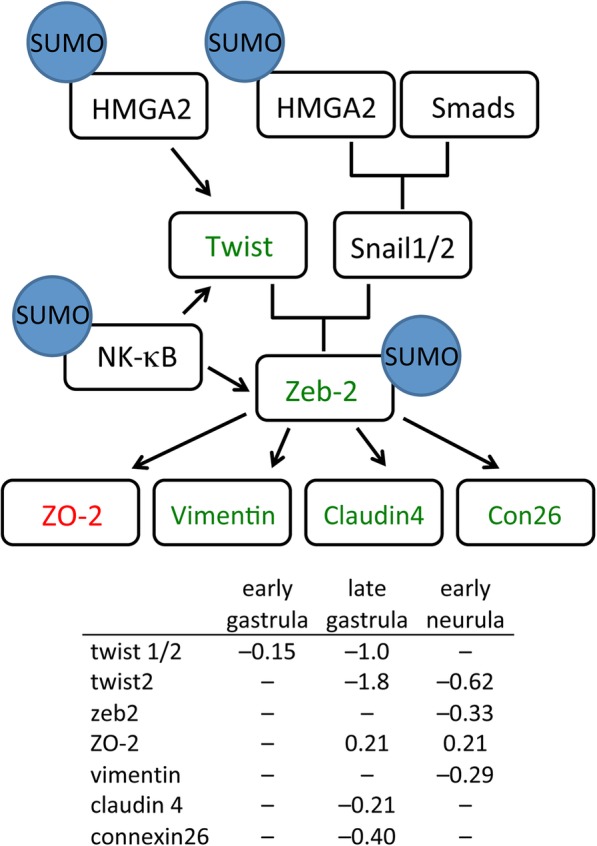
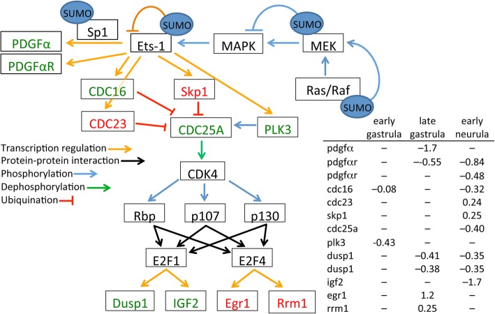
Results: We find that SUMOylation-deficient embryos consistently exhibit defects in neural tube and heart development. We have measured differences in gene expression between control and embryos injected with Gam1 mRNA at three developmental stages: early gastrula (immediately following the initiation of zygotic transcription), late gastrula (completion of the formation of the three primary germ layers), and early neurula (appearance of the neural plate). Although changes in gene expression are widespread and can be linked to many biological processes, three pathways, non-canonical Wnt/PCP, snail/twist, and Ets-1, are especially sensitive to the loss of SUMOylation activity and can largely account for the predominant phenotypes of Gam1 embryos. SUMOylation appears to generate different pools of a given transcription factor having different specificities with this post-translational modification involved in the regulation of more complex, as opposed to housekeeping, processes.

Conclusions: We have identified changes in gene expression that underlie the neural tube and heart phenotypes resulting from depressed SUMOylation activity. Notably, these developmental defects correspond to the two most frequently occurring congenital birth defects in humans, strongly suggesting that perturbation of SUMOylation, either globally or of a specific protein, may frequently be the origin of these pathologies.

Keywords: Congenital birth defects; Heart development; Neural tube development; Non-canonical Wnt signaling; Planar cell polarity; SUMO.


Conflict of interest statement

The authors declare that they have no competing interests.

Figures

Fig. 1
Fig. 1
Depletion of SUMOylation activity in embryos injected with Gam1 mRNA. a Expression levels of Gam1 protein during embryogenesis. One-cell embryos were injected with mRNA (0.5 ng) encoding Gam1 with an N-terminal myc tag. Whole cell protein extract was prepared from embryos at the indicated Nieuwkoop–Faber stage and 25 μg taken for western blot analysis using anti-myc antibody. Lanes: 1, midblastula; 2, late blastula; 3, early gastrula; 4, early neurula; 5, late neurula; 6, water injected control. b In vitro SUMOylation assay. All assays contained Ubc9 (E2 enzyme), SUMO1, ATP, and substrate peptide (25 kDa) with (lane 1) or without (lane 2) purified E1 enzyme (500 nM) added. Samples were analyzed by western blot using an antibody specific for a 25 kDa SUMO substrate peptide. c Assays for E1 activity. One-cell embryos were injected with the indicated amount of Gam1 mRNA and allowed to develop to the indicated stage. Whole cell extract was prepared from 20 embryos and an equivalent amount of protein (25 μg) was used as a source of E1 enzyme for each assay
Fig. 2
Fig. 2
Developmental defects of Gam1-injected embryos. a Control (left) and Gam1-injected (right) late gastrula stage embryos showing delayed closure of the blastopore (arrow) in the latter. b Stage 35 (50 hpf) control embryo (left), Gam1 embryo with shortened A–P axis (center), Gam1 embryo with an open neural tube and edema around the heart (right). c An example of an eye defect (cyclopia). d Failure of the heart tube to initiate looping. (Left) A control embryo (96 hpf) in which the two atria (red) and ventricle (black) have fully developed compared to (right) a Gam1 embryo in which the heart tube has failed to undergo looping
Fig. 3
Fig. 3
Gam1 disrupts convergence and extension. a Examples of Keller sandwich explants prepared from embryos injected with (i) water, (ii) 2.5 ng Gam1 mRNA, (iii) 5 ng Gam1 mRNA. Explants from embryos injected with water (n = 17) or 2.5 ng Gam1 mRNA (n = 18) were measured to quantify effects on b convergence and c extension. The red lines designate the lengths measured in the explants. Center lines in plots show the medians; box limits indicate the 25th and 75th percentiles; whiskers extend 1.5 times the interquartile range from the 25th and 75th percentiles
Fig. 4
Fig. 4
Biological processes most affected by loss of SUMOylation activity. Functional enrichment analysis of differentially expressed genes at each time point based on GeneGo categorization of annotated protein function. Only selected groups containing the largest number of genes are presented
Fig. 5
Fig. 5
Changes in gene expression in the non-canonical Wnt/PCP signaling pathway. Decreased mRNA levels in Gam1 embryos relative to control embryos is represented by green and increased levels by red. Documented targets of SUMOylation are indicated. Transcriptional regulation is denoted by blue arrows and signaling through protein-protein interactions by black arrows. Log (2) changes in gene expression are listed for the three experimental time points
Fig. 6
Fig. 6
Changes in gene expression in the Twist/Snail pathway. Decreased mRNA levels in Gam1 embryos relative to control embryos is represented by green and increased levels by red. Documented targets of SUMOylation are indicated. Transcriptional regulation is denoted by black arrows. Log (2) changes in gene expression are listed for the three experimental time points
Fig. 7
Fig. 7
Changes in gene expression in the Ets-1 pathway. Decreased mRNA levels in Gam1 embryos relative to control embryos is represented by green and increased levels by red. Documented targets of SUMOylation are indicated. Connections by arrows represent activation and T-bars represent inhibition. Log (2) changes in gene expression are listed for the three experimental time points

Similar articles

Cited by

References

    1. Chymkowitch P, Nguea PA, Enserink JM. SUMO-regulated transcription: challenging the dogma. BioEssays. 2015;37:1095–1105. doi: 10.1002/bies.201500065. - DOI - PubMed
    1. Eifler K, Vertegaal AC. Mapping the SUMOylated landscape. FEBS J. 2015;282:3669–3680. doi: 10.1111/febs.13378. - DOI - PMC - PubMed
    1. Flotho A, Melchior F. Sumoylation: a regulatory protein modification in health and disease. Annu Rev Biochem. 2013;82:357–385. doi: 10.1146/annurev-biochem-061909-093311. - DOI - PubMed
    1. Hendriks IA, D'Souza RC, Yang B, Verlaan-de Vries M, Mann M, Vertegaal AC. Uncovering global SUMOylation signaling networks in a site-specific manner. Nat Struct Mol Biol. 2014;21:927–936. doi: 10.1038/nsmb.2890. - DOI - PMC - PubMed
    1. Makhnevych T, Sydorskyy Y, Xin X, Srikumar T, Vizeacoumar FJ, Jeram SM, Li Z, Bahr S, Andrews BJ, Boone C, et al. Global map of SUMO function revealed by protein-protein interaction and genetic networks. Mol Cell. 2009;33:124–135. doi: 10.1016/j.molcel.2008.12.025. - DOI - PubMed