. 2019 May 17;10(6):389.
doi: 10.1038/s41419-019-1620-3.

MALAT1 regulates miR-34a expression in melanoma cells

Affiliations

MALAT1 regulates miR-34a expression in melanoma cells

Fei Li et al. Cell Death Dis. .

Abstract

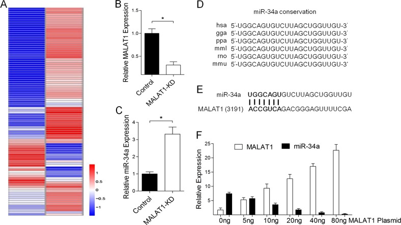
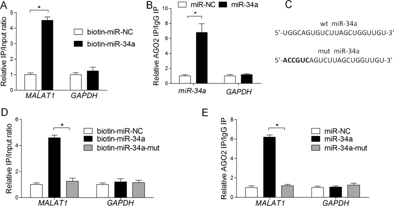
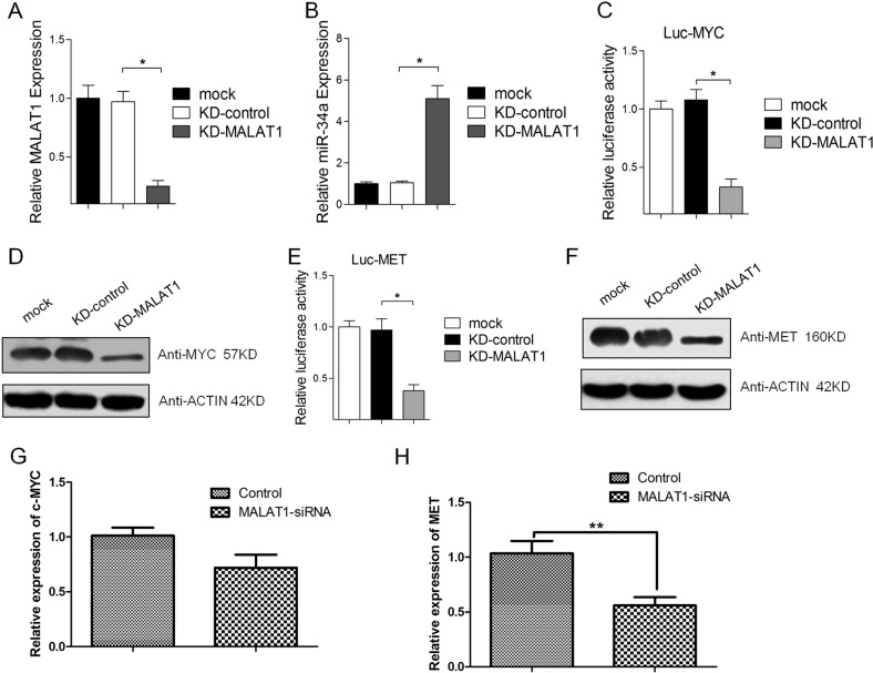
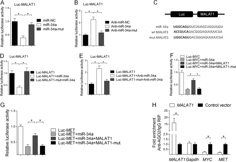
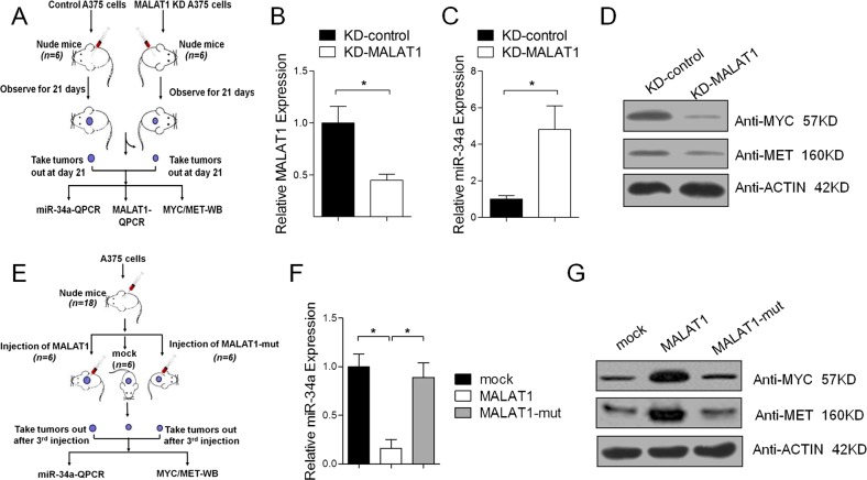
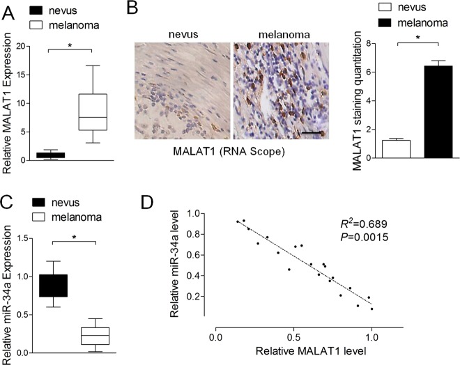
Melanoma is one of the most common skin malignancies. Both microRNAs and long non-coding RNAs (lncRNAs) have critical roles in the progression of cancers, including melanoma. However, the underlying molecular mechanism has not been fully characterized. We demonstrated that miR-34a is negatively correlated with MALAT1 in melanoma cells and tumor specimens. Interestingly, MALAT1, which contains functional sequence-specific miR-34a-binding sites, regulates miR-34a stability in melanoma cells and in vivo. Importantly, MALAT1 was significantly enriched in the Ago2 complex, but not when the MALAT1-binding site of miR-34a was mutated. Furthermore, MALAT1 could be shown to regulate c-Myc and Met expression by functioning as a miR-34a sponge. Our results reveal an unexpected mode of action for MALAT1 as an important regulator of miR-34a.


Conflict of interest statement

The authors declare that they have no conflict of interest.

Figures

Fig. 1
Fig. 1. miR-34a is negatively correlated with MALAT1 in melanoma cells.
a Heat map of differentially expressed miRNAs in A375 cells transfected with the negative control siRNA vector and MALAT1 siRNA (MALAT1-KD). A375 cells were transfected with 20 nM control siRNA or MALAT1 siRNA, and after a 48 h incubation, b MALAT1 and c miR-34a expression levels were analyzed in a quantitative real-time polymerase chain reaction (qRT-PCR) assay, with the expression data normalized against that of the control. d Aligned miR-34a sequences from nine species. e Bioinformatics analyses predicted the binding sites between MALAT1 and miR-34a. f A375 cells were transfected with different concentrations of MALAT1 expression vectors (0, 5, 10, 20, 40 , and 80 ng), after which miR-34a expression was analyzed in a qRT-PCR assay
Fig. 2
Fig. 2. MALAT1 directly binds to miR-34a in melanoma cells.
a A streptavidin-capture assay was performed for A375 cells transfected with biotin-miR-NC or biotin-miR-34a, followed by a quantitative real-time polymerase chain reaction (qRT-PCR) assay to analyze MALAT1 and GAPDH mRNA levels. b An Ago2 immunoprecipitation experiment was completed for A375 cells transfected with control miRNA (miR-NC) or miR-34a, followed by a qRT-PCR assay to analyze the MALAT1 associated with Ago2. c Schematic representation of the wild-type miR-34a and mutated miR-34a. d Streptavidin -capture assay was performed for A375 cells transfected with biotin-miR-NC, biotin-miR-34a, or mutated biotin-miR-34a, followed by a qRT-PCR assay to analyze MALAT1 and GAPDH mRNA levels. e An Ago2 immunoprecipitation experiment was performed for A375 cells transfected with control miRNA (miR-NC), miR-34a, or mutated miR-34a, followed by a qRT-PCR assay to analyze the MALAT1 associated with Ago2
Fig. 3
Fig. 3. miR-34a target genes are regulated by MALAT1 in melanoma cell.
A375 cells were transfected with 20 nM control siRNA or MALAT1 siRNA. After a 48-h incubation, a MALAT1 and b miR-34a expression levels were analyzed in a quantitative real-time polymerase chain reaction (qRT-PCR) assay, with the expression data normalized against that of the control. After a 72-h incubation post-transfection, the luciferase activities of c Luc-c-Myc and e Luc-Met were analyzed in a luciferase reporter assay. After a 72-h incubation post-transfection, d c-Myc and f Met protein levels were analyzed in a western blot, and g) c-Myc and (h) Met mRNA levels were analyzed in a qRT-PCR assay
Fig. 4
Fig. 4. MALAT1 functions as a miR-34a sponge in A375 melanoma cells.
a Luciferase reporter constructs: Wild-type MALAT1 (wt-MALAT1) and MALAT1 with mutations in the miR-34a-binding sites (Luc-MALAT1-mut) were inserted into the psiCHECK-2 vector. Letters in bold font represent mutation sites. b Relative luciferase activity of A375 cells with psiCHECK-2 containing wt-MALAT1 co-transfected with control miRNA (miR-NC), miR-34a, or mutated miR-34a. c Relative luciferase activity of A375 cells with psiCHECK-2 containing wt-MALAT1 co-transfected with control miRNA (anti-miR-NC), anti-miR-34a, or mutated miR-34a (anti-miR-34a-mut). d Relative luciferase activity of A375 cells with psiCHECK-2 containing wt-MALAT1 and mutated MALAT1 (Luc-MALAT1-mut) co-transfected with miR-34a. e Relative luciferase activity of A375 cells with psiCHECK-2 containing wt-MALAT1 and mutated MALAT1 (Luc-MALAT1-mut) co-transfected with anti-miR-34a. f Relative luciferase activity of A375 cells with psiCHECK-2 containing c-Myc co-transfected with miR-34a and wt-MALAT1 or mutated MALAT1 (MALAT1-mut) expression vectors. g Relative luciferase activity of A375 cells with psiCHECK-2 containing Met co-transfected with miR-34a and wt-MALAT1 or mutated MALAT1 (MALAT1-mut) expression vectors. h An Ago2 immunoprecipitation experiment was performed for A375 cells transfected with the control vector or MALAT1 expression vector, followed by a quantitative real-time polymerase chain reaction assay to analyze the MALAT1, GAPDH, c-Myc, and Met associated with Ago2
Fig. 5
Fig. 5. In vivo confirmation that MALAT1 functions as a miR-34a sponge.
a Schematic diagram of the animal experimental design. The A375 cells stably expressing either sh-lncRNA-MALAT1 or sh-NC were injected subcutaneously into the dorsal flanks of the indicated numbers of BALB/c nude mice. (bc) At 21 days after injection, the xenografts were dissected and examined in a quantitative real-time polymerase chain reaction (qRT-PCR) assay to analyze MALAT1 (b) and miR-34a (c) expression levels. d c-Myc and Met protein levels were analyzed in a western blot. e Schematic diagram of the animal experimental design. Stable A375 cells with pLVX-IRES-Puro-MALAT1 or pLVX-IRES-Puro-MALAT1-mut were injected subcutaneously into the dorsal flanks of 5-week-old male BALB/c nude mice. f Xenografts were dissected 21 days after injections, and MALAT1 and MALAT1-mut expression levels were analyzed in a qRT-PCR assay. g c-Myc and Met protein levels were analyzed in a western blot
Fig. 6
Fig. 6. The expression of miR-34a is inversely associated with MALAT1 in melanoma tissues.
a Relative MALAT1 expression levels in melanoma tissues (n = 20) and benign nevi (n = 20) were analyzed in a quantitative real-time polymerase chain reaction (qRT-PCR) assay. b RNAscope detection of MALAT1 expression in melanoma tissues (n = 20) and benign nevi (n = 20). Left panel: representative images. Scale bars: 100 µm. Right panel: data analysis. c Relative miR-34a expression levels in melanoma tissues (n = 20) and benign nevi (n = 20) were analyzed in a qRT-PCR assay. d miR-34a expression is inversely associated with MALAT1 in melanoma tissues (r2 = 0.689, P = 0.0015)

Similar articles

Cited by

References

    1. Siegel RL, Miller KD, Jemal A. Cancer statistics, 2017. CA Cancer J. Clin. 2017;67:7–30. doi: 10.3322/caac.21387. - DOI - PubMed
    1. Chen W, et al. Cancer statistics in China, 2015. CA Cancer J. Clin. 2016;66:115–132. doi: 10.3322/caac.21338. - DOI - PubMed
    1. De Vito C, et al. A TARBP2-dependent miRNA expression profile underlies cancer stem cell properties and provides candidate therapeutic reagents in ewing sarcoma. Cancer Cell. 2012;21:807–821. doi: 10.1016/j.ccr.2012.04.023. - DOI - PubMed
    1. Lin A, et al. The LINK-A lncRNA activates normoxic HIF1α signalling in triple-negative breast cancer. Nat. Cell. Biol. 2016;18:213–224. doi: 10.1038/ncb3295. - DOI - PMC - PubMed
    1. Melo SA, et al. Cancer exosomes perform cell-independent microRNA biogenesis and promote tumorigenesis. Cancer Cell. 2014;26:707–721. doi: 10.1016/j.ccell.2014.09.005. - DOI - PMC - PubMed

Publication types

MeSH terms