Review
. 2019 Aug;76(15):2933-2956.
doi: 10.1007/s00018-019-03140-2. Epub 2019 May 17.

Cellular consequences of arginine methylation

Affiliations
Review

Cellular consequences of arginine methylation

Benjamin M Lorton et al. Cell Mol Life Sci. 2019 Aug.

Abstract

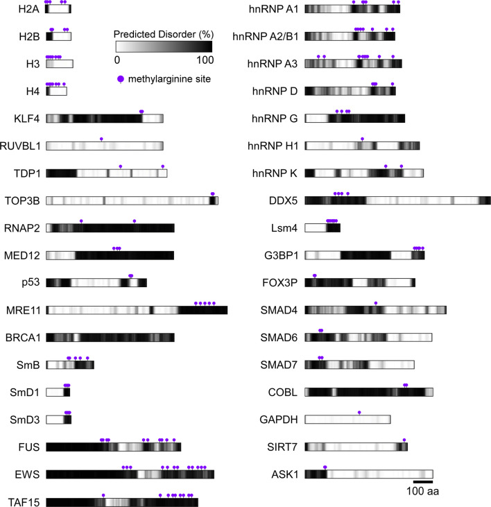
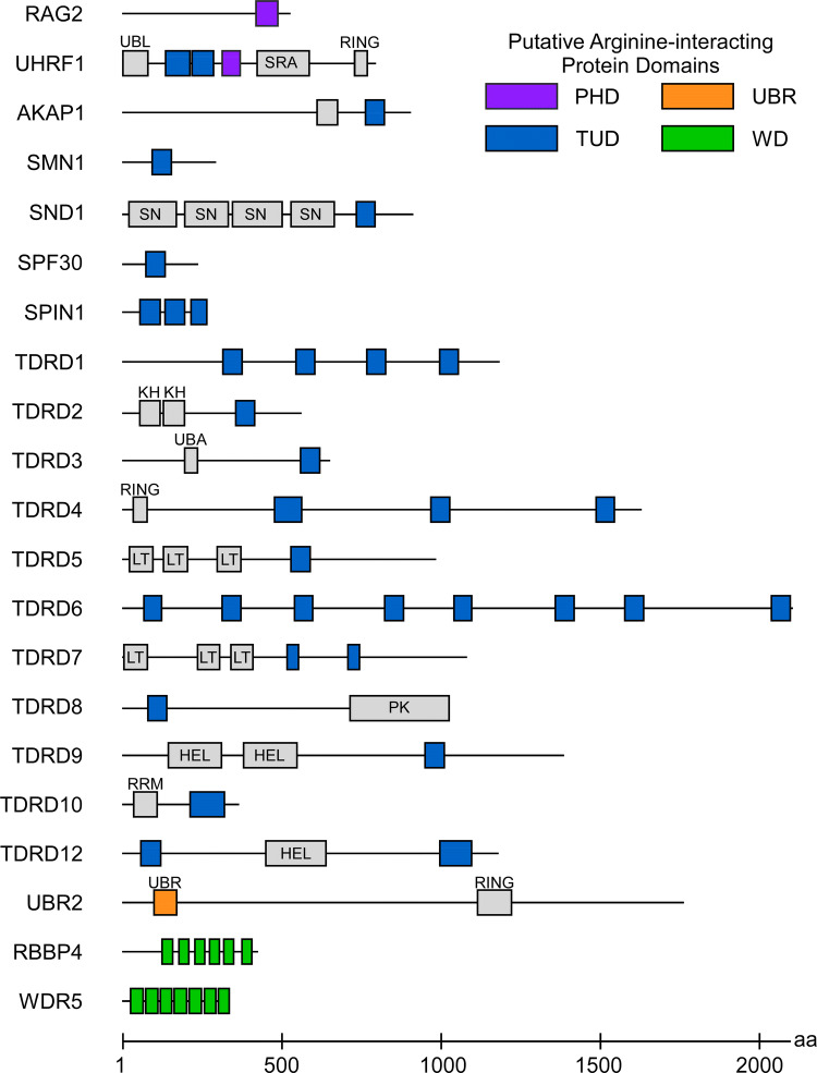
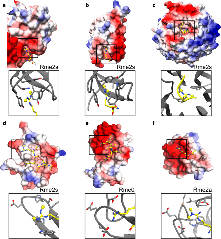
Arginine methylation is a ubiquitous post-translational modification. Three predominant types of arginine-guanidino methylation occur in Eukarya: mono (Rme1/MMA), symmetric (Rme2s/SDMA), and asymmetric (Rme2a/ADMA). Arginine methylation frequently occurs at sites of protein-protein and protein-nucleic acid interactions, providing specificity for binding partners and stabilization of important biological interactions in diverse cellular processes. Each methylarginine isoform-catalyzed by members of the protein arginine methyltransferase family, Type I (PRMT1-4,6,8) and Type II (PRMT5,9)-has unique downstream consequences. Methylarginines are found in ordered domains, domains of low complexity, and in intrinsically disordered regions of proteins-the latter two of which are intimately connected with biological liquid-liquid phase separation. This review highlights discoveries illuminating how arginine methylation affects genome integrity, gene transcription, mRNA splicing and mRNP biology, protein translation and stability, and phase separation. As more proteins and processes are found to be regulated by arginine methylation, its importance for understanding cellular physiology will continue to grow.

Keywords: Crosstalk; Histones; Liquid–liquid phase separation; Protein arginine methyltransferase; Ribonucleoprotein.


Figures

Fig. 1
Fig. 1
Arginine methylations and their chemical states. a All PRMTs catalyze the addition of one methyl group to one of the terminal ω nitrogens of the guanidinium side chain, producing monomethylarginine (Rme1 or MMA). Type I enzymes (PRMT1,2,3,4,6,8) catalyze a second methylation to the same ω nitrogen, producing asymmetric dimethylarginine (Rme2a or ADMA). Type II enzymes (PRMT5, 9) catalyze a second methylation to the ω′ nitrogen, producing symmetric dimethylarginine (Rme2s or SDMA). b Electrostatic potential maps of L-arginine and its methylated derivatives reveal a diffuse, positive characteristic. Red = electron dense, blue = electron poor
Fig. 2
Fig. 2
Arginine methylations are frequently found on protein intrinsically disordered regions. All arginine methylated proteins discussed in this review were analyzed with DISOPRED3. Predicted disordered regions are shown in grayscale on a per-amino acid basis (white = no predicted disorder, black = 100% predicted disorder). Known sites of arginine methylation are indicated with a purple flag. All proteins are full-length except MED12 (1618–2176 aa); RNAP2 (C-terminal 500aa); ASK1 (N-terminal 500aa); BRCA (400–900 aa); and COBL (C-terminal 500aa) All sequences shown to scale, as indicated at the bottom right
Fig. 3
Fig. 3
Selection of proteins with putative arginine-interacting domains. Proteins known to specifically bind to arginine are shown. Domains characterized to interact with arginine are colored as indicated; accessory domains are shown in gray. Helicase (HEL); K homology (KH); LOTUS (LT); (PK); Plant homeodomain (PHD) in purple; (RING); RNA-recognition motif (RRM); Staphylococcal nuclease (SN); SET and Ring-finger associated (SRA); Tudor and tudor-like (TUD) in blue; Ubiquitin-associated (UBA); Ubiquitin-like (UBL); Ubiquitin E3 ligase n-recognin (UBR) in orange; WD40 repeat (WD) in green. All sequences are shown to scale, as indicated at the bottom
Fig. 4
Fig. 4
Representative examples of characterized arginine-interacting domains. Coloumbic potential surface maps are depicted with interacting ligands. Black squares indicate arginine residues. Binding site details are shown in zoomed boxes. a Tudor domain, SND1 (anti-syn me2s conformation, PDB: 3OMC) b Tudor domain, SMN (syn-syn me2s conformation, PDB: 4A4E). c WD40 Repeat domain, WDR5 (PDB: 4A7J). d RAG2-PHD (PDB: 2V88). e UHRF1-PHD (PDB:3SOW). f UBR1 (PDB:5TDB)

References

    1. Minguez P, et al. Deciphering a global network of functionally associated post-translational modifications. Mol Syst Biol. 2012;8:599. doi: 10.1038/msb.2012.31. - DOI - PMC - PubMed
    1. Paik WK, Kim S. Protein methylase. I purification and properties of the enzyme. J Biol Chem. 1968;243(9):2108–2114. - PubMed
    1. Paik WK, Kim S. Enzymatic methylation of histones. Arch Biochem Biophys. 1969;134(2):632–637. doi: 10.1016/0003-9861(69)90327-0. - DOI - PubMed
    1. Evich M, et al. Effect of methylation on the side-chain pKa value of arginine. Protein Sci. 2016;25(2):479–486. doi: 10.1002/pro.2838. - DOI - PMC - PubMed
    1. Fuhrmann J, Clancy KW, Thompson PR. Chemical biology of protein arginine modifications in epigenetic regulation. Chem Rev. 2015;115(11):5413–5461. doi: 10.1021/acs.chemrev.5b00003. - DOI - PMC - PubMed