Review
. 2019 Aug;17(8):1229-1239.
doi: 10.1111/jth.14487. Epub 2019 Jun 17.

Blood coagulation factor Va's key interactive residues and regions for prothrombinase assembly and prothrombin binding

Affiliations
Review

Blood coagulation factor Va's key interactive residues and regions for prothrombinase assembly and prothrombin binding

Mark Schreuder et al. J Thromb Haemost. 2019 Aug.

Abstract

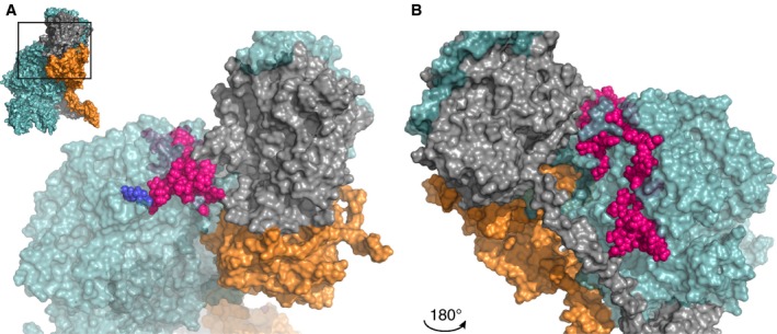
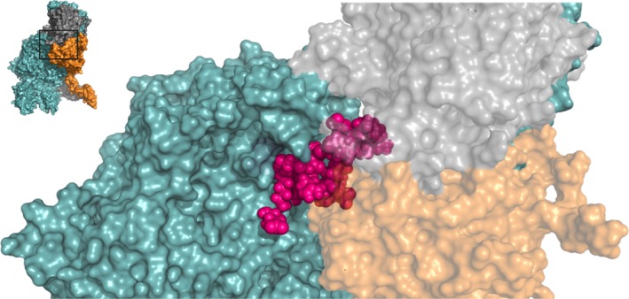
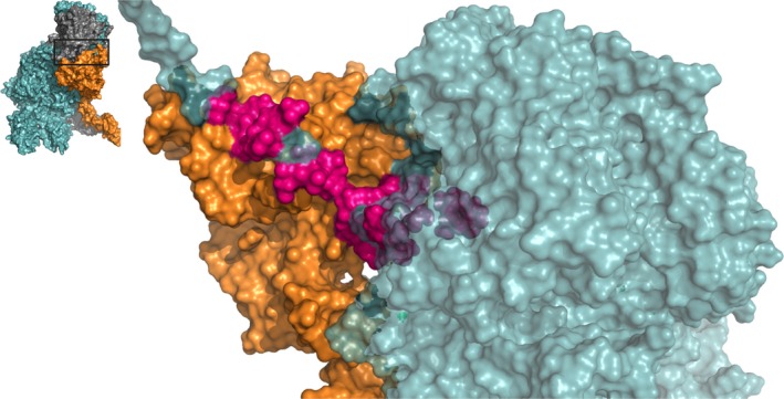
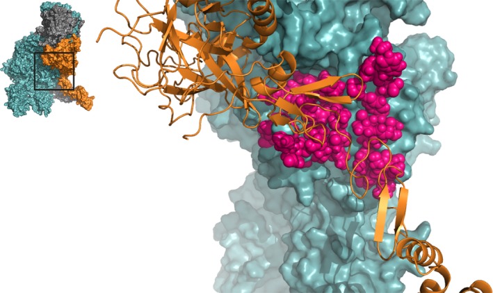
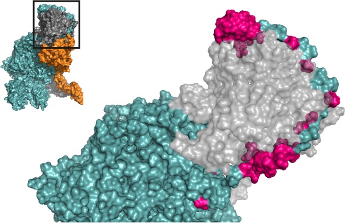
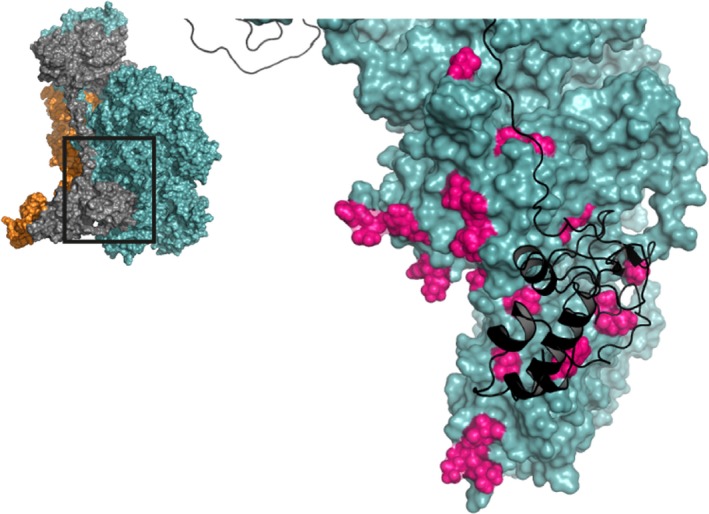
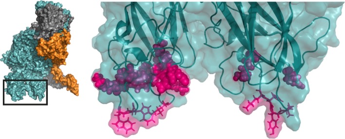
Blood coagulation factor Va serves an indispensable role in hemostasis as cofactor for the serine protease factor Xa. In the presence of an anionic phospholipid membrane and calcium ions, factors Va and Xa assemble into the prothrombinase complex. Following formation of the ternary complex with the macromolecular zymogen substrate prothrombin, the latter is rapidly converted into thrombin, the key regulatory enzyme of coagulation. Over the years, multiple binding sites have been identified in factor Va that play a role in the interaction of the cofactor with factor Xa, prothrombin, or the anionic phospholipid membrane surface. In this review, an overview of the currently available information on these interactive sites in factor Va is provided, and data from biochemical approaches and 3D structural protein complex models are discussed. The structural models have been generated in recent years and provide novel insights into the molecular requirements for assembly of both the prothrombinase and the ternary prothrombinase-prothrombin complexes. Integrated knowledge of functionally important regions in factor Va will allow for a better understanding of factor Va cofactor activity.

Keywords: binding sites; coagulation factor V; coagulation factor Xa; prothrombin activation; prothrombinase complex.


Conflict of interest statement

P. H. Reitsma owns equity in VarmX B.V. The remaining authors declare no competing financial interests.

Figures

Figure 1
Figure 1
Interface between the factor Va A2 domain Arg306 region and factor Xa or prothrombin. A zoomed‐in region of the human ternary model by Shim et al. is shown38; the insert in panel A outlines the specific region depicted. Panel B displays the same region after a 180° rotation around the Y‐axis. The region (Asn307‐Lys386, specified in Tables S1 and S3) surrounding the Arg306 site (indicated in blue) is in close proximity to prothrombin, suggesting that this specific region is a binding site for prothrombin rather than for FXa. FVa amino acids implicated as contact residues are highlighted in pink; FVa (teal), FXa (orange), and prothrombin (gray) are indicated. See Movie S1 for a 3D overview. Abbreviations: FVa, factor Va; FXa, factor Xa
Figure 2
Figure 2
The factor Va A2 domain Arg506 region functions as a binding site for both factor Xa and prothrombin. A zoomed‐in region of the human ternary model by Shim et al. is shown38; the insert outlines the specific region depicted. A cluster of amino acids between Glu467‐Ile514, specified in Tables S1 and S3, forms a binding site for factor Xa, while residues Leu503, Arg505, and Arg506 interact with both FXa and prothrombin. FVa amino acids implicated as contact residues are indicated in pink; FVa (teal), FXa (orange), and prothrombin (gray) are indicated. See Movie S2 for a 3D overview. Abbreviations: FVa, factor Va; FXa, factor Xa
Figure 3
Figure 3
The factor Xa binding region in the A2 domain C‐terminus of factor Va. A zoomed‐in region of the human ternary model by Shim et al. is shown38; the insert outlines the specific region depicted. Several residues in the A2‐domain C‐terminus, spanning from Cys575 to Pro670, as specified in Table S1, are involved in the assembly of the prothrombinase complex by interacting with the heparin‐binding site of FXa. FVa amino acids implicated as contact residues are indicated in pink; FVa (teal) and FXa (orange) are indicated; prothrombin has been removed for clarity. See Movie S3 for a 3D overview. Abbreviations: FVa, factor Va; FXa, factor Xa
Figure 4
Figure 4
The factor Va A3 domain interacts with the serine protease and EGF domains of factor Xa. A zoomed‐in region of the human ternary model by Shim et al. is shown38; the insert outlines the specific region depicted. All highlighted residues (pink spheres) in FVa, spanning from Asn1547 to Lys1725 (specified in Table S2), are located close to FXa and may be directly involved in the formation of the prothrombinase complex. FVa (teal) and FXa (orange cartoon) are indicated; prothrombin has been removed for clarity. See Movie S4 for a 3D overview. Abbreviations: FVa, factor Va; FXa, factor Xa
Figure 5
Figure 5
The factor Va A2 domain C‐terminus is predicted to wrap around the serine protease domain of prothrombin. A zoomed‐in region of the human ternary model by Shim et al. is shown38; the insert outlines the specific region depicted. Asp628‐Arg709 (highlighted in pink, specified in Table S3) within the A2 domain C‐terminus has been implicated to function as a prothrombin binding site. This region forms an extended arm that likely facilitates the docking of prothrombin onto the prothrombinase complex. FVa (teal) and prothrombin (gray) are indicated; FXa has been removed for clarity. See Movie S5 for a 3D overview. Abbreviations: FVa, factor Va; FXa, factor Xa
Figure 6
Figure 6
The interaction of the factor Va A3 and C1 domains with prothrombin. A zoomed‐in region of the human ternary model by Shim et al. is shown38; the insert outlines the specific region depicted. Several prothrombin‐interactive regions have been suggested for the FVa A3 and C1 domains (highlighted in pink, specified in Tables S4 and S5) and are proposed to interact with the Kringle 2 and Gla domains of prothrombin. FVa (teal) and prothrombin (black cartoon) are indicated; FXa has been removed for clarity. See Movie S6 for a 3D overview. Abbreviations: FVa, factor Va; FXa, factor Xa
Figure 7
Figure 7
The factor Va C1 and C2 domains facilitate interaction with the anionic lipid membrane surface. A zoomed‐in region of the human ternary model by Shim et al. is shown38; the insert outlines the specific region depicted. The hydrophobic protruding spikes of the C1 and C2 domains (shown in stick configuration) insert into the anionic lipid membrane layer. Additional interactions are provided by the groove between the C domains (residues spanning Lys1954‐Arg2027 and Lys2060‐Arg2187, specified at Tables S6 and S7). FVa amino acids implicated as contact residues are indicated in pink, FVa is indicated in teal. See Movie S7 for a 3D overview. Abbreviations: FVa, factor Va; FXa, factor Xa

References

    1. Esmon CT, Owen WG, Duiguid DL, Jackson CM. The action of thrombin on blood clotting factor V: conversion of factor V to a prothrombin‐binding protein. Biochim Biophys Acta. 1973;310:289–94. - PubMed
    1. Suzuki K, Dahlback B, Stenflo J. Thrombin‐catalyzed activation of human coagulation factor V. J Biol Chem. 1982;257:6556–64. - PubMed
    1. Camire RM, Bos MH. The molecular basis of factor V and VIII procofactor activation. J Thromb Haemost. 2009; 7: 1951–61. - PMC - PubMed
    1. Mann KG, Nesheim ME, Church WR, Haley P, Krishnaswamy S. Surface‐dependent reactions of the vitamin K‐dependent enzyme complexes. Blood. 1990;76:1–16. - PubMed
    1. Kalafatis M, Mann KG. Factor V: Dr. Jeckyll and Mr. Hyde. Blood. 2003;101:20–30. - PubMed

Publication types