Review
. 2019 Jul;110(7):2080-2089.
doi: 10.1111/cas.14069. Epub 2019 Jun 18.

Regulatory T (Treg) cells in cancer: Can Treg cells be a new therapeutic target?

Affiliations
Review

Regulatory T (Treg) cells in cancer: Can Treg cells be a new therapeutic target?

Yoshihiro Ohue et al. Cancer Sci. 2019 Jul.

Abstract

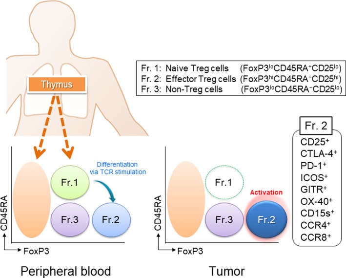
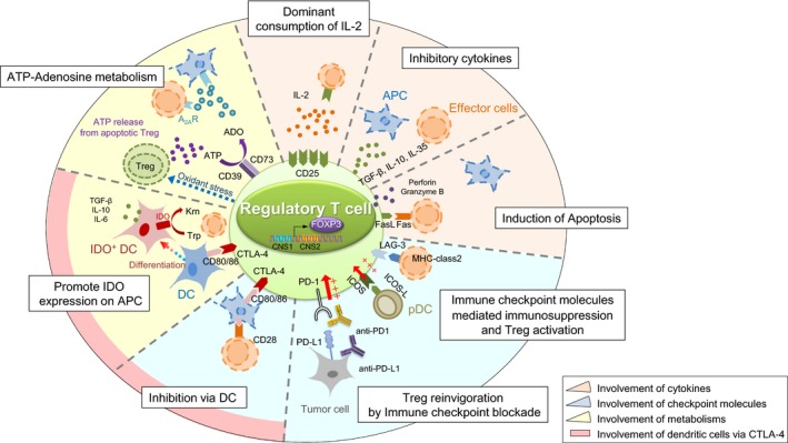
Regulatory T (Treg) cells suppress abnormal/excessive immune responses to self- and nonself-antigens to maintain immune homeostasis. In tumor immunity, Treg cells are involved in tumor development and progression by inhibiting antitumor immunity. There are several Treg cell immune suppressive mechanisms: inhibition of costimulatory signals by CD80 and CD86 expressed by dendritic cells through cytotoxic T-lymphocyte antigen-4, interleukin (IL)-2 consumption by high-affinity IL-2 receptors with high CD25 (IL-2 receptor α-chain) expression, secretion of inhibitory cytokines, metabolic modulation of tryptophan and adenosine, and direct killing of effector T cells. Infiltration of Treg cells into the tumor microenvironment (TME) occurs in multiple murine and human tumors. Regulatory T cells are chemoattracted to the TME by chemokine gradients such as CCR4-CCL17/22, CCR8-CCL1, CCR10-CCL28, and CXCR3-CCL9/10/11. Regulatory T cells are then activated and inhibit antitumor immune responses. A high infiltration by Treg cells is associated with poor survival in various types of cancer. Therefore, strategies to deplete Treg cells and control of Treg cell functions to increase antitumor immune responses are urgently required in the cancer immunotherapy field. Various molecules that are highly expressed by Treg cells, such as immune checkpoint molecules, chemokine receptors, and metabolites, have been targeted by Abs or small molecules, but additional strategies are needed to fine-tune and optimize for augmenting antitumor effects restricted in the TME while avoiding systemic autoimmunity. Here, we provide a brief synopsis of these cells in cancer and how they can be controlled to achieve therapeutic outcomes.

Keywords: Treg; immune checkpoint; immune suppression; tolerance; tumor.


Conflict of interest statement

No potential conflict of interest was disclosed by YO. HN has received honoraria and grants from Bristol‐Myers Squibb, Chugai, and Ono and grants from Astellas, BD Japan, Daiichi Sankyo, Kyowa Hakko Kirin, Sysmex, Taiho, Asahikasei, and Zenyaku Kogyo.

Figures

Figure 1
Figure 1
Classification of human regulatory T (Treg) cells. Human Treg cells can be classified into the following 3 subfractions: Fraction (Fr.) 1, naive/resting Treg cells, defined by FoxP3loCD45RA+CD25lo cells; Fr. 2, effector/activated Treg (eTreg) cells, defined by FoxP3hiCD45RACD25hi cells; and Fr. 3, non‐Treg cells, defined by FoxP3loCD45RACD25lo cells. Naive/resting Treg cells that have just left the thymus have a weak immune suppressive function and differentiate into effector/activated Treg cells following T‐cell receptor (TCR) stimulation. eTreg cells are the terminal differentiation state and harbor strong immune suppressive activity. Non‐Treg cells do not possess immune suppressive activity, but produce inflammatory cytokines. Typical staining pattern of CD4+ T cells in peripheral blood and lung cancer tissue. In general, the frequency of eTreg cells in humans is 1%‐5% in peripheral blood but approximately 10%‐50% in the TME. eTreg cells predominantly express various activation cell surface markers including cytotoxic T‐lymphocyte antigen‐4 (CTLA)‐4, programmed cell death (PD)‐1, inducible T‐cell costimulator (ICOS), glucocorticoid‐induced tumor necrosis factor receptor‐related protein (GITR), OX40, CD15s, CCR4, and CCR8. Naive/resting Treg cells are hardly detected in the tumor microenvironment
Figure 2
Figure 2
Suppressive mechanism of regulatory T (Treg) cells. Treg cells exert their immunosuppressive function through several mechanisms. The first immunosuppressive mechanisms involving cytokines include consumption of interleukin (IL)‐2 by Treg cells highly expressing CD25 (IL‐2 receptor α‐chain), suppression by inhibitory cytokines, such as transforming growth factor (TGF)‐β, IL‐10, and IL‐35, and direct killing of effector or antigen‐presenting cells (APC) by perforin, granzyme B, or Fas/Fas ligand (FasL) interaction. The second immunosuppressive mechanisms involving immune checkpoint molecules include inhibition of effector T cells by the lymphocyte activation gene‐3 (LAG‐3)‐MHC class II pathway and Treg activation through the inducible T‐cell costimulator (ICOS)‐ICOS ligand (ICOSL) and programmed cell death (PD)‐1/PD‐ligand (PD‐L)1 pathways. The third immunosuppressive mechanisms include metabolic modulation by indoleamine 2,3‐dioxygenase (IDO) expression in dendritic cells (DC), which exhausts T cells because critical amino acids for survival are depleted. Furthermore, the generation of adenosine from ATP, which is metabolized by CD39 and CD73 expressed in activated Treg cells, results in T cell suppression from the induction of negative signaling to effector T cells and APCs. The fourth immunosuppressive mechanism involves DCs through cytotoxic T‐lymphocyte antigen‐4 (CTLA‐4) and decreased CD80/86 expression by APCs by binding to CTLA‐4 expressed by activated eTreg cells, which causes impairment of APC maturation, downregulation of CD80/86 molecules on APCs, and attenuation of T cell stimulation. A2AR, A2A receptor; CNS, conserved noncoding sequence; Krn, kyneurenine; pDC, plasmacytoid dendritic cell; Trp, tryptophan

Similar articles

Cited by

References

    1. Carey TE, Takahashi T, Resnick LA, Oettgen HF, Old LJ. Cell surface antigens of human malignant melanoma: mixed hemadsorption assays for humoral immunity to cultured autologous melanoma cells. Proc Natl Acad Sci USA. 1976;73:3278‐3282. - PMC - PubMed
    1. Sakaguchi S. Regulatory T cells: key controllers of immunologic self‐tolerance. Cell. 2000;101:455‐458. - PubMed
    1. Gabrilovich DI. Myeloid‐derived suppressor cells. Cancer Immunol Res. 2017;5:3‐8. - PMC - PubMed
    1. Mantovani A, Marchesi F, Malesci A, Laghi L, Allavena P. Tumour‐associated macrophages as treatment targets in oncology. Nat Rev Clin Oncol. 2017;14:399‐416. - PMC - PubMed
    1. Ishida Y, Agata Y, Shibahara K, Honjo T. Induced expression of PD‐1, a novel member of the immunoglobulin gene superfamily, upon programmed cell death. EMBO J. 1992;11:3887‐3895. - PMC - PubMed

MeSH terms