. 2019 Jun 4;116(11):2161-2171.
doi: 10.1016/j.bpj.2019.04.018. Epub 2019 Apr 22.

Mobility of Molecular Motors Regulates Contractile Behaviors of Actin Networks

Affiliations

Mobility of Molecular Motors Regulates Contractile Behaviors of Actin Networks

Atsushi Matsuda et al. Biophys J. .

Abstract

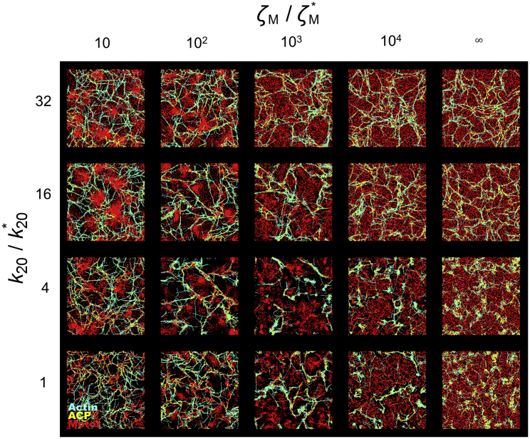
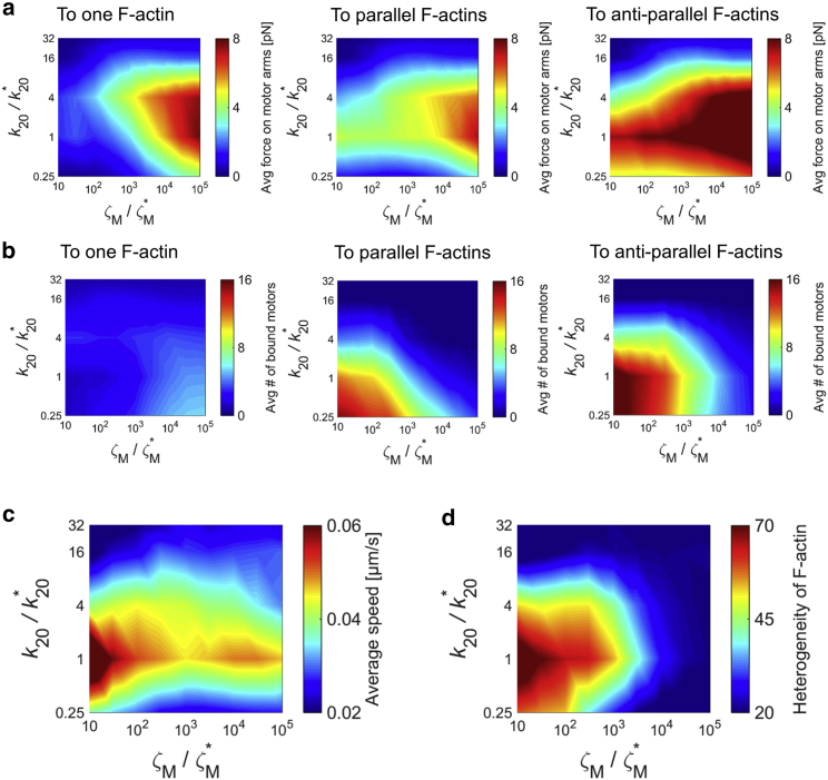
Cells generate mechanical forces primarily from interactions between F-actin, cross-linking proteins, myosin motors, and other actin-binding proteins in the cytoskeleton. To understand how molecular interactions between the cytoskeletal elements generate forces, a number of in vitro experiments have been performed but are limited in their ability to accurately reproduce the diversity of motor mobility. In myosin motility assays, myosin heads are fixed on a surface and glide F-actin. By contrast, in reconstituted gels, the motion of both myosin and F-actin is unrestricted. Because only these two extreme conditions have been used, the importance of mobility of motors for network behaviors has remained unclear. In this study, to illuminate the impacts of motor mobility on the contractile behaviors of the actin cytoskeleton, we employed an agent-based computational model based on Brownian dynamics. We find that if motors can bind to only one F-actin like myosin I, networks are most contractile at intermediate mobility. In this case, less motor mobility helps motors stably pull F-actins to generate tensile forces, whereas higher motor mobility allows F-actins to aggregate into larger clustering structures. The optimal intermediate motor mobility depends on the stall force and affinity of motors that are regulated by mechanochemical rates. In addition, we find that the role of motor mobility can vary drastically if motors can bind to a pair of F-actins. A network can exhibit large contraction with high motor mobility because motors bound to antiparallel pairs of F-actins can exert similar forces regardless of their mobility. Results from this study imply that the mobility of molecular motors may critically regulate contractile behaviors of actin networks in cells.


Figures

Figure 1
Figure 1
Agent-based computational model. (a) A schematic diagram showing a network consisting of F-actin (cyan), actin cross-linking protein (ACP, yellow), and motor (red) is given. Each element is simplified by cylindrical segments. Bending (κb) and extensional stiffnesses (κs) maintain equilibrium angles formed by adjacent cylindrical segments (indicated by bent arrows) and equilibrium lengths of cylindrical segments (indicated by springs), respectively. Details of the model and the list of all model parameters are in the Supporting Materials and Methods. (b) An example of networks formed by self-assembly of the three elements in a very thin computational domain (10 × 10 × 0.1 μm) with a periodic boundary condition in x and y directions is given. To see this figure in color, go online.
Figure 2
Figure 2
Effects of mobility of motors and mechanochemical rate with one-arm motors. (a) Average tensile force exerted by motor arms, (b) fraction of motors bound to F-actins, (c) average speed of F-actin, and (d) heterogeneity of F-actin spatial distribution are shown. ATP-dependent unbinding rate of myosin heads (k20) and motor mobility modulated by the drag coefficient of motors (ζM) are shown. Forces exerted by motor arms and the average speed of F-actins are larger at higher ζM and intermediate levels of k20. More motors are bound to F-actins with smaller ζM and k20. Distribution of F-actins is the most heterogeneous at intermediate levels of ζM and k20. ζM = 8.10 × 10−8 kg/s and k20 = 20 s−1 are reference values of ζM and k20, respectively. To see this figure in color, go online.
Figure 3
Figure 3
Morphology of networks with one-arm motors depends on mobility of motors and mechanochemical rate. Network morphology measured in all cases at the last time point at which steady state is reached, t = 100 s, is shown, with various ATP-dependent unbinding rates of myosin heads (k20) and motor mobility modulated by the drag coefficient of motors (ζM). F-actins, ACPs, and motors are visualized by cyan, yellow, and red, respectively. ζM = 8.10 × 10−8 kg/s and k20 = 20 s−1 are reference values of ζM and k20, respectively. To see this figure in color, go online.
Figure 4
Figure 4
Impacts of motor mobility and mechanochemical rate on network contraction with two-arm motors. (a) Average tensile force exerted by motor arms, (b) fraction of motors bound to F-actins, (c) average speed of F-actin, and (d) heterogeneity of F-actin spatial distribution are shown, with various ATP-dependent unbinding rate of myosin heads (k20) and motor mobility modulated by the drag coefficient of motors (ζM). In (a) and (b), the left, center, and right plots correspond to motors bound to one F-actin, motors bound to relatively parallel F-actins, and motors bound to relatively anti-parallel F-actins, respectively. The average force exerted by two-arm motors bound to parallel F-actins shows weak dependence on ζM, and the number of those motors increases as ζM decreases. Thus, the total forces exerted by all motors are higher with lower ζM. F-actins move faster, and the distribution of F-actins is much more heterogeneous with lower ζM. ζM = 8.10 × 10−8 kg/s and k20 = 20 s−1 are reference values of ζM and k20, respectively. To see this figure in color, go online.
Figure 5
Figure 5
Morphology of networks with two-arm motors depending on the mobility and mechanochemical rate of motors. Network morphology was measured in all cases at the last time point at wide ranges of ATP-dependent unbinding rate of myosin heads (k20) and motor mobility modulated by the drag coefficient (ζM). F-actins, ACPs, and motors are visualized by cyan, yellow, and red, respectively. The last time point is 100 s unless it is indicated otherwise. ζM = 8.10 × 10−8 kg/s and k20 = 20 s−1 are reference values of ζM and k20, respectively. To see this figure in color, go online.
Figure 6
Figure 6
Schematic diagrams summarizing results. (a and b) Comparison between one-arm motor and two-arm motor is shown. Because one-arm motors can bind to only one F-actin, they rely mostly on resistance originating from drag force (Fd) when they pull F-actin. For example, one-arm motors with a higher drag coefficient (ζM) feel a larger drag force when they try to move. Thus, they have less mobility, enabling them to exert a larger force on F-actin. If two-arm motors pull two F-actins in relatively opposite directions, they do not need to rely on Fd. (c and d) Initial state (top) and final states of networks (the rest) with one-arm motors or two-arm motors with different ζM are shown. Note that all two-arm motors in the diagram are initially located in antiparallel F-actins to show a case opposite to that with one-arm motors. In (c) and (d), it is assumed that a periodic boundary condition exists on the left and right boundaries. To see this figure in color, go online.

Similar articles

Cited by

References

    1. Gardel M.L., Kasza K.E., Weitz D.A. Chapter 19: mechanical response of cytoskeletal networks. Methods Cell Biol. 2008;89:487–519. - PMC - PubMed
    2. Gardel, M. L., K. E. Kasza, …, D. A. Weitz. 2008. Chapter 19: mechanical response of cytoskeletal networks. Methods Cell Biol. 89:487-519. - PMC - PubMed
    1. Murrell M., Oakes P.W., Gardel M.L. Forcing cells into shape: the mechanics of actomyosin contractility. Nat. Rev. Mol. Cell Biol. 2015;16:486–498. - PMC - PubMed
    2. Murrell, M., P. W. Oakes, …, M. L. Gardel. 2015. Forcing cells into shape: the mechanics of actomyosin contractility. Nat. Rev. Mol. Cell Biol. 16:486-498. - PMC - PubMed
    1. Schliwa M., Woehlke G. Molecular motors. Nature. 2003;422:759–765. - PubMed
    2. Schliwa, M., and G. Woehlke. 2003. Molecular motors. Nature. 422:759-765. - PubMed
    1. McConnell R.E., Tyska M.J. Leveraging the membrane - cytoskeleton interface with myosin-1. Trends Cell Biol. 2010;20:418–426. - PMC - PubMed
    2. McConnell, R. E., and M. J. Tyska. 2010. Leveraging the membrane - cytoskeleton interface with myosin-1. Trends Cell Biol. 20:418-426. - PMC - PubMed
    1. Spudich J.A. The myosin swinging cross-bridge model. Nat. Rev. Mol. Cell Biol. 2001;2:387–392. - PubMed
    2. Spudich, J. A. 2001. The myosin swinging cross-bridge model. Nat. Rev. Mol. Cell Biol. 2:387-392. - PubMed

Publication types

LinkOut - more resources