. 2019 Jun 18;50(6):1381-1390.e5.
doi: 10.1016/j.immuni.2019.04.009. Epub 2019 May 15.

Cytomegalovirus Infection Drives Avidity Selection of Natural Killer Cells

Affiliations

Cytomegalovirus Infection Drives Avidity Selection of Natural Killer Cells

Nicholas M Adams et al. Immunity. .

Abstract

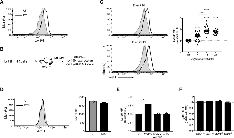
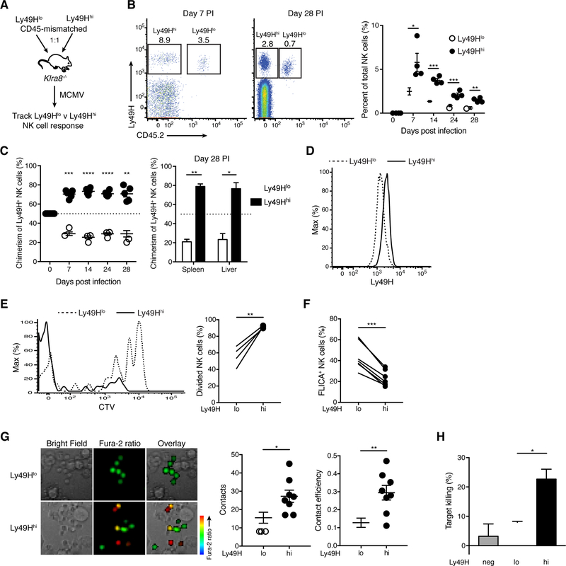
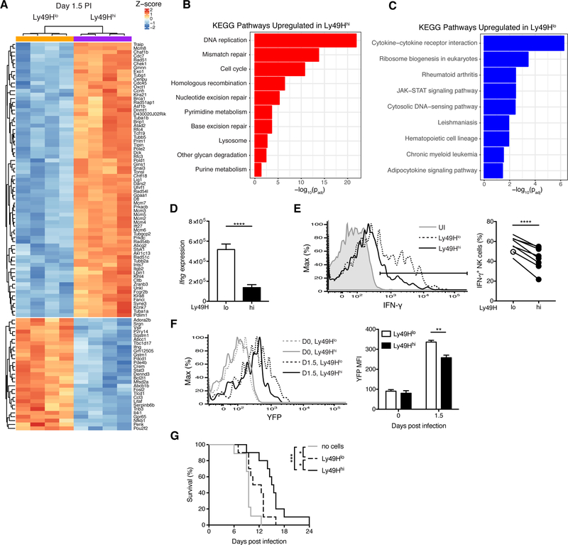
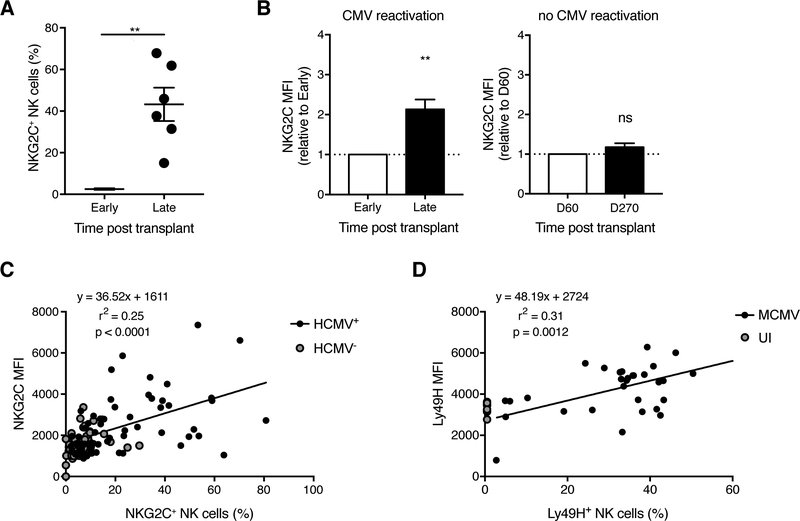
The process of affinity maturation, whereby T and B cells bearing antigen receptors with optimal affinity to the relevant antigen undergo preferential expansion, is a key feature of adaptive immunity. Natural killer (NK) cells are innate lymphocytes capable of "adaptive" responses after cytomegalovirus (CMV) infection. However, whether NK cells are similarly selected on the basis of their avidity for cognate ligand is unknown. Here, we showed that NK cells with the highest avidity for the mouse CMV glycoprotein m157 were preferentially selected to expand and comprise the memory NK cell pool, whereas low-avidity NK cells possessed greater capacity for interferon-γ (IFN-γ) production. Moreover, we provide evidence for avidity selection occurring in human NK cells during human CMV infection. These results delineate how heterogeneity in NK cell avidity diversifies NK cell effector function during antiviral immunity, and how avidity selection might serve to produce the most potent memory NK cells.

Keywords: NK cells; cytomegalovirus; heterogeneity; selection; viral infection.


Conflict of interest statement

Declaration of Interests

The authors declare no competing interests.

Figures

Figure 1.
Figure 1.. MCMV-driven changes in Ly49H expression within the antiviral NK cell pool are m157-dependent
(A) Histograms of Ly49H expression on splenic Ly49H+ NK cells (gating strategy in Figure S1A) from uninfected (UI) and MCMV-infected WT mice at day 7 PI (D7). Data are representative of at least five independent experiments with 5–10 mice per experiment. (B-C) Experimental schematic (B). Ly49H+ NK cells were transferred into Klra8−/− mice, and their phenotype analyzed following MCMV infection of the recipients. (C) Histograms of Ly49H expression on Ly49H+ NK cells from blood of UI WT mice (shaded gray) versus at day 7 PI (top left) and day 29 PI (bottom left) (black lines). Quantification of Ly49H median fluorescence intensity (MFI) on Ly49H+ NK cells at indicated days PI relative to UI WT mice bled on the same day (right). Data are representative of three independent experiments with 10–25 mice per experiment. (D) As in (C), histograms (left) and MFI (right) of NK1.1 on Ly49H+ NK cells from blood of UI WT mice and at day 28 PI. Data are representative of at least three independent experiments with 3–9 mice per experiment. (E) WT mice were infected with MCMV, MCMV-Δm157, Listeria monocytogenes (L.m.) or UI. Data is represented as Ly49H MFI on Ly49H+ NK cells from blood at day 7 PI for indicated infections relative to UI. Data are representative of two independent experiments with 2–5 mice per group. (F) As in (B), except splenocytes from WT and indicated knock-out (KO) mice were co-transferred. Data is represented as Ly49H MFI on KO Ly49H+ NK cells relative to WT from blood at day 7 PI. Data are representative of two to four independent experiments with 2–5 mice per group. Groups with a ratio < 1 were compared against 1 using a one sample t test. Groups were compared using an unpaired, two-tailed Student’s t test (D) or against 1 using a one sample t test (E). In (C), each timepoint was compared against 1 using a one sample t test, and against each other using a paired two-tailed t test. Data are presented as the mean ± SEM. *p < 0.05; ****p < 0.0001. See also Figure S1.
Figure 2.
Figure 2.. Ly49H+ NK cells undergo avidity selection during MCMV infection
(A-D) Experimental schematic (A). Equal numbers of splenic Ly49Hlo and Ly49Hhi NK cells, purified from congenically distinct WT mice, were co-transferred into Klra8−/− mice, and their responses tracked following MCMV infection of the recipients. (B) Flow plots gated on NK cells from blood at day 7 PI (left) and spleen at day 28 PI (middle). Quantification of percent Ly49Hlo and Ly49Hhi NK cells within total NK cells in blood at indicated days PI (right). (C) The percentage of Ly49Hlo and Ly49Hhi NK cells within transferred Ly49H+ NK cells in blood at indicated days PI (left) and in indicated organs at day 28 PI (right). (D) Histograms of Ly49H expression on Ly49Hlo and Ly49Hhi NK cells from blood at day 7 PI. Data are representative of two independent experiments with 3–5 mice per experiment. (E) WT splenocytes were labeled with CTV and transferred into congenically distinct WT mice prior to MCMV infection. Histograms of CTV in splenic Ly49Hlo and Ly49Hhi NK cells at day 7 PI (left). Quantification of indicated NK cell populations that have divided at least once (right). Data are representative of three independent experiments with 4 mice per experiment. (F) Percent of splenic Ly49Hlo and Ly49Hhi NK cells staining positive for FLICA at day 4 PI. Data are pooled from two independent experiments with 3–4 mice per experiment. (G) Purified splenic Ly49Hlo and Ly49Hhi NK cells were loaded with Fura-2AM and co-cultured with Ba/F3–m157 cells. Live-cell imaging was then performed. Representative images from the time-lapse analysis (left). Scale bar = 10 μM. Quantification of the number of contacts between NK cells and target cells (middle) and contact efficiency (right), defined as the proportion of target cell contacts that resulted in an NK cell Ca2+ flux (change in NK cell color from green to yellow or red). Data are representative of two experiments with 4 replicates per group per experiment. (H) Purified splenic Ly49H, Ly49Hlo, and Ly49Hhi NK cells were incubated with Ba/F3–m157 target cells (CTVhi) and Ba/F3 control cells (CTVlo) at 10:1:1 ratio (effector: target: control) for 6 hours. Quantification of target cell killing by indicated NK cell populations compared to control wells lacking NK cells. Data are representative of three independent experiments with 3 replicates per group per experiment. Ly49Hlo and Ly49Hhi NK cells were compared using an unpaired, two-tailed Student’s t test. Groups were compared using a paired, two-tailed t test (B, E, F), an unpaired, two-tailed Student’s t test (G) or against 50 using a one sample t test (C). Data are presented as the mean ± SEM. *p < 0.05, **p < 0.01, ***p < 0.001, ****p < 0.0001. See also Figures S1 and S2.
Figure 3.
Figure 3.. Ly49Hlo NK cells produce more IFN-γ during early MCMV infection
(A-D) Splenic Ly49Hlo and Ly49Hhi NK cells were sorted for RNA-seq at day 1.5 PI (4 replicates each). (A) Heat map and hierarchical clustering of top 100 differentially expressed genes by p-value. (B-C) Gene ontology analysis of differential KEGG pathways for genes significantly (padj < 0.05) upregulated in Ly49Hhi NK cells (B) and upregulated in Ly49Hlo NK cells (C). Their respective p values are shown. (D) Quantification of RNA-seq reads mapping to the Ifng locus. P value was calculated in DESeq2 and adjusted for testing multiple hypotheses. (E) Histograms of intracellular IFN-γ expression in splenic Ly49H+ NK cells from UI and MCMV-infected WT mice at day 1.5 PI (left). Quantification of percent IFN-γ+ NK cells within indicated NK cell populations (right). Data are representative of at least five independent experiments with 3–15 mice per experiment. (F) As in (E), except UI or MCMV-infected Ifng-IRES-YFP mice at day 1.5 PI. Histograms (left) and quantification of YFP MFI (right) before and after MCMV infection. Data are representative of two independent experiments with 2–3 mice per time point per experiment. (G) Kaplan-Meier survival curves of Rag2−/− Il2rg−/− mice that received either no cells, 50,000 purified Ly49Hlo NK cells, or 50,000 purified Ly49Hhi NK cells 2 days prior to MCMV infection. Data are pooled from two independent experiments with 4–5 mice per group per experiment. Groups were compared using a paired, two-tailed t test (E, F) or the Log-rank (Mantel-Cox) test with correction for testing multiple hypotheses (G). Data are presented as the mean ± SEM. *p < 0.05, **p < 0.01, ***p < 0.001, ****p < 0.0001. See also Figures S2 and S3 and Table S1.
Figure 4.
Figure 4.. HCMV reactivation in HSCT recipients drives higher NKG2C expression on human NKG2C+ NK cells
(A-B) Blood was drawn from T cell-depleted HSCT recipients in the year following transplant (every ~30–90 days). HCMV infection status was determined by viral qPCR. “Early” and “Late” are paired samples from the same HCMV-reactivating transplant recipients. “Early” refers to 15–60 days post transplant depending on the recipient, and “Late” includes the window between 200 days to 1 year post transplant. The data are presented as such to synchronize the timing of adaptive NKG2C+ NK cell appearance, which was recipient-dependent. It was expansion of the NKG2C+ NK cell population that dictated the observed kinetic profile of NKG2C MFI. (A) Percentage of NKG2C+ NK cells among CD3CD56+ cells (gating strategy in Figure S4). (B) Quantification of NKG2C MFI on NKG2C+ NK cells from transplant recipients who reactivated (left) or did not reactivate (right) HCMV. Groups were compared using a paired, two-tailed t test (A) or against 1 using a one sample t test (B). Data are presented as the mean ± SEM. ns, not significant; **p < 0.01. (C) Blood was drawn from healthy HCMV-seropositive and HCMV-seronegative donors. Correlation between NKG2C MFI and percentage of NKG2C+ NK cells. Linear regression was performed on HCMV+ samples. P value represents the likelihood of a non-zero slope. (D) Experimental design as in Figure 1B. Correlation between Ly49H MFI and the Ly49H+ NK cell expansion (i.e. percentage of Ly49H+ NK cells among total NK cells) at day 7 PI. UI WT mice were assigned 0% expansion. Linear regression was performed on MCMV-infected samples. P value represents the likelihood of a non-zero slope. See also Figure S4.

Comment in

References

    1. Aguilar OA, Berry R, Rahim MM, Reichel JJ, Popović B, Tanaka M, Fu Z, Balaji GR, Lau TN, Tu MM, et al. (2017). A viral immunoevasin controls innate immunity by targeting the prototypical natural killer cell receptor family. Cell 169, 58–71. - PubMed
    1. Arase H, Mocarski ES, Campbell AE, Hill AB, and Lanier LL (2002). Direct recognition of cytomegalovirus by activating and inhibitory NK cell receptors. Science 296, 1323–1326. - PubMed
    1. Biron CA, Byron KS, and Sullivan JL (1989). Severe herpesvirus infections in an adolescent without natural killer cells. N. Engl. J. Med 320, 1731–1735. - PubMed
    1. Biron CA, and Tarrio ML (2015). Immunoregulatory cytokine networks: 60 years of learning from murine cytomegalovirus. Med. Microbiol. Immunol 204, 345–354. - PMC - PubMed
    1. Bolger AM, Lohse M, and Usadel B (2014). Trimmomatic: a flexible trimmer for Illumina sequence data. Bioinformatics 30, 2114–2120. - PMC - PubMed

Publication types

MeSH terms

Substances