. 2019 Jul;189(7):1386-1401.
doi: 10.1016/j.ajpath.2019.04.008. Epub 2019 May 17.

CD55 Is Essential for CD103+ Dendritic Cell Tolerogenic Responses that Protect against Autoimmunity

Affiliations

CD55 Is Essential for CD103+ Dendritic Cell Tolerogenic Responses that Protect against Autoimmunity

Michael G Strainic et al. Am J Pathol. 2019 Jul.

Abstract

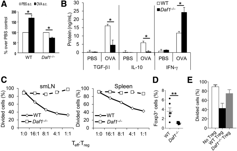
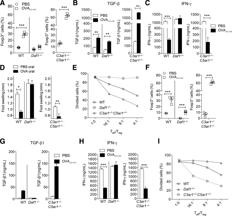
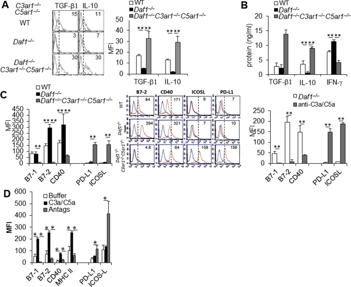
Recent studies traced inflammatory bowel disease in some patients to deficiency of CD55 [decay-accelerating factor (DAF)], but the mechanism underlying the linkage remained unclear. Herein, we studied the importance of DAF in enabling processes that program tolerance in the gut and the eye, two immune-privileged sites where immunosuppressive responses are continuously elicited. Unlike oral feeding or ocular injection of ovalbumin in wild-type (WT) mice, which induced dominant immune tolerance, identical treatment of DAF-/- mice or DAF-/- to WT bone marrow chimeras did not. While 10% to 30% of mesenteric and submandibular lymph node CD4+ cells became robust T-regulatory cells (Tregs) in WT forkhead box P3 (Foxp3)-green fluorescent protein mice, few in either site became Tregs with little suppressor activity in DAF-/- Foxp3-green fluorescent protein mice. Phenotyping of CD103+ dendritic cells (DCs) from the ovalbumin-fed DAF-/- mice showed impaired expression of inducer of costimulation (ICOS) ligand, programmed death receptor 1-ligand 1 (PD1-L1), CxxxC chemokine receptor 1 (Cx3CR1), CCR7, and CCR9. Analyses of elicited DAF-/- Foxp3+ Tregs showed reduced expression of interferon regulatory factor 8 (IRF-8)/aldehyde dehydrogenase 1 family member A2 (Aldh1a2) and glycoprotein A repetitions predominant/latency-associated protein associated with Treg transforming growth factor-β production and presentation, as well as integrin β6/integrin β8 associated with Treg and CD103+ DC transforming growth factor-β release. Thus, DAF is required for the properties of CD103+ DCs and their naïve CD4+ cell partners that together program tolerance.


Figures

Figure 1
Figure 1
Decay-accelerating factor (DAF) is required for the ocular tolerogenic response to an anterior chamber (a.c.) antigen. A:Daf1+/+ and Daf1–/– mice were preinjected in the a.c. with 50 μg ovalbumin (OVA) or phosphate-buffered saline (PBS). Seven days later, the mice were challenged subcutaneously with 12.5 μg OVA in IFA in the right leg and with PBS in IFA in the left leg; and 48 hours later, delayed-type hypersensitivity (DTH) size was measured. B: Representative DTH lesions from wild-type (WT) and Daf1–/– mice. C: Splenocytes (6 × 105) from Daf1+/+ and Daf1–/– mice preinjected a.c. with OVA or with PBS (7 days earlier, as in the above protocol) were assayed on interferon (IFN)-γ ELISPOT plates in the presence of a varying amount of whole OVA protein or OVA323-339 peptide. Arrows point to the assays with peptide rather than the whole protein. D: Lymph node cells [effectors (E)] from mice preinjected a.c. with OVA or PBS were mixed with OVA-transfected EL-4 targets at the designated effector to target (E/T) ratios. Percentage cytolysis was determined using Alamar blue. E:Daf1+/+ and Daf1–/– mice were preinjected in the a.c. with OVA or PBS and challenged with OVA in CFA subcutaneously at day 7 (as in AC). Sera harvested at day 17 were assayed for anti-OVA antibody isotypes by isotype enzyme-linked immunosorbent assays. F: IFN-γ ELISPOT assays were performed on splenocytes from groups of Daf1+/+ and CD97–/– mice 7 days after a.c. preinjection with OVA or PBS. G and H: IFN-γ ELISPOT (G) and cytotoxicity (H) assays were performed on splenocytes from WT bone marrow (BM) → WT recipients (left panels) or Daf1–/– BM → WT recipients (right panels) on day 7 after s.c. injection with OVA (day 17 after a.c. injection). I: (F12) Daf1–/– and (F12) Daf1–/–C3ar1–/–C5ar1–/– mice were studied using the protocol described in F. Data are shown as ratios of IFN-γ–producing cells by OVA a.c. protected mice normalized to PBS a.c. protected controls. n = 6 (A and F); n = 3 per group (GI). P < 0.05, ∗∗P < 0.01; P < 0.05 versus Daf1–/–. Original magnification, ×40 (B).
Figure 2
Figure 2
Ocular tolerance requires immunosuppression of the decay-accelerating factor (DAF)–dependent regulatory T cell (Treg). A: Ocular explants from anterior chamber (a.c.)–preinjected mice were placed in RPMI 1640 medium for 48 hours. Supernatants were assayed for transforming growth factor (TGF)-β by enzyme-linked immunosorbent assay (ELISA). B: Wild-type (WT) or Daf1–/– mice were preinjected a.c. with ovalbumin (OVA) or phosphate-buffered saline (PBS), followed by s.c. challenge with OVA in IFA in the right leg 7 days later. Splenocytes were cultured at 37°C for 48 hours with 100 μg/mL of OVA, and culture supernatants were assayed for TGF-β1, IL-10, and interferon (IFN)-γ by ELISAs. C: Wild-type (WT) or Daf1–/– mice were preinjected a.c. with OVA or PBS, followed 7 days later by s.c. challenge with OVA in IFA in the right leg. After another 7 days, CD4+ cells from draining submandibular lymph node (smLN) and from the spleen were isolated. Sorted Foxp3+ cells from each site were incubated for 5 days in increasing ratios with mixtures of dendritic cells (DCs), OVA323-339, and OT-II cells (prelabeled with CellTracker Red), after which suppression of proliferation was assessed by determining percentage dividers. D: A total of 4 × 106 sorted green fluorescent protein (GFP) CD4+ cells from (Thy1.2) WT or Daf1–/– Foxp3-GFP mice were injected intravenously into WT Thy1.1 mice. Two days thereafter, the mice were preinjected a.c. with OVA or PBS and challenged 7 days later with OVA in IFA subcutaneously in the right leg. Seven days after the s.c. challenge, Foxp3+ Thy1.2+ cells in the spleen were quantified by flow cytometry. E: Foxp3+ cells from D were sorted, and the sorted cells were incubated for 5 days at a 4:1 ratio with mixtures of DCs, OVA323-339, and OT-II cells (prelabeled with CellTracker Red). Suppression of proliferation was assessed by determining percentage dividers. n = 3 (C and E); n = 6 GFP CD4+ cells from (Thy1.2) WT or Daf1–/– Foxp3-GFP mice and Foxp3+ Thy1.2+ cells in the spleen (D). P < 0.05, ∗∗P < 0.01.
Figure 3
Figure 3
Oral tolerance requires immunosuppression of decay-accelerating factor (DAF)–dependent regulatory T cell (Treg). AE: Data are organized showing wild-type (WT) versus Daf1–/– mice on the left, and for side-by-side comparison, C3ar1–/–C5ar1–/– mice on the right. WT, Daf1–/–, and C3ar1–/–C5ar1–/– OT II mice were fed 20 mg/mL ovalbumin (OVA) or phosphate-buffered saline (PBS) in their drinking water for 5 days, after which mesenteric lymph nodes (mLNs) were isolated and CD4+ and CD11c+ cells were separated via sorting. A: Percentage of CD4+ cells that were CD25+ Foxp3+ in the initial isolate were quantified by flow cytometry. B and C: After overnight culture of the remainder of the cells with OVA (100 μg/mL), amounts of transforming growth factor (TGF)-β1 (B) and interferon (IFN)-γ (C) released into culture supernatants were assayed by enzyme-linked immunosorbent assays (ELISAs). D: In other animals, delayed-type hypersensitivity responses were measured 48 hours after OVA injection into the footpad. E: Sorted CD25+ cells that were Foxp3+ in other aliquots from A were incubated for 5 days at increasing T-effector (Teff)/Treg ratios with mixtures of dendritic cells (DCs), OVA323-339, and WT OT-II cells prelabeled with CellTracker Red. Suppression of CellTracker Red–labeled OT-II cell proliferation was assessed by quantifying percentage dividers. FI: Sorted Foxp3 OT-II cells from WT, Daf1–/–, and C3ar1–/–C5ar1–/– OT-II Foxp3–green fluorescent protein mice were adoptively transferred into Thy1.1 recipients. The Thy1.1 recipients were fed 20 mg/mL OVA or an equal volume of PBS in their drinking water for 5 days and immunized subcutaneously with OVA in IFA in the hind leg. Seven days later, mLNs were harvested and the cells were isolated. F: Percentage of CD4+ cells that were Foxp3+ CD25+ in the initial mLN isolate were quantified by flow cytometry. G and H: After 48 hours of culture of the remainder of the cells with OVA, the amounts of TGF-β1 and IFN-γ released into the supernatants were quantified by ELISAs. I: Sorted Foxp3+ cells in the initial isolate (F) were incubated for 5 days with mixtures of DCs, OVA323-339, and sorted WT OT-II cells (prelabeled with CellTracker Red) at varying ratios. Suppression of proliferation was assessed by quantifying percentage dividers. n = 6 mice for each genotype, OVA group, and PBS group (FI); n = 12 for each recipient group (FI). P < 0.05, ∗∗P < 0.01, and ∗∗∗P < 0.001.
Figure 4
Figure 4
Decay-accelerating factor (DAF) is required for the tolerogenic properties of CD103+ dendritic cells (DCs) and regulatory T cells. AC: CD11c+ DCs harvested from the eyes (A), draining submandibular lymph nodes (smLNs; B), and mesenteric lymph nodes (mLNs; C) of wild-type (WT), Daf1–/–, and Daf1–/–C3ar1–/–C5ar1–/– mice were assayed for CD40, B7.1, B7.2, major histocompatibility complex (MHC) II, PD-L1, and ICOS-L by flow cytometry (Daf1–/– versus WT and Daf1–/–C3ar1–/–C5ar1–/– versus WT). DF: CD11b+F4/80+ macrophages harvested from the eyes (D), draining smLNs (E), and mLNs (F) of WT, Daf1–/–, and Daf1–/–C3ar1–/–C5ar1–/– mice were assayed for the same markers. Cells were also assayed for C5ar1 and C5L2. The fact that differences in ICOS-L levels between Daf1−/− and WT cells reach statistical significance reflects mouse numbers as other studies in the laboratory (M.G.S., J.L., F.A., and M.E.M., unpublished data) reproducibly have documented statistical significance. G and H: Human plasmacytoid DCs (pDCs; G) and CD4 T cells (H) were treated with siRNA targeting DAF or C3ar1 and C5ar1 for 72 hours, after which expression of CD40, B7.1, B7.2, CCR7, CCR9, PD-L1, and ICOS-L on pDCs (G) and expression of CD28, CD40 ligand, CCR7, CCR9, PD-1, and ICOS on CD4+ T cells (H) was assessed by flow cytometry. n = 10 per group (AF); n = 3 (G and H). ∗∗P < 0.01. MFI, mean fluorescent intensity.
Figure 5
Figure 5
The expression of T-cell and dendritic cell (DC) decay-accelerating factor (DAF) regulates cytokine and surface protein expression. A: A total of 1 × 106 CD11c+ cells from the submandibular lymph node (smLN) of wild-type (WT), Daf1–/–, and Daf1–/–C3ar1–/–C5ar1–/– mice were incubated in RPMI 1640 media for 24 hours, after which IL-10 and transforming growth factor (TGF)-β1 were assayed by internal staining. B: A total of 2.5 × 105 WT dendritic cells (DCs) were incubated for 24 hours with 1 × 106 CD4+ T cells from WT, Daf1–/–, and Daf1–/–C3ar1–/–C5ar1–/– mice and anti-CD3/28 (1 μg/mL each), after which IL-10 and TGF-β1 were assayed by enzyme-linked immunosorbent assays. C: A total of 2.5 × 105 WT DCs were incubated for 24 hours with 1 × 106 WT, Daf1–/–, and Daf1–/–C3ar1–/–C5ar1–/– CD4+ T cells and anti-CD3/28, after which B7-1, B7-2, CD40, PD-L1, and ICOS-L expression levels on the DCs were assayed by flow cytometry (Supplemental Figure S2B). D: A total of 1 × 106 CD62LhiCD25 CD4+ cells from WT, ICOS–/–, or PD-1–/– mice were incubated for 3 days with 1 × 105 CD11c+ WT DCs + anti-CD3/28 (1 μg/mL each) or IL-2 and C3aR-A/C5aR-A (10 μg/mL each), after which percentage of Foxp3+ cells was assayed by intracellular staining. E: A total of 1 × 105 CD11c+ WT or PD-L1–/– DCs were incubated for 3 days with 1 × 106 CD62LhiCD25 CD4+ cells from WT mice + anti-CD3/28 (1 μg/mL each) or IL-2 and C3aR-A/C5aR-A (10 μg/mL each), after which percentage Foxp3+ cells was assayed by intracellular staining. F: A total of 1 × 106 sorted Daf1−/−, WT, and C3ar1–/–C5ar1−/− Foxp3-GFP CD4+ cells were incubated for 2 hours without or with anti-CD3/CD28 Dynabeads in the presence of IL-2 and Shp-1, and Shp-2 mRNA level was quantitated by real-time quantitative PCR. The results are shown in comparison to WT cells set as 100%. As in the above studies, each group contained three mice and experiments were repeated three times. n = 6 (A and C); n = 5 (B). ∗∗P < 0.01. KO, knockout; MFI, mean fluorescent intensity.
Figure 6
Figure 6
CD103+ dendritic cell (DC) expression of decay-accelerating factor (DAF) is required for tolerance. A: CD11c+ CD103+ DCs were harvested from mesenteric lymph nodes (mLNs) of wild-type (WT) and Daf1–/– Foxp3–green fluorescent protein mice and assayed for expression of CD40, B7.1, B7.2, ICOS-L, and PD-L1 by flow cytometry. B: CD11c+CD103+ DCs harvested from mesenteric LNs of WT, Daf1–/–, and C3ar1–/–C5ar1–/– mice were assayed for expression of CxCR3, CCR7, and CCR9 by flow cytometry. C: CD11c+CD103+ DCs were harvested from submandibular lymph nodes (LNs) of WT, Daf1–/–, and C3ar1–/–C5ar1–/– mice and assayed for expression of CD40, B7.1, B7.2, ICOS-L, and PD-L1 by flow cytometry. D: CD11c+CD103+ DCs harvested from submandibular LNs of WT, Daf1–/–, and C3ar1–/–C5ar1–/– mice were assayed for expression of CxCR3, CCR7, and CCR9 by flow cytometry. E: A total of 2.5 × 105 WT DCs were incubated for 24 hours with 1 × 106 CD4+ T cells from WT and Daf1–/–C3ar1–/–C5ar1–/– mice and anti-CD3/28 (1 μg/mL each), after which cells were assayed for TGFB2, ALDH1A2, and IRF8 mRNA expression by real-time quantitative PCR (qPCR). F: Foxp3+ CD4+ T cells from WT, Daf1–/–, and C3ar1–/–C5ar1–/– mice fed ovalbumin or phosphate-buffered saline were assayed for glycoprotein A repetitions predominant (GARP) expression by flow cytometry. G: Sorted Foxp3 CD4+ T cells from WT, Daf1–/–, and C3ar1–/–C5ar1–/– mice were activated for 3 days with anti-CD3/28 Dynabeads + C3aR-A/C5aR-A (10 μg/mL each), after which sorted Foxp3+ cells were assayed for latency-associated protein (LAP) expression by flow cytometry. H: Sorted Foxp3 CD4+ T cells from WT, Daf1–/–, and C3ar1–/–C5ar1–/– mice were activated for 3 days with anti-CD3/28 Dynabeads + C3aR-A/C5aR-A (10 μg/mL each), after which sorted Foxp3+ cells were assayed for integrin β6 (Itg-β6) and Itg-β8 mRNA expression by qPCR. I: CD103+CD11c+ cells from the mLN of WT, Daf1–/–, and C3ar1–/–C5ar1–/– mice were assayed for Itg-β6 expression by flow cytometry. n = 10 (AD). P < 0.05, ∗∗P < 0.01. MFI, mean fluorescent intensity.
Supplemental Figure S1
Representative flow cytometry plots. A: Gating strategy for isolating cells for assaying regulatory T cells. B: Top panel: Gating strategy for analyzing dendritic cells (DCs) from the mesenteric lymph nodes (mLNs). Arrows designate the cells gated to eliminate doublets that were analyzed. Bottom panels: Representative flow plots for CD11c+ DCs harvested from the mLN of wild-type (WT) and Daf1–/–C3ar1–/–C5ar1–/– mice assayed for expression of CD40, B7.1, B7.2, major histocompatibility complex (MHC) II, PD-L1, and ICOS-L by flow cytometry. C: CD11c+ DCs were harvested from the inguinal LN of WT, Daf1–/–, and Daf1–/–C3ar1–/–C5ar1–/– mice and assayed for expression of CD40, B7.1, B7.2, MHC II, PD-L1, and ICOS-L by flow cytometry. D: CD11b+F4/80+ macrophages harvested from mesenteric LNs of WT, Daf1–/–, and C3ar1–/–C5ar1–/– mice were assayed for expression of CxCR3, CCR7, and CCR9 by flow cytometry. n = 10 (BD). P < 0.05. FSC, forward scatter; MFI, mean fluorescent intensity; SSC, side scatter.
Supplemental Figure S2
Comparative transforming growth factor (TGF)-β, IL-10, and interferon (IFN)-γ production by Daf1−/−, wild-type (WT), and C3ar1–/–C5ar1−/− CD4+ cells and effects of C3a/C5a signaling on the phenotypes of dendritic cells (DCs) from each genotype. A and B: A total of 1 × 106 splenocytes from WT, Daf1–/–, and Daf1–/–C3ar1–/–C5ar1–/– mice were incubated in RPMI 1640 media for 24 hours, after which IFN-γ, IL-10, and TGF-β1 expression levels were assayed by internal staining. A: Left panel: Representative histograms. Right panel: Mean fluorescent intensity (MFI). C: Phenotypes of DCs from each genotype homeostatically (left and middle panels) and the inability of anti-C3a/anti-C5a monoclonal antibody blockade to reverse the maturity of Daf1−/− DCs (right panel). D: Effects of C3a/C5a or C3ar1/C5ar1 antagonists on costimulatory and coinhibitory molecule expression by WT CD11c+ F40 DCs. n = 6 (A, right panel, B, left and right panels, and D). P < 0.05, ∗∗P < 0.01. MHC, major histocompatibility complex.
Supplemental Figure S3
Representative flow cytometry plots. Representative flow plots from CD103+CD11c+ (A) and wild-type (WT), Daf1–/–, and C3ar1–/–C5ar1–/– (B) dendritic cells assayed for B7-1, B7-2, CD40, PD-L1, and ICOS-L expression levels by flow cytometry. smLN, submandibular lymph node.

Similar articles

Cited by

References

    1. Kaplan H.J., Stevens T.R., Streilein J.W. Transplantation immunology of the anterior chamber of the eye. J Immunol. 1975;115:800–804. - PubMed
    2. Kaplan HJ, Stevens TR, Streilein JW: Transplantation immunology of the anterior chamber of the eye. J Immunol 1975, 115:800-804. - PubMed
    1. Niederkorn J., Streilein J.W., Shadduck J.A. Deviant immune responses to allogeneic tumors injected intracamerally and subcutaneously in mice. Invest Ophthalmol Vis Sci. 1981;20:355–363. - PubMed
    2. Niederkorn J, Streilein JW, Shadduck JA: Deviant immune responses to allogeneic tumors injected intracamerally and subcutaneously in mice. Investigative ophthalmology & visual science 1981, 20:355-363. - PubMed
    1. Niederkorn J.Y., Streilein J.W. Alloantigens placed into the anterior chamber of the eye induce specific suppression of delayed-type hypersensitivity but normal cytotoxic T lymphocyte and helper T lymphocyte responses. J Immunol. 1983;131:2670–2674. - PubMed
    2. Niederkorn JY, Streilein JW: Alloantigens placed into the anterior chamber of the eye induce specific suppression of delayed-type hypersensitivity but normal cytotoxic T lymphocyte and helper T lymphocyte responses. Journal of immunology 1983, 131:2670-2674. - PubMed
    1. McKenna K.C., Xu Y., Kapp J.A. Injection of soluble antigen into the anterior chamber of the eye induces expansion and functional unresponsiveness of antigen-specific CD8+ T cells. J Immunol. 2002;169:5630–5637. - PubMed
    2. McKenna KC, Xu Y, Kapp JA: Injection of soluble antigen into the anterior chamber of the eye induces expansion and functional unresponsiveness of antigen-specific CD8+ T cells. Journal of immunology 2002, 169:5630-5637. - PubMed
    1. Streilein J.W., Niederkorn J.Y., Shadduck J.A. Systemic immune unresponsiveness induced in adult mice by anterior chamber presentation of minor histocompatibility antigens. J Exp Med. 1980;152:1121–1125. - PMC - PubMed
    2. Streilein JW, Niederkorn JY, Shadduck JA: Systemic immune unresponsiveness induced in adult mice by anterior chamber presentation of minor histocompatibility antigens. J Exp Med 1980, 152:1121-1125. - PMC - PubMed

Publication types

MeSH terms