

Retinal Pathologic Features on OCT among Eyes of Older Adults Judged Healthy by Color Fundus Photography
- PMID: 31103641
- PMCID: PMC6718291
- DOI: 10.1016/j.oret.2019.03.019
Retinal Pathologic Features on OCT among Eyes of Older Adults Judged Healthy by Color Fundus Photography
Abstract
Purpose: OCT has revealed many details of retinal disease that were not available with older imaging technologies. In eyes of adults older than 60 years with healthy maculas as determined by color fundus photography (CFP) and a validated grading system, we screened for pathologic features using OCT. We also tested visual function to assess potential impact of the observed pathologic features on patients.
Design: Cross-sectional study.
Participants: Persons recruited from primary ophthalmology care clinics.
Methods: Color fundus photographs were assessed by the 9-step Age-Related Eye Disease Study scale. OCT macular volumes of participants at step 1 on the Age-Related Eye Disease Study scale, considered healthy, were reviewed by a retina specialist masked to other participant characteristics. Participants were tested for 6 different cone- and rod-mediated visual functions.
Main outcome measures: Percentage of participants with disorders detected on OCT review and visual function measures.
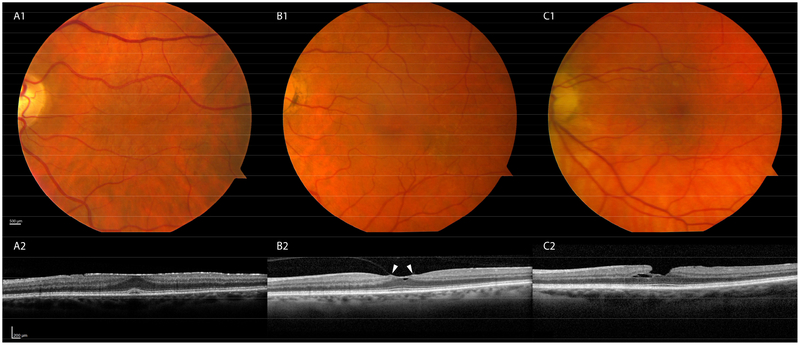
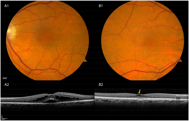
Results: In 138 of 984 eyes (14%) considered healthy by CFP, pathologic features were detectable by OCT, with 8.4% having vitreomacular interface disorders. Among the low-prevalence disorders found, 5 eyes (0.5%) showed macular telangiectasia type 2. Relative to eyes lacking detectable chorioretinal pathologic features, eyes with any pathologic features were associated with poorer low-luminance visual acuity and rod-mediated dark adaptation. In eyes with epiretinal membranes, the largest single entity identified (n = 61 [6.2%]), significantly worse visual functions were best-corrected visual acuity (P = 0.0444), low-luminance visual acuity (P = 0.0151), and light sensitivity (central 3° and 9°; P = 0.0035 and P = 0.0097, respectively).
Conclusions: Macular pathologic features with functional visual implications not identified by clinical examination or CFP are detectable with OCT. Vitreomacular interface disorders often are visually significant and treatable conditions that are visible on OCT, but are easily missed on CFP and clinical examination. Another such condition best seen on OCT is macular telangiectasia type 2, an untreatable disorder for which a clinical trial is in progress. OCT has a potential role in primary eye care clinics to screen for retinal pathologic features, especially in eyes with decreased visual acuity and otherwise normal examination results.
In 14.0% of 984 eyes of adults ≥60 years, considered normal by color fundus photography, retinal pathology affecting visual function was detected by optical coherence tomography; 8.4% involved vitreomacular interface disorders, and 6.2%, epiretinal membranes.
Copyright © 2019 American Academy of Ophthalmology. Published by Elsevier Inc. All rights reserved.
Figures
References
-
- Smith W, Assink J, Klein R, et al. Risk factors for age-related macular degeneration. Pooled findings from three continents. Ophthalmology. 2001;108:697–704. - PubMed
-
- Duker JS, Kaiser PK, Binder S, et al. The International Vitreomacular Traction Study Group classification of vitreomacular adhesion, traction, and macular hole. Ophthalmology. 2013;120(12):2611–2619. - PubMed
-
- Gattoussi S, Buitendijk GHS, Peto T, et al. The European Eye Epidemiology spectral-domain optical coherence tomography classification of macular diseases for epidemiological studies. Acta Ophthalmol. 2018. - PubMed
Publication types
MeSH terms
Grants and funding
LinkOut - more resources
Full Text Sources
Medical
Miscellaneous
