Review
. 2019 May 18;21(2):47.
doi: 10.1007/s10544-019-0389-6.

Advanced implantable drug delivery technologies: transforming the clinical landscape of therapeutics for chronic diseases

Affiliations
Review

Advanced implantable drug delivery technologies: transforming the clinical landscape of therapeutics for chronic diseases

Fernanda P Pons-Faudoa et al. Biomed Microdevices. .

Abstract

Chronic diseases account for the majority of all deaths worldwide, and their prevalence is expected to escalate in the next 10 years. Because chronic disorders require long-term therapy, the healthcare system must address the needs of an increasing number of patients. The use of new drug administration routes, specifically implantable drug delivery devices, has the potential to reduce treatment-monitoring clinical visits and follow-ups with healthcare providers. Also, implantable drug delivery devices can be designed to maintain drug concentrations in the therapeutic window to achieve controlled, continuous release of therapeutics over extended periods, eliminating the risk of patient non-compliance to oral treatment. A higher local drug concentration can be achieved if the device is implanted in the affected tissue, reducing systemic adverse side effects and decreasing the challenges and discomfort of parenteral treatment. Although implantable drug delivery devices have existed for some time, interest in their therapeutic potential is growing, with a global market expected to reach over $12 billion USD by 2018. This review discusses implantable drug delivery technologies in an advanced stage of development or in clinical use and focuses on the state-of-the-art of reservoir-based implants including pumps, electromechanical systems, and polymers, sites of implantation and side effects, and deployment in developing countries.

Keywords: Implants; Long-acting formulations; MEMS; NEMS; Non-biodegradable polymers.


Conflict of interest statement

Compliance with ethical standards J.S and A.G. disclose a financial interest in NanoMedical systems, Inc. F.P.P. and A.B. disclose no competing financial interest.

Figures

Fig. 1
Fig. 1
Top ten global chronic diseases by prevalence (Bertolote 2005; Ferkol and Schraufnagel 2014; Goldberg and McGee 2011; Steel et al. 2014; The Global Cancer Observatory 2018; Vos et al. 2015; World Health Organization 2016)
Fig. 2
Fig. 2
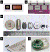
FDA-approved and experimental non-biodegradable reservoir-based polymer systems. a Non-biodegradable polymer schematic depicting an outer polymer coating encompassing an inner drug core. b Drug-versatile3D-printedBiocage device (a)Magnifiedlight microscopy image to detect porosity with 100-μm scale bar. (b) Biocage device boxed in orange in relation to pencil tip and dime to appreciate its minute size and how it can be inserted using a 22-gauge needle. (Image 2B adapted from (Son et al. 2017) licensed under CC BY 4.0). c Vantas® and SUPPRELIN® LA 50 mg histrelin acetate implants for prostate cancer symptom relief and childhood central precocious puberty treatment, respectively (Image reproduced from (Rudlang and Brasso 2016). d Probuphine® 80 mg buprenorphine hydrochloride implant for opioid dependence treatment (Image used with permission from Titan Pharmaceuticals Inc.). e Retisert® implant design consists on a platform for suturing device and drug core with 0.59 mg fluocinolone acetonide enclosed in silicone elastomer cup with a PVA membrane outlet for treating chronic noninfectious uveitis. f Intraocular ILUVIEN® 0.19 mg fluocinolone acetonide device for diabetic macular edema treatment in relation to a grain of rice to demonstrate its size (Image is courtesy of Alimera Sciences Inc.)
Fig. 3
Fig. 3
Osmotic pump drug release schematics and FDA-approved osmotic pump implant. a Osmotic pump drug release mechanism for liquid drug formulations use a high salt concentration osmotic engine driven by osmotic flow through semipermeable membrane to move piston and displace drug through orifice. b Osmotic pumps with an inner solid drug reservoir encompassed by a high osmolyte concentration osmotic engine surrounded by semipermeable membrane osmotically displace solubilized drug through orifice. c Intravesical GemRIS™ implant loaded with solid gemcitabine for bladder cancer treatment (Image adapted from (Cima et al. 2014))
Fig. 4
Fig. 4
Peristaltic pump drug release mechanism and design. a Peristaltic pump drug release mechanism: a central rotor with rollers attached to its circumference rotates, compressing the flexible tube, trapping liquid drug doses between rollers and displacing it through the catheter. b General outer schematic of implantable pumps: a discoidal-shaped implant with a central reservoir fill port that can be accessed percutaneously, a catheter port that connects the catheter and implant, and suture loops to securely anchor the implant in the abdominal pump pocket
Fig. 5
Fig. 5
Infusion pump drug release schematic. The infusion pump is divided into two chambers: a collapsible drug reservoir and a propellant chamber. At body temperature the propellant changes from liquid to gas compressing the drug reservoir thus forcing the drug out through the restrictor filter into the pump catheter. The drug reservoir is refilled with a designated needle that closes the safety valve avoiding drug release while refilling
Fig. 6
Fig. 6
30-ml Micro-CHIP device used for leuprolide release in dogs. a Silicon wafer with 100 30 μl drug reservoirs capped by gold membranes. b Assembled 30-ml Micro CHIP device. c Micro-CHIP drug release schematic: an electrical potential to the gold membrane permits selective opening of specific reservoir for drug release. d Internal circuitry of the implant. (Image 6A, 6B, 6D adapted from (Prescott et al. 2006))
Fig. 7
Fig. 7
Replenish MicroPump schematic and drug release mechanism. a Replenish MicroPump implanted on the sclera beneath the conjunctiva. b Replenish MicroPump drug release mechanism: electrodes in the electrolysis chamber generate an electric potential electrolyzing H2O into H2 and O2 when the device is turned on. This creates pressure on the diaphragm that shifts drug in drug reservoir and displaces it through the cannula
Fig. 8
Fig. 8
NEMS translational research devices. a Schematic of nanoportal membrane from Nano Precision Medical showing how the nanotubes are the rate-limiting step for drug release from the reservoir (Image used with permission from Nano Precision Medical). b Drug-versatile Delpor Inc. implant with two membranes with NANOPOR™ technology fixed at each end (Image used with permission from Delpor Inc.)
Fig. 9
Fig. 9
nDS. a Differently sized mechanically robust silicon microfabricated nDS membranes, which house a defined number of densely packed slit-nanochannels to achieve constant and sustained delivery of therapeutics over extended periods of time. b Drug release diffusion path through nDS membrane: first from drug reservoir to perpendicular microchannels, then rate-limiting horizontal nanochannels, and then out through perpendicular microchannels. c The membrane is conveniently mounted on a drug delivery reservoir with a size and shape that can be optimized for the therapeutic application, drug, duration of treatment, and site of implantation. d Image of NDES with nDS membrane next to ruler to illustrate its small size. e Dynamically controlled nDS membrane mounted on polyether ether ketone, sized 24 × 34× 4.5 mm3, with an 800-μl drug reservoir chamber and a circuitry chamber with the electronics and battery
Fig. 10
Fig. 10
Sites of implantation for FDA-approved implants and devices in clinical trials
Fig. 11
Fig. 11
Implantation procedures. a ILUVIEN® intravitreal insertion. b Retisert® sutured in posterior chamber. c Replenish MicroPump episcleral placement. d Implanon®, Nexplanon®, SUPPRELIN® LA, and Vantas® insertion in the inner arm with personalized applicator. e Four Probuphine® implants positioned in fan-shaped distribution in the inner arm. f SynchroMed™ II, Codman® 3000, and Prometra® pump surgery for abdominal subcutaneous placement of the pump and intrathecal catheter. g SynchroMed™ II pump surgery for abdominal subcutaneous placement of the pump and intravenous catheter. h GemRIS™ and LiRIS intravesical insertion with a catheter-like tool

Similar articles

Cited by

References

    1. Alfaqawi F, Lip PL, Elsherbiny S, Chavan R, Mitra A, Mushtaq B, Eye 31, 650 (2017) - PMC - PubMed
    1. Ali M, Akin A, Bahamondes L, Brache V, Habib N, Landoulsi S, Hubacher D, Hum. Reprod 31, 2491 (2016) - PMC - PubMed
    1. Allen EE, Instrument for transfusion of blood with patent 249,285. 8 November 1881.
    1. Allen RH, Kaunitz AM, Hickey M, in Williams Textb. Endocrinol, 13th edn, ed. by Melmed S, Polonsky KS, Larsen PR, Kronenberg HM. (Elsevier, Philadelphia, 2016), pp. 664–693
    1. Almeida DRP, Chin EK, Mears K, Russell SR, Mahajan VB, Retin. Cases Brief Rep 9, 142 (2015) - PubMed

Publication types