

Induction of Angiogenesis by Malarial Infection through Hypoxia Dependent Manner
- PMID: 31104403
- PMCID: PMC6526210
- DOI: 10.3347/kjp.2019.57.2.117
Induction of Angiogenesis by Malarial Infection through Hypoxia Dependent Manner
Abstract
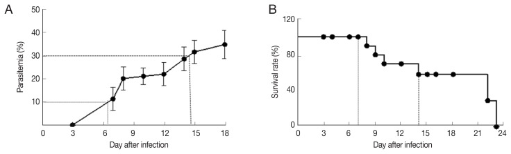
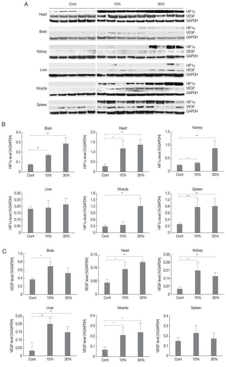
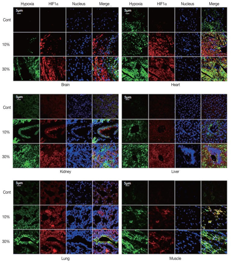
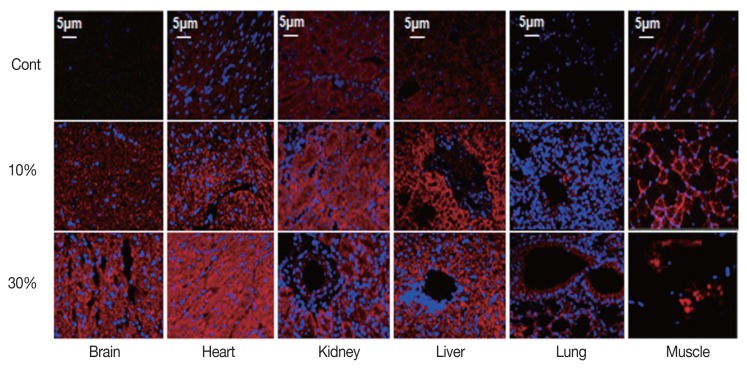
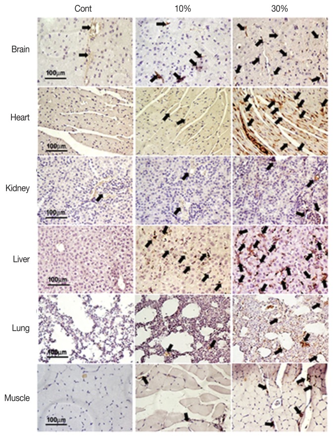
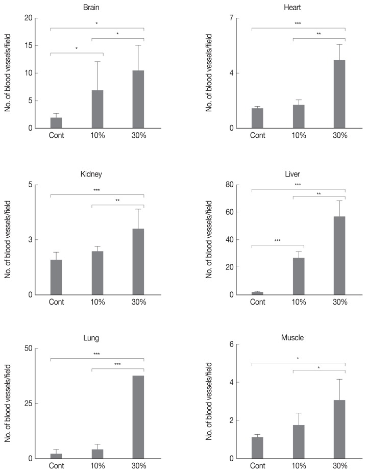
Malarial infection induces tissue hypoxia in the host through destruction of red blood cells. Tissue hypoxia in malarial infection may increase the activity of HIF1α through an intracellular oxygen-sensing pathway. Activation of HIF1α may also induce vascular endothelial growth factor (VEGF) to trigger angiogenesis. To investigate whether malarial infection actually generates hypoxia-induced angiogenesis, we analyzed severity of hypoxia, the expression of hypoxia-related angiogenic factors, and numbers of blood vessels in various tissues infected with Plasmodium berghei. Infection in mice was performed by intraperitoneal injection of 2×10⁶ parasitized red blood cells. After infection, we studied parasitemia and survival. We analyzed hypoxia, numbers of blood vessels, and expression of hypoxia-related angiogenic factors including VEGF and HIF1α. We used Western blot, immunofluorescence, and immunohistochemistry to analyze various tissues from Plasmodium berghei-infected mice. In malaria-infected mice, parasitemia was increased over the duration of infection and directly associated with mortality rate. Expression of VEGF and HIF1α increased with the parasitemia in various tissues. Additionally, numbers of blood vessels significantly increased in each tissue type of the malaria-infected group compared to the uninfected control group. These results suggest that malarial infection in mice activates hypoxia-induced angiogenesis by stimulation of HIF1α and VEGF in various tissues.
Keywords: HIF1α; Plasmodium berghei; VEGF; angiogenesis; hypoxia; malaria.
Conflict of interest statement
The authors declare no conflict of interest related to this study.
Figures
Similar articles
-
Genistein inhibited retinal neovascularization and expression of vascular endothelial growth factor and hypoxia inducible factor 1alpha in a mouse model of oxygen-induced retinopathy.J Ocul Pharmacol Ther. 2005 Apr;21(2):107-13. doi: 10.1089/jop.2005.21.107. J Ocul Pharmacol Ther. 2005. PMID: 15857276
-
VEGF and LPS synergistically silence inflammatory response to Plasmodium berghei infection and protect against cerebral malaria.Pathog Glob Health. 2015 Sep;109(6):255-65. doi: 10.1179/2047773215Y.0000000018. Epub 2015 Sep 21. Pathog Glob Health. 2015. PMID: 26392042 Free PMC article.
-
The role of IL-18 in blood-stage immunity against murine malaria Plasmodium yoelii 265 and Plasmodium berghei ANKA.J Immunol. 2002 May 1;168(9):4674-81. doi: 10.4049/jimmunol.168.9.4674. J Immunol. 2002. PMID: 11971017
-
Impact of Galectin-Receptor Interactions on Liver Pathology During the Erythrocytic Stage of Plasmodium berghei Malaria.Front Immunol. 2021 Nov 24;12:758052. doi: 10.3389/fimmu.2021.758052. eCollection 2021. Front Immunol. 2021. PMID: 34899708 Free PMC article.
-
Blood-Stage Plasmodium Berghei ANKA Infection Promotes Hepatic Fibrosis by Enhancing Hedgehog Signaling in Mice.Cell Physiol Biochem. 2018;50(4):1414-1428. doi: 10.1159/000494604. Epub 2018 Oct 24. Cell Physiol Biochem. 2018. PMID: 30355912
Cited by
-
Cytoadhesion of Plasmodium falciparum-Infected Red Blood Cells Changes the Expression of Cytokine-, Histone- and Antiviral Protein-Encoding Genes in Brain Endothelial Cells.Mol Microbiol. 2024 Dec;122(6):948-967. doi: 10.1111/mmi.15331. Epub 2024 Dec 4. Mol Microbiol. 2024. PMID: 39630601 Free PMC article.
-
Altered Cytokine Response of Human Brain Endothelial Cells after Stimulation with Malaria Patient Plasma.Cells. 2021 Jul 1;10(7):1656. doi: 10.3390/cells10071656. Cells. 2021. PMID: 34359826 Free PMC article.
-
Gymnema inodorum Leaf Extract Improves Cardiac Function in Experimental Mice Infected with Plasmodium Berghei.J Evid Based Integr Med. 2023 Jan-Dec;28:2515690X221150526. doi: 10.1177/2515690X221150526. J Evid Based Integr Med. 2023. PMID: 36617811 Free PMC article.
-
Role of TAM Receptors in Antimalarial Humoral Immune Response.Pathogens. 2024 Apr 2;13(4):298. doi: 10.3390/pathogens13040298. Pathogens. 2024. PMID: 38668253 Free PMC article. Review.
-
Understanding the significance of oxygen tension on the biology of Plasmodium falciparum blood stages: From the human body to the laboratory.PLoS Pathog. 2024 Sep 19;20(9):e1012514. doi: 10.1371/journal.ppat.1012514. eCollection 2024 Sep. PLoS Pathog. 2024. PMID: 39298535 Free PMC article. Review.
References
-
- Engwerda CR, Beattie L, Amante FH. The importance of the spleen in malaria. Trends Parasitol. 2005;21:75–80. - PubMed
-
- Deininger MH, Winkler S, Kremsner PG, Meyermann R, Schluesener HJ. Angiogenic proteins in brains of patients who died with cerebral malaria. J Neuroimmunol. 2003;142:101–111. - PubMed
MeSH terms
Substances
LinkOut - more resources
Full Text Sources
Medical
