. 2019 May 30;177(6):1553-1565.e16.
doi: 10.1016/j.cell.2019.04.035. Epub 2019 May 16.

Human Neonatal Fc Receptor Is the Cellular Uncoating Receptor for Enterovirus B

Affiliations

Human Neonatal Fc Receptor Is the Cellular Uncoating Receptor for Enterovirus B

Xin Zhao et al. Cell. .

Abstract

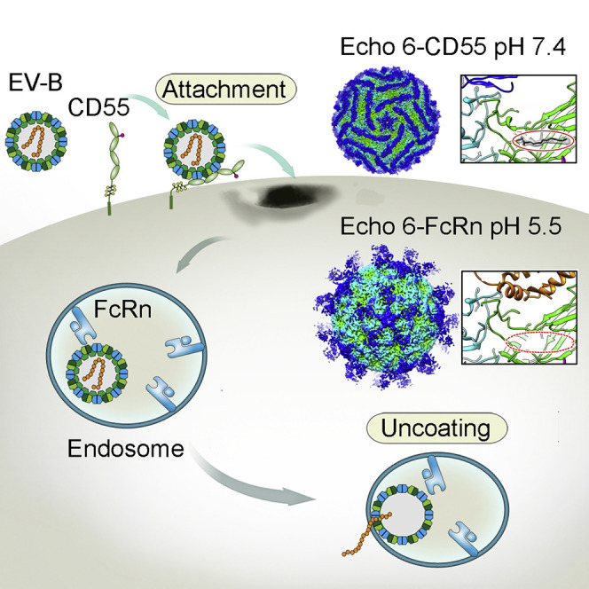
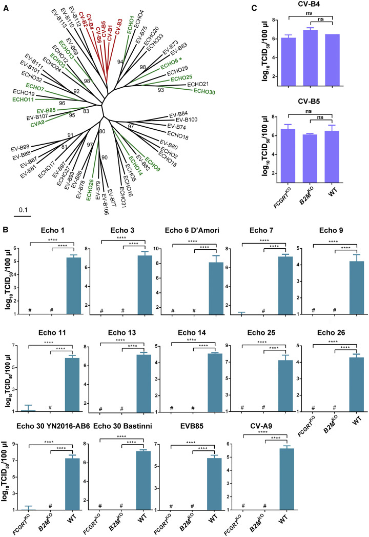
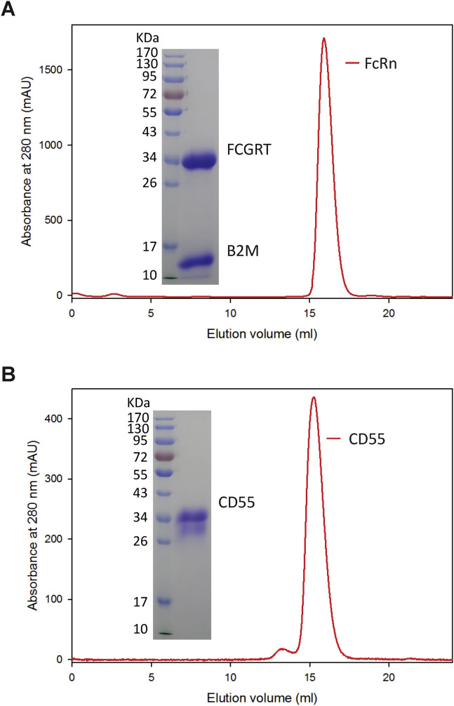
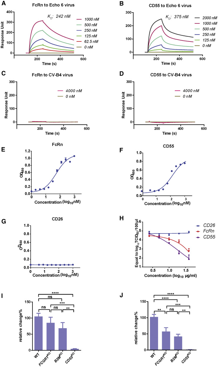
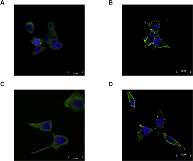
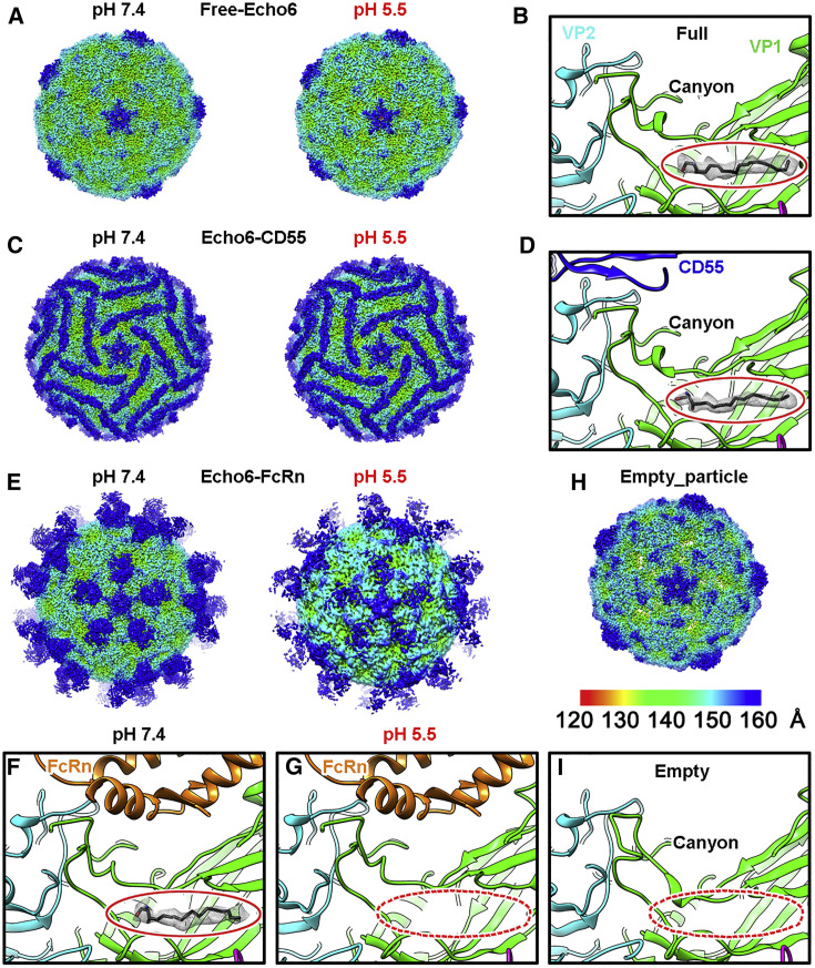
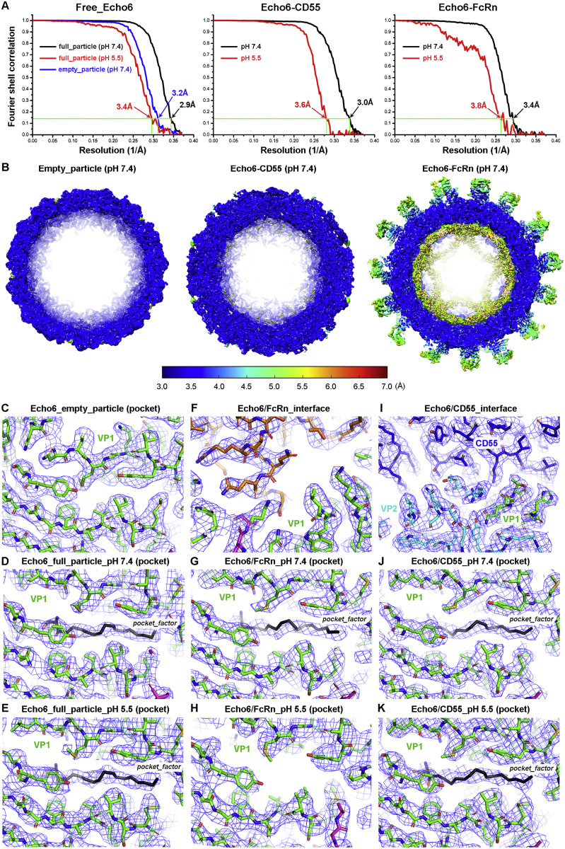
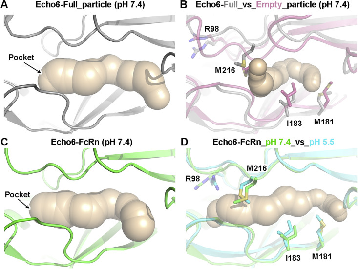
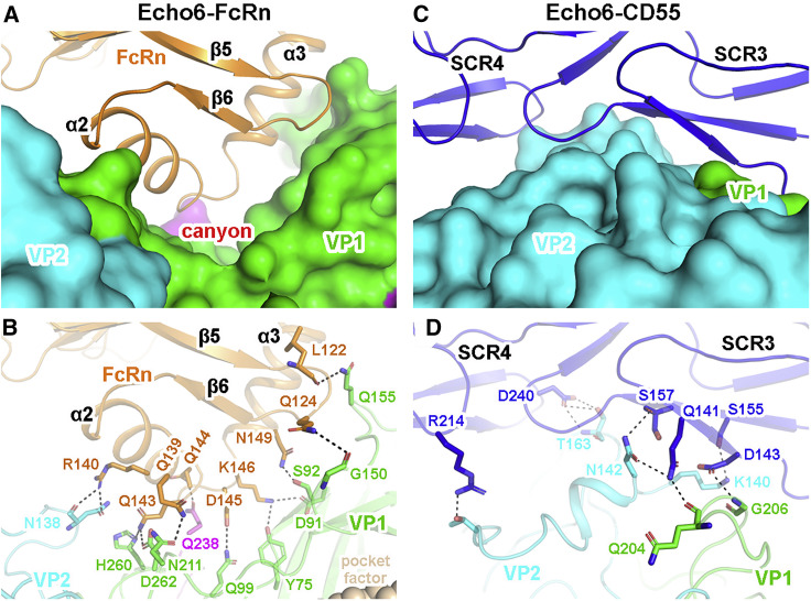
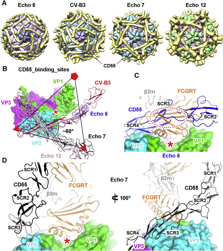
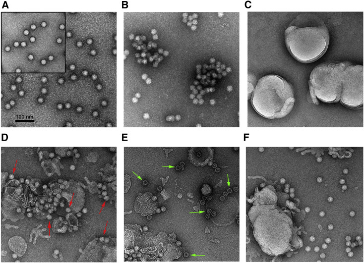
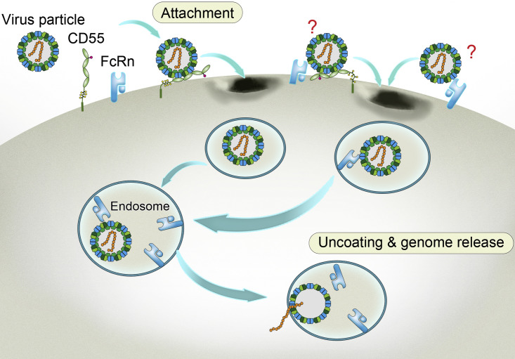
Enterovirus B (EV-B), a major proportion of the genus Enterovirus in the family Picornaviridae, is the causative agent of severe human infectious diseases. Although cellular receptors for coxsackievirus B in EV-B have been identified, receptors mediating virus entry, especially the uncoating process of echovirus and other EV-B remain obscure. Here, we found that human neonatal Fc receptor (FcRn) is the uncoating receptor for major EV-B. FcRn binds to the virus particles in the "canyon" through its FCGRT subunit. By obtaining multiple cryo-electron microscopy structures at different stages of virus entry at atomic or near-atomic resolution, we deciphered the underlying mechanisms of enterovirus attachment and uncoating. These structures revealed that different from the attachment receptor CD55, binding of FcRn to the virions induces efficient release of "pocket factor" under acidic conditions and initiates the conformational changes in viral particle, providing a structural basis for understanding the mechanisms of enterovirus entry.

Keywords: FcRn; attachment; cryo-EM; echovirus; enterovirus; human neonatal Fc receptor; receptor; uncoating.


Conflict of interest statement

W.W. serves as a scientific advisor for EdiGene.

Figures

None
Graphical abstract
Figure 1
Figure 1
CRISPR Screening Uncovered FcRn, a Functional Receptor for Echo 6 Infection (A) Dot plot illustrating the enriched genes from the CRISPR screen. Genes were rank-ordered by robust rank aggregation (RRA) scores based on MAGeCK analysis. The height and size of the top three genes, differently colored, was proportional to their RRA scores. (B) Schematic diagram of sgRNA library construction and Echo 6 receptor screening. (C and G) Echo 6 infection in FCGRTKO, B2MKO, CD55KO, and wild type (WT) HEK293T cells (C). Echo 6 infection in CD55KO (G), with the above three cell lines in (C) as controls. TCID50 was calculated at 48 h post infection (h.p.i). “+” and “-” indicate the presence or absence of FcRn or CD55 in the KO cell lines. (D and E) Echo 6 infection in FCGRTKO cells complemented with lentivirus expressing FCGRT (D) and B2MKO cells complemented with lentivirus expressing β2 m (E). KO cell lines with lentivirus expressing GFP was used as a control. Cells were infected with Echo 6 (MOI 0.01), and supernatants were harvested for virus titration at 24 h.p.i. (F) Light microscopy images showing CPE of FCGRTKO, B2MKO, CD55KO, and WT HEK293T cells at 24 h.p.i with Echo 6 (MOI 1). Immunofluorescence images of KO and WT cells infected by Echo 6 (MOI 10) for 8 h and then stained by anti-Echo 6 antibodies. The scale bar represents 50 μm. (H) Growth curve of Echo 6 in FCGRTKO, B2MKO, CD55KO, and WT HEK293T cells. (I) Echo 6 was inoculated onto non-susceptible CHO (left) and BHK (right) cells with ectopic expression of human FcRn (FCGRT+β2 m), CD55, or both. The data depict means with SEM. TCID50 values were calculated by the Reed-Muench method. Experiments were repeated three times. # stands for undetected; ∗∗∗∗p < 0.0001; ns, not significant. See also Figures S1 and S2 and Tables S1 and S2.
Figure S1
Figure S1
FCGRT or B2M KO Has No Effect on CD55 Expression, Related to Figure 1 (A) Diagram of FcRn. The α chain expressed by FCGRT contains a transmembrane domain and three extracellular domains (α1, α2 and α3). The β2 m chain is expressed by B2M. (B) Sanger sequencing of FCGRT, B2M and CD55 in KO cells. Sequencing data shows an alignment of WT sequence with the KO sequence. (C) western blot of CD55 expression in WT, FCGRTKO, B2MKO and CD55KO cells. (D) Flow cytometry analysis of CD55 expression in WT, FCGRTKO, B2MKO (left panel: fixed cell staining, middle panel: cell surface expression) and CD55KO cells (right panel).
Figure S2
Figure S2
FcRn Complementation Can Restore Susceptibility; Ectopic Expression Makes Non-permissive Cells Susceptible to Echovirus, Related to Figure 1 (A and B) western blot of FCGRT (A) and β2 m (B) expression in WT, KO and supplementary cells. (C and D) qPCR analysis of Echo 6 infection in FCGRTKO cells complemented with lentivirus expressing FCGRT (C). B2MKO cells complemented with lentivirus expressing β2 m (D). KO cell lines with lentivirus expressing GFP were used as a control. Cells were infected with 0.01 MOI Echo 6, and supernatants were taken for virus titration after 24 h. (E and F) western blot of FCGRT and CD55 expression in WT, human FcRn, human CD55 and human FcRn+ CD55 ectopic expression CHO (E) or BHK (F) cells. (G and H) qPCR analysis of Echo 6 in non-susceptible CHO (G) and BHK (H) cells with ectopic expression of human FcRn (FCGRT+B2M), CD55 or both. (I) Echovirus infection in CHO cells with the ectopic expression of FCGRT only. (The data depict means with SEM. C, D, G and H: one-way ANOVA with multiple comparisons; I: unpaired two-tailed t test, with a Welch post-correction. # stands for undetected. ∗∗∗∗p < 0.0001; and ns, not significant.)
Figure 2
Figure 2
FcRn Is Essential for Major Serotypes of EV-Bs (A) Phylogenetic tree of all Enterovirus B serotypes. The representative serotypes proven to use FcRn as a receptor are shown in green, and the ones proven in previous works to use CAR as their uncoating receptor are shown in red. The remaining untested serotypes are shown in black. (B) Infection of 12 echoviruses, EV-B85 and CVA-9 in FCGRTKO, B2MKO, and WT HEK293T cells. TCID50 was calculated at 48 h.p.i. (C) Infection of CV-B4 and CV-B5 in FCGRTKO, B2MKO, and WT HEK293T cells. TCID50 was calculated at 48 h.p.i. The data depict means with SEM. Experiments were repeated three times. # stands for undetected; ∗∗∗∗p < 0.0001; ns, not significant.
Figure S3
Figure S3
FcRn and CD55 Soluble Protein Expression and Purification, Related to Figure 3 (A) FcRn soluble protein was expressed by co-transfection of constructs encoding the FCGRT extracellular domain and B2M in HEK293T cells. The protein was eluted as a single peak from gel filtration on Superdex 200 column (GE Healthcare), and two bands showing FCGRT and B2M were evident by SDS-PAGE. (B) Gel filtration and SDS-PAGE of CD55 soluble protein purification.
Figure 3
Figure 3
Direct Binding of FcRn to Echo 6 Virus (A and B) BIAcore diagram of soluble FcRn protein (A) or CD55 protein (B) bound to Echo 6 virus. The KD values were calculated by the BIAcore 3000 analysis software (BIAevaluation version 4.1). (C and D) The binding affinity of CV-B4 virus with FcRn (C) or CD55 protein (D) was measured by SPR, which served as a control. (E–G) Binding of FcRn (E), CD55 (F), or CD26 (G, control) in 2-fold serial dilutions with Echo 6 virus was assayed using ELISAs. (H) Dose dependence of soluble FcRn or CD55 protein to block Echo 6 virus infection. CD26 was used as a control protein. (I and J) Echo 6 was incubated with FCGRTKO, B2MKO, CD55KO, or WT HEK293T cells at 4°C for 1 h (I) or plus 37°C for 30 min (J). Cells were collected, and RNA was extracted for qPCR analysis. The data depict means with SEM. ∗∗p < 0.01; ∗∗∗p < 0.001; ∗∗∗∗p < 0.0001; ns, not significant. See also Figures S3 and S4.
Figure S4
Figure S4
Confocal Analysis of FcRn and CD55 Expressed in HEK293T and RD Cells, Related to Figure 3 (A and C) Immunofluorescence staining of FcRn in HEK293T cells (A) and RD cells (C). FcRn is expressed both on the cell surface and in the cytoplasm. (B and D) Immunofluorescence staining of CD55 in HEK293T cells (B) and RD cells (D). CD55 was mostly expressed on the cell surface.
Figure 4
Figure 4
Cryo-EM Structures of the Echo 6 Viral Particle and Its Complex with Attachment (CD55) or Uncoating (FcRn) Receptor (A) The density maps of free Echo 6 full particles at pH 7.4 (2.9 Å) and pH 5.5 (3.4 Å). (B) The close-up view of the hydrophobic pocket and the “pocket factor” inside. (C and D) Cryo-EM maps (C) of Echo 6 virus in complex with CD55 at pH 7.4 (3.0 Å) and pH 5.5 (3.6 Å), as well as the close-up view (D) of the “pocket factor” with the receptor binding site shown in the same style as (B). (E–G) Cryo-EM maps (E) of Echo 6 virus in complex with FcRn at pH 7.4 (3.4 Å) and pH 5.5 (3.8 Å), as well as the close-up view showing the hydrophobic pocket of Echo 6 virus in complex with FcRn at pH 7.4 (F) and pH 5.5 (G). The receptor binding sites are shown in the same style as in (B) and (D). (H and I) Cryo-EM map of Echo 6 virus empty particle (H) at pH 7.4 (3.2 Å) and its collapsed pocket without “pocket factor” (I). All density maps are colored by radius as shown in the legend in (H). Atomic models of proteins are shown in ribbons and colored by chains with VP1, VP2, VP3, CD55, and FcRn heavy chain in green, cyan, magenta, blue, and orange, respectively. The “pocket factor” is shown in sticks, and the corresponding density map is shown at the 18σ contour level, which is highlighted by red ovals in close-up views. In the structures of the Echo 6 virus-FcRn complex at pH 5.5 and free Echo 6 virus empty particle, the pocket factor is absent, and its supposed positions are indicated by red dashed ovals. See also Figures S5, S6, and Table S3.
Figure S5
Figure S5
Cryo-EM Analysis of Echo 6 Virus and Its Complex with Receptors, Related to Figure 4 (A) Gold-standard Fourier shell correlation (FSC) curves of the structures of Echo 6 virus alone or in complex with its receptors. The 0.143 cut-off value is shown to indicate the resolution of each reconstruction. (B) Local resolution maps of representative density maps of Echo 6 virus or its complex with receptors. In all of these structures, most regions reach 3.0 Å and allowed the atomic details to be resolved. (C-K) Representative density maps and atomic models of the pocket region or receptor binding interface. Most side chains of key residues within the pocket are clearly resolved. The “pocket factor” is preserved in structures of free Echo 6 full-particle or in complex with CD55 at both pH 7.4 and pH 5.5. In the structures with FcRn binding, it is well accommodated at pH 7.4, but is released at pH 5.5.
Figure S6
Figure S6
Conformational Change Surrounding the “Pocket” of Echo 6 Virus and Echo 6 Virus with Its Receptor FcRn, Related to Figure 4 (A) Backbone traces of atomic models showing the conformation of the “pocket” in Echo 6 full particles in pH 7.4. Backbone of Echo 6 full particles is shown in gray. The inner pocket is shown in surface model. (B) Conformational difference of the protein surrounding the “pocket” region of Echo 6 full-particle with empty-particle at pH 7.4. The backbone of Echo 6 full particle is shown in gray, and empty particle is shown in pink. The remarkable residues involved in conformational changes are shown as sticks and colored by elements. The pocket in the empty-particle is obviously collapsed as compared to that in the full viral particle. (C) Backbone traces of atomic models showing the conformation of the “pocket.” Backbone of Echo 6 particle in the Echo 6-FcRn complex at pH 7.4 is shown in green. The pocket is highly similar to that in the full viral particle and allows the accommodation of the “pocket factor.” (D) The conformational difference of the Echo 6-FcRn complex at pH 7.4 and pH 5.5. Backbones of the Echo 6 particle in the Echo 6-FcRn complex at pH 7.4 and pH 5.5 are shown in green and cyan, respectively. The “pocket” in the Echo 6-FcRn complex at pH 5.5 began to shrink compared to that at pH 7.4, but is not fully collapsed as observed in the empty particle. The remarkable residues involved in the conformational change are shown as sticks and colored by elements.
Figure 5
Figure 5
Molecular Interactions between Echo 6 Virus and FcRn/CD55 Receptors (A) The atomic model of Echo 6-FcRn in the main contacting interface. The viral proteins are shown in surface models, and the receptor is represented as ribbon model. VP1, VP2, and VP3 of Echo 6 are shown in green, cyan, and magenta, respectively. The interacting FCGRT subunit of FcRn is colored in orange. The “canyon” is indicated by a red asterisk. (B) The interaction details between Echo 6 virus and FcRn receptor. The proteins are colored by chains as in (A), and the contacting residues are shown as sticks and colored by elements. Hydrogen bonds are represented by black dashed lines. (C) The atomic model of the Echo 6-CD55 in the main contacting interface. The viral proteins (colored by chains) are shown in surface models, and CD55 (blue) is represented as ribbons. The interaction is mainly mediated by the SCR3 and SCR4 domains. (D) The interaction details between Echo 6 virus and CD55 in the same style of FcRn in (B). See also Figure S7.
Figure S7
Figure S7
Comprehensive Comparison of CD55 and FcRn Binding Sites on Different Enteroviruses, Related to Figure 5 (A) Cartoon diagrams of CD55 bound to different enteroviruses determined by cryo-EM. Each component is represented with a unique color, and CD55 is colored in gold. (B) Superimposition of the atomic models of CD55 bound to different enteroviruses within an asymmetric unit. The icosahedral axes are shown as triangles and pentangles. The binding site of CD55 on Echo 6 virus is similar to that on CV-B3, but different from those on the Echo 7 and Echo 12 viruses. The “canyon” is indicated by a red asterisk. (C) Comparison of the binding sites of CD55 and FcRn on Echo 6 virus. The viral proteins are shown in surface models, and the receptors are shown as ribbons. The position of the “canyon” is indicated by a red asterisk. The steric clash between CD55 and FcRn is highlighted by a dashed oval. (D) Comparison of the binding sites of CD55 and FcRn on Echo 7 virus. The two receptors could bind simultaneously on the virus without clashing. Two different views of the superimposition are shown to reveal the compatibility of the two receptors in space. VP1, VP2 and VP3 of the virus are shown in green, cyan and magenta, respectively. CD55 molecules bound to Echo 6, Echo 7, Echo 12 and CV-B3 are shown in blue, black, pink and red, respectively. The FCGRT subunit of FcRn is shown in orange, and β2 m is in gray.
Figure 6
Figure 6
FcRn-Decorated Liposomes Induce Echo 6 Uncoating Negative staining EM detection of: (A) Purified Echo 6 full particles (upper left) and Echo 6-CD55 complexes. (B) Echo 6-FcRn complexes. (C) Nickel-charged liposomes. (D) Echo 6 incubated with CD55-decorated liposomes. Red arrows indicate the particles that attached to the surface of the liposomes. (E) Echo 6 incubated with FcRn-decorated liposomes. Green arrows indicate empty particles. (F) Echo 6 incubated with nickel-charged liposomes. All samples were treated for 10 min at 37°C, pH 5.5. The scale bar represents 100 nm.
Figure 7
Figure 7
Model of Echovirus Entry into the Cell The four structural proteins (VP1, VP2, VP3, and VP4) comprising of viral capsid are indicated in blue, dark olive green, forest green, and pigment green, respectively. The viral RNA genome is shown in orange. CD55 and FcRn are displayed in light green and sky blue, respectively. The two undefined receptor binding modes on the viral surface are labeled with question marks.

References

    1. Abedi G.R., Watson J.T., Nix W.A., Oberste M.S., Gerber S.I. Enterovirus and Parechovirus Surveillance - United States, 2014-2016. MMWR Morb. Mortal. Wkly. Rep. 2018;67:515–518. - PMC - PubMed
    1. Abzug M.J. The enteroviruses: problems in need of treatments. J. Infect. 2014;68(Suppl 1):S108–S114. - PubMed
    1. Adams P.D., Afonine P.V., Bunkóczi G., Chen V.B., Davis I.W., Echols N., Headd J.J., Hung L.W., Kapral G.J., Grosse-Kunstleve R.W. PHENIX: a comprehensive Python-based system for macromolecular structure solution. Acta Crystallogr. D Biol. Crystallogr. 2010;66:213–221. - PMC - PubMed
    1. Baggen J., Hurdiss D.L., Zocher G., Mistry N., Roberts R.W., Slager J.J., Guo H., van Vliet A.L.W., Wahedi M., Benschop K. Role of enhanced receptor engagement in the evolution of a pandemic acute hemorrhagic conjunctivitis virus. Proc. Natl. Acad. Sci. USA. 2018;115:397–402. - PMC - PubMed
    1. Bergelson J.M., Coyne C.B. Picornavirus entry. Adv. Exp. Med. Biol. 2013;790:24–41. - PubMed

Publication types

MeSH terms

Substances