. 2017 Jul 14;2(3):13.
doi: 10.3390/biomimetics2030013.

A Bacteria-Based Self-Healing Cementitious Composite for Application in Low-Temperature Marine Environments

Affiliations

A Bacteria-Based Self-Healing Cementitious Composite for Application in Low-Temperature Marine Environments

Damian Palin et al. Biomimetics (Basel). .

Abstract

The current paper presents a bacteria-based self-healing cementitious composite for application in low-temperature marine environments. The composite was tested for its crack-healing capacity through crack water permeability measurements, and strength development through compression testing. The composite displayed an excellent crack-healing capacity, reducing the permeability of cracks 0.4 mm wide by 95%, and cracks 0.6 mm wide by 93% following 56 days of submersion in artificial seawater at 8 °C. Healing of the cracks was attributed to autogenous precipitation, autonomous bead swelling, magnesium-based mineral precipitation, and bacteria-induced calcium-based mineral precipitation in and on the surface of the bacteria-based beads. Mortar specimens incorporated with beads did, however, exhibit lower compressive strengths than plain mortar specimens. This study is the first to present a bacteria-based self-healing cementitious composite for application in low-temperature marine environments, while the formation of a bacteria-actuated organic⁻inorganic composite healing material represents an exciting avenue for self-healing concrete research.

Keywords: bacteria-actuated; low-temperature; marine; organic–inorganic composite; self-healing concrete.


Conflict of interest statement

The authors declare no conflicts of interest.

Figures
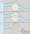
Figure 1
Figure 1
Schematic diagram illustrating the proposed healing mechanism: (a) in the event of cracking and water ingress; (b) the bacteria-based beads incorporated in the composite will swell, this swelling will clog the cracks, and concomitantly “free up” the bacteria, yeast extract and magnesium acetate contained in the beads; (c) the magnesium will precipitate as magnesium-based minerals, the spores will germinate as a result of being exposed to the solubilized yeast extract, and metabolize the acetate, inducing calcium-based mineral precipitation in and on the surface of the beads, healing the crack.
Figure 2
Figure 2
Scheme showing the experimental program used to quantify the crack-healing capacity of the bacteria-based self-healing cementitious composite. The program consisted of four series: (1h) to establish the initial permeability of unhealed cracked mortar specimens; (2h) to determine the autogenous healing capacity of cracked mortar specimens after 56 days of submersion in artificial seawater at 8 °C, and the same specimens after drying; (3h) to determine the autonomous healing capacity of cracked mortar specimens incorporated with beads containing mineral precursor compounds after 56 days of submersion in artificial seawater at 8 °C, and the same specimens after drying; and (4h) to determine the autonomous healing capacity of cracked bacteria-based self-healing cementitious specimens tested after 56 days of submersion in artificial seawater at 8 °C, and the same specimens after drying. Each series consisted of seven specimens with cracks 0.4 mm and 0.6 mm wide.
Figure 3
Figure 3
Schematic showing the setup used to test the bio-functionality of the bacteria-based beads. The setup consisted of: an insulated tank, oxygen microsensor, and water cooler. The tank was made up of an experiment chamber containing an artificial marine concrete crack solution (AMCCS) and cooling chamber containing freshwater. A cooling tube attached to a water cooler was used to cool the water of the cooling chamber, which was in turn used to cool the AMCCS of the experiment chamber to 8 °C. Specimens were submerged in the AMCCS of the experiment chamber, and oxygen measurements made with a microsensor mounted to a motorized micromanipulator.
Figure 4
Figure 4
(a) A schematic of the water column tested above the specimens; and (bd) graphs showing the dissolved oxygen (DO) microprofiles above (b) a cement paste specimen; (c) a cement paste specimen embedded with beads without bacterial spores; and (d) a cement paste specimen embedded with bacteria-based beads, all submerged in an artificial marine concrete crack solution (AMCCS) at 8 °C. The height of the schematic diagram corresponds to the height of the graph profiles. 0d–3d represent profiles taken after zero, one, two, and three days, respectively.
Figure 5
Figure 5
Box plot graphs depicting the permeability data for each series. Graph (a) shows the permeability data for specimens with cracks 0.4 mm wide and graph (b) shows the permeability data for specimens with cracks 0.6 mm wide. 1h: Initial permeability for cracked unhealed mortar specimens; 2h: Permeability of cracked mortar specimens after 56 days of submersion in seawater at 8 °C; 2hd: Permeability of the 2h specimens after drying; 3h: Permeability of cracked mortar specimens with beads containing mineral precursor compounds after 56 days of submersion in seawater at 8 °C; 3hd: Permeability of the 3h specimens after drying; 4h: Permeability of cracked bacteria-based self-healing cementitious specimens after 56 days of submersion in seawater at 8 °C; 4hd: Permeability of the 4h specimens after drying. Each box plot represents the permeability data for seven separate specimens. The square symbol of the boxes represents the mean permeability; the whiskers the minimum and maximum permeability values; and the top, middle and bottom lines the 75th, 50th and 25th percentiles, respectively.
Figure 6
Figure 6
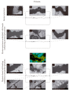
Environmental scanning electron microscopy (ESEM) and energy dispersive spectroscopy (EDS) images of specimens with 0.4 mm wide cracks. (ac) ESEM images of a mortar specimen following 56 days of submersion in seawater at 8 °C: (a) a crack mouth; (b) an area towards the center of the crack; and (c) the other crack mouth. (df) ESEM images of a mortar specimen incorporated with beads containing mineral precursor compounds after 56 days of submersion in seawater at 8 °C: (d) a crack mouth; (e) an area towards the center of the crack; and (f) the other crack mouth. (gl) ESEM images of a bacteria-based self-healing cementitious composite specimen following 56 days of submersion in seawater at 8 °C: (g) a crack mouth; (h) an area towards the centre of the crack; (i) the other crack mouth; (j) a section along the crack of a bacteria-based bead; and (k,l) successive close-ups of the bead. The EDS elemental map (m) corresponding to ESEM image (h). Green in the EDS maps represents calcium, yellow represents magnesium, and blue represents silicate.
Figure 7
Figure 7
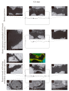
Environmental scanning electron microscopy (ESEM) and energy dispersive spectroscopy (EDS) images of specimens with 0.6 mm wide cracks. (ac) ESEM images of a mortar specimen following 56 days of submersion in seawater at 8 °C: (a) a crack mouth; (b) an area towards the center of the crack; and (c) the other crack mouth. (df) ESEM images of a mortar specimen incorporated with beads containing mineral precursor compounds after 56 days of submersion in seawater at 8 °C: (d) a crack mouth; (e) an area towards the centre of the crack; and (f) the other crack mouth. (gm) ESEM images of a bacteria-based self-healing cementitious composite specimen following 56 days of submersion in seawater at 8 °C: (g,h) a crack mouth, which had split into two smaller cracks; (i) an area towards the centre of the crack; (j) the other crack mouth; (k) a section along the crack including a bacteria-based bead; and successive close-ups (l, m) of a bacteria-based bead. The EDS elemental map (n) corresponding to ESEM image (i). Green of the EDS maps represents calcium, yellow represents magnesium and blue represents silicate.
Figure 8
Figure 8
Compressive strength development of mortar cubes and mortar cubes incorporated with beads containing mineral precursor compounds, cured for 28 days and subsequently submerged in artificial seawater at 8 °C.

References

    1. Marsell R., Einhorn T.A. The biology of fracture healing. Injury. 2011;42:551–555. doi: 10.1016/j.injury.2011.03.031. - DOI - PMC - PubMed
    1. Guimard N.K., Oehlenschlaeger K.K., Zhou J., Hilf S., Schmidt F.G., Barner-Kowollik C. Current trends in the field of self-healing materials. Macromol. Chem. Phys. 2012;213:131–143. doi: 10.1002/macp.201100442. - DOI
    1. Van Breugel K. Is there a market for self-healing cement-based materials; Proceedings of the First International Conference on Self-Healing Materials; Noordwijk aan Zee, The Netherlands. 18–20 April 2007.
    1. Jonkers H.M. Self Healing Materials. Springer; Doordercht, The Netherlands: 2008. Self healing concrete: A biological approach; pp. 195–204.
    1. Jonkers H.M., Thijssen A., Muyzer G., Copuroglu O., Schlangen E. Application of bacteria as self-healing agent for the development of sustainable concrete. Ecol. Eng. 2010;36:230–235. doi: 10.1016/j.ecoleng.2008.12.036. - DOI

LinkOut - more resources