Review
. 2018 Jul 10;3(3):15.
doi: 10.3390/biomimetics3030015.

Hydrogel Actuators and Sensors for Biomedical Soft Robots: Brief Overview with Impending Challenges

Affiliations
Review

Hydrogel Actuators and Sensors for Biomedical Soft Robots: Brief Overview with Impending Challenges

Hritwick Banerjee et al. Biomimetics (Basel). .

Abstract

There are numerous developments taking place in the field of biorobotics, and one such recent breakthrough is the implementation of soft robots-a pathway to mimic nature's organic parts for research purposes and in minimally invasive surgeries as a result of their shape-morphing and adaptable features. Hydrogels (biocompatible, biodegradable materials that are used in designing soft robots and sensor integration), have come into demand because of their beneficial properties, such as high water content, flexibility, and multi-faceted advantages particularly in targeted drug delivery, surgery and biorobotics. We illustrate in this review article the different types of biomedical sensors and actuators for which a hydrogel acts as an active primary material, and we elucidate their limitations and the future scope of this material in the nexus of similar biomedical avenues.

Keywords: biomedical sensors; hydrogel robots; soft actuators; stimuli-responsive hydrogels.


Conflict of interest statement

The authors declare no conflict of interest.

Figures

Figure 1
Figure 1
Different regimes of soft actuators and natural muscle. Optimized hydrogel actuators closely mimic artificial muscles’ tissue profile.
Figure 2
Figure 2
Thermo-responsive hydrogel actuators (HAs). (A) Basic diagram and mechanism for a unidirectional procession of L-shaped symmetric poly(N-isopropylacrylamide) (pNIPAM)/titanate(IV) nanosheet HAs (5 mm thick). (B) In the absence of substantial water uptake and release, distance between the nanosheets rapidly expands and contracts on heating and cooling, respectively, even in air, for which a hydrogel walker achieved a good forward motion. (C) Images of elastin-like polypeptide (ELP)–reduced graphene oxide (rGO) HAs reversibly curling in response to cooling or heating of the surrounding solution. (D) Photothermally actuated hygromorphic crawler. A hydrogel molded with a slight curvature was placed with porous side facing down. The laser was applied so as to induce gel curling. Subsequent uncurling during recovery after the laser was removed pushed the gel forward (1 mm tick marks). (E) Programmable cubes: folding cubes based on thermo-responsive HAs. (a) Fabrication scheme for folding cubes based on single-walled carbon nanotube (SWNT)–pNIPAM/low-density polyethylene (LDPE) bilayer actuators. (b) Cube folding by thermal actuation in 48 °C water. (c) Cube reversibly unfolded by cooling down the water bath in which the cube was immersed. (F) Programmable flower made by heterogeneous integration of pNIPAM and SWNT–pNIPAM bilayer actuators. (a) Fabrication scheme for making a programmable flower, consisting of two layers of actuators. (b) Flower folded (i.e., closed) when heated in a water bath to 50 °C. (c) Flower bloomed by cooling down in the water bath. (A,B) Adapted with permission from [79]. Copyright 2015, Macmillan Publishers Ltd. (C,D) Reprinted (adapted) with permission from [80]. Copyright 2013, American Chemical Society. (E,F) Reprinted (adapted) with permission from [64]. Copyright 2011, American Chemical Society.
Figure 3
Figure 3
Biomedical soft robotic application areas from a materials’ perspective. The denoted region is best suited for hydrogel soft robotic applications in biology and medicine. Concept adapted with permission from [95]. Copyright 2018, Springer Nature Ltd.
Figure 4
Figure 4
Ionic-hydrogel-based actuators. (A) Basic diagram of mechanism with (a) anionic and (b) cationic gels in the same solutions; each could achieve opposite bending directions under the same applied field. (B) Walker achieved unidirectional motion using both anionic and cationic gels. A field of 5 V/cm was applied; electrode field directions are shown in the images. (C) Locomotion of an anionic gel (a) octopus and (b) walker under electric fields in solution; the applied voltage signals were alternating (+7 V/−15 V) and ±15 V, respectively. (D,E) Patterning of hydrogel using ionoprinting for directional embedding of ions. (D) A gel gripper was able to grasp and release items. (E) The imprinted area size and location affected the degree of bending. (A,B) Adapted with permission from [52]. Copyright 2014, The Royal Society of Chemistry. (C) Reproduced with permission from [105]. Copyright 2008, John Wiley & Sons Inc. (D,E) Adapted with permission from [106]. Copyright 2013, Macmillan Publishers Ltd. The entire figure adapted with permission from [107]. Copyright 2017, John Wiley & Sons Inc.
Figure 5
Figure 5
Magneto-responsive hydrogel-based actuators. (A) Reconfigurable body plans for soft hydrogel-based micromachines inspired by microorganisms. (a) A variety of hydrogel-based body designs and propeller mechanisms. (b) Anisotropic swelling behavior controlled by the alignment of magnetic nanoparticles (MNPs) along prescribed three-dimensional (3D) pathways and selective patterning of supporting layers results in 3D functional micromachines. The folding axes 1 and 2 denote the direction of folding for each compartment. The micromachine possesses multiple different magnetic axes, which determine the motility when the magnetic field (MF) is applied. The flagellated micromachine, which contains self-assembled MNPs, performs controllable swimming in the 3D space under a homogeneous rotating MF. (ce) Optical images of flagellated soft micromachines with complex body plans. Magnetic axis (MA1 and MA3) denote the magnetic axes in the head and tail, respectively. Scale bars: 500 μm. (B) The active hydrogel scaffold undergoes a large deformation and volume change via a moderate MF. (a) A cylinder of nanoporous hydrogel was reduced by nearly 5% of its height when subjected to a vertical MF gradient of 38 Am2. (b) The corresponding macroporous hydrogel deformed by nearly 70% under the same MF. (c) Scanning electron microscopy (SEM) images of a free-dried macroporous hydrogel in the undeformed and deformed states. Scale bar: 500 μm. (A) Adapted with permission from [124]. Copyright 2016, Springer Nature Ltd. (B) Reproduced with permission from [125]. Copyright 2011, National Academy of Sciences.
Figure 6
Figure 6
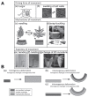
(A) Examples of reversible movement in plants with respect to the driving force: (a) turgor, and (b) swelling of cell walls; mechanism: (c) swelling, and (d) snap-buckling; and trajectory: (e) bending, (f) twisting, and (g) change of two-dimensional (2D) curvature. (B) Different scenarios of swelling of hydrogels: (a) homogeneous deformation of homogeneous hydrogel; (b) inhomogeneous deformation of homogeneous hydrogel; (c) inhomogeneous deformation of inhomogeneous hydrogel. (A,B) Adapted with permission from [8]. Copyright 2013, John Wiley & Sons Inc.
Figure 7
Figure 7
Overall schematic of a hydrogel-based sensor architecture. Concept adapted with permission from [162].
Figure 8
Figure 8
A touch sensor using stretchable and ionically conductive hydrogel electrodes, which project electric field above the sensor to couple with and sense a finger. (A) Working principle and properties of the touch sensor; (B) touching the sensor while sensing and bending; and (C) multi-touch, swipe, and augmented bend detection. Reproduced with permission from [190]. Copyright 2017, AAAS.
Figure 9
Figure 9
Living materials’ design and devices based on stretchable, robust, and biocompatible hydrogel–elastomer hybrids that host various types of genetically engineered bacterial cells. (A) Schematic illustration of cell suspension injection and sealing of injection points; (B) deformation of agar-based living devices; (C) functional living device under large uniaxial stretch; and (D) living hydrogel-based wearable devices. Reproduced with permission from [209]. Copyright 2017, National Academy of Sciences.
Figure 10
Figure 10
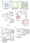
Schematic representation of different types of hydrogel polymer internal structures. (A) Representation of an alginate gel for which G blocks of ionic polymeric chains cross-link via Ca2+ ions (red circle). (B) Internal structure configuration of a polyacrylamide gel for which N,N-methylene-bis-acrylamide (MBA) (blue square) acts as a covalent cross-linker. (C) An example of alginate–polyacrylamide hybrid gel, where both alginate and polyacrylamide networks are integrated through a strong covalent cross-linker (black triangle): carboxyl groups for alginate gel and amine groups for polyacrylamide gel. The components required for this biocompatible alginate–polyacrylamide hybrid gel are deionised (DI) water, acrylamide (AA), sodium alginate (SA); 2-hydroxy-4-(2-hydroxyethoxy)-2-methylpropiophenone (photoinitiator); N, N-methylene-bis-acrylamide (MBA), customized ultraviolet (UV) light exposure, and resting time. (D) Schematic representation of two types of ionic hydrogels. (E) Schematic representation of hydrogel formation with chemical modification process inherited for hydrophobic polymers. (F) Schematic of hydrogel preparation though cross-linker and free radical reaction. (G) Schematic representation for formation of hydrogel cross-linked polymer through condensation reaction of diverse reactants.
Figure 11
Figure 11
Hydrogel network formation and mechanical strength testing. (A) A typical example of double-network (DN) hydrogel formation. (B) Representation of the transition from brittle to ductile structures in a polymer network. Adapted with permission from [4]. Copyright 2009, John Wiley & Sons, Inc. (C) Compression of (a) single-network gel, and (b) DN gel illustrating how DN gel can sustain high compression. Reproduced with permission from [91]. Copyright 2003, John Wiley & Sons, Inc.
Figure 12
Figure 12
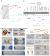
Steps in making robust, tougher, and stretchable hydrogels and their mechanical properties. (A) The transition from soft to rigid materials. (B) Young’s modulus of various materials. Biocompatible soft hydrogel stiffness fits in the cellular and human skin zone. LCEs: Liquid-crystal polymer elastomers; LCNs: Liquid-crystal polymer networks; LCPs: Liquid-crystal polymers; SMPs: Shape-memory polymers. (C) An example of morphing of various “frozen” shapes of tough gels. (D) Three-dimensional folding of the Fe3+ ion-patterned Ca–alginate/polyacrylamide (PAAm) tough hydrogels. Adapted with permission from [305]. Copyright 2017, The Royal Society of Chemistry.
Figure 13
Figure 13
Hydrogel-based 3D printing for artificial organs, and soft biosensors. (A) Advanced bioinks for 3D printing, (B) interpenetrating network (IPN) bioinks for 3D printing, (C) nanoengineered hydrogel-based bioinks for 3D printing, and (D) multi-material bioinks for 3D bioprinting using an artificial support bath. Reproduced with permission from [311]. Copyright 2016, Springer Nature Ltd.
Figure 14
Figure 14
Highly elastic, transparent, and conductive 3D printed ionic composite hydrogels. (A) (a) 3D printed transparent, conductive ionic composite hydrogel bear, (b) a light-emitting diode (LED) lights up when electric potential is applied across the object, and (ce) demonstration of resilience of a 3D printed Eiffel tower after repeatable extensive deformation. (B) (a) Printed ionic composite of different surface area/volume ratios with different swellings, (b) a high-surface area/volume ratio ionic composite hydrogel before and after water absorption, (c) design of a multi-armed hydrogel gripper, (d) before and after swelling in blue dyed water. Reproduced with permission from [326]. Copyright 2017, John Wiley & Sons, Inc.
Figure 15
Figure 15
Self-healing poly(acrylic acid) (PAAc) hydrogels for potential biorobotic applications. (A) (a) Schematic illustration of the structure of the self-healing hydrogel. (be) Self-healing of a cylindrical hydrogel (Entry 3) at room temperature: (b) the original hydrogel, (c) the hydrogel after being cut, (d) the hydrogel after the two parts were brought into contact with each other, (e) the hydrogel after healing for 6 h. (f) Stretching the self-healed hydrogel up to 200%. (B) The use of the self-healing nature of the hydrogel to produce complex architectures. (a) The original hydrogel. (b) The hydrogel after being cut. (c–e) The hydrogel segments arranged in the form of letters: U, M, and D, after (c) 1, (d) 3, and (e) 6 h of self-healing. Reproduced with permission from [337]. Copyright 2013, The Royal Society of Chemistry.

Similar articles

Cited by

References

    1. Haque M.A., Kurokawa T., Gong J.P. Super tough double network hydrogels and their application as biomaterials. Polymer. 2012;53:1805–1822. doi: 10.1016/j.polymer.2012.03.013. - DOI
    1. Zhang Y.S., Khademhosseini A. Advances in engineering hydrogels. Science. 2017;356:eaaf3627. doi: 10.1126/science.aaf3627. - DOI - PMC - PubMed
    1. Sun J.Y., Zhao X., Illeperuma W.R., Chaudhuri O., Oh K.H., Mooney D.J., Vlassak J.J., Suo Z. Highly stretchable and tough hydrogels. Nature. 2012;489:133. doi: 10.1038/nature11409. - DOI - PMC - PubMed
    1. Calvert P. Hydrogels for soft machines. Adv. Mater. 2009;21:743–756. doi: 10.1002/adma.200800534. - DOI
    1. Haraguchi K., Takehisa T. Nanocomposite hydrogels: A unique organic–inorganic network structure with extraordinary mechanical, optical, and swelling/de-swelling properties. Adv. Mater. 2002;14:1120–1124. doi: 10.1002/1521-4095(20020816)14:16<1120::AID-ADMA1120>3.0.CO;2-9. - DOI

LinkOut - more resources