

Intermittent Theta-Burst Stimulation Reverses the After-Effects of Contralateral Virtual Lesion on the Suprahyoid Muscle Cortex: Evidence From Dynamic Functional Connectivity Analysis
- PMID: 31105511
- PMCID: PMC6491879
- DOI: 10.3389/fnins.2019.00309
Intermittent Theta-Burst Stimulation Reverses the After-Effects of Contralateral Virtual Lesion on the Suprahyoid Muscle Cortex: Evidence From Dynamic Functional Connectivity Analysis
Abstract
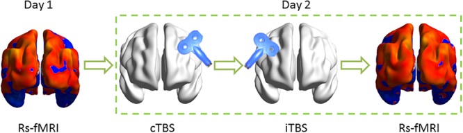
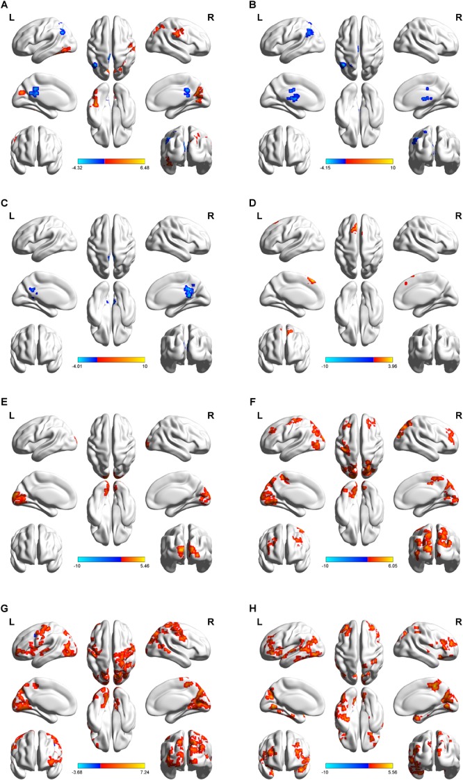
Contralateral intermittent theta burst stimulation (iTBS) can potentially improve swallowing disorders with unilateral lesion of the swallowing cortex. However, the after-effects of iTBS on brain excitability remain largely unknown. Here, we investigated the alterations of temporal dynamics of inter-regional connectivity induced by iTBS following continuous TBS (cTBS) in the contralateral suprahyoid muscle cortex. A total of 20 right-handed healthy subjects underwent cTBS over the left suprahyoid muscle motor cortex and then immediately afterward, iTBS was applied to the contralateral homologous area. All of the subjects underwent resting-state functional magnetic resonance imaging (Rs-fMRI) pre- and post-TBS implemented on a different day. We compared the static and dynamic functional connectivity (FC) between the post-TBS and the baseline. The whole-cortical time series and a sliding-window correlation approach were used to quantify the dynamic characteristics of FC. Compared with the baseline, for static FC measurement, increased FC was found in the precuneus (BA 19), left fusiform gyrus (BA 37), and right pre/post-central gyrus (BA 4/3), and decreased FC was observed in the posterior cingulate gyrus (PCC) (BA 29) and left inferior parietal lobule (BA 39). However, in the dynamic FC analysis, post-TBS showed reduced FC in the left angular and PCC in the early windows, and in the following windows, increased FC in multiple cortical areas including bilateral pre- and postcentral gyri and paracentral lobule and non-sensorimotor areas including the prefrontal, temporal and occipital gyrus, and brain stem. Our results indicate that iTBS reverses the aftereffects induced by cTBS on the contralateral suprahyoid muscle cortex. Dynamic FC analysis displayed a different pattern of alteration compared with the static FC approach in brain excitability induced by TBS. Our results provide novel evidence for us in understanding the topographical and temporal aftereffects linked to brain excitability induced by different TBS protocols and might be valuable information for their application in the rehabilitation of deglutition.
Keywords: dynamic functional connectivity analysis; magnetic resonance imaging; repetitive transcranial magnetic stimulation; swallowing; theta-burst stimulation.
Figures
Similar articles
-
The After-Effects of Theta Burst Stimulation Over the Cortex of the Suprahyoid Muscle on Regional Homogeneity in Healthy Subjects.Front Behav Neurosci. 2019 Mar 1;13:35. doi: 10.3389/fnbeh.2019.00035. eCollection 2019. Front Behav Neurosci. 2019. PMID: 30881294 Free PMC article.
-
Alterations of the amplitude of low-frequency fluctuation in healthy subjects with theta-burst stimulation of the cortex of the suprahyoid muscles.Neuroscience. 2017 Dec 4;365:48-56. doi: 10.1016/j.neuroscience.2017.09.027. Epub 2017 Sep 23. Neuroscience. 2017. PMID: 28947393
-
Intermittent Theta-Burst Stimulation Over the Suprahyoid Muscles Motor Cortex Facilitates Increased Degree Centrality in Healthy Subjects.Front Hum Neurosci. 2020 Jun 16;14:200. doi: 10.3389/fnhum.2020.00200. eCollection 2020. Front Hum Neurosci. 2020. PMID: 32612517 Free PMC article.
-
The effects of theta burst stimulation (TBS) targeting the prefrontal cortex on executive functioning: A systematic review and meta-analysis.Neuropsychologia. 2018 Mar;111:344-359. doi: 10.1016/j.neuropsychologia.2018.02.004. Epub 2018 Feb 10. Neuropsychologia. 2018. PMID: 29438672
-
Efficacy and Time Course of Theta Burst Stimulation in Healthy Humans.Brain Stimul. 2015 Jul-Aug;8(4):685-92. doi: 10.1016/j.brs.2015.03.004. Epub 2015 Mar 26. Brain Stimul. 2015. PMID: 26014214 Review.
Cited by
-
High-Frequency rTMS of the Motor Cortex Modulates Cerebellar and Widespread Activity as Revealed by SVM.Front Neurosci. 2020 Mar 19;14:186. doi: 10.3389/fnins.2020.00186. eCollection 2020. Front Neurosci. 2020. PMID: 32265624 Free PMC article.
-
A reexamination of motor and prefrontal TMS in tobacco use disorder: Time for personalized dosing based on electric field modeling?Clin Neurophysiol. 2021 Sep;132(9):2199-2207. doi: 10.1016/j.clinph.2021.06.015. Epub 2021 Jul 10. Clin Neurophysiol. 2021. PMID: 34298414 Free PMC article.
-
rTMS regulates homotopic functional connectivity in the SCD and MCI patients.Front Neurosci. 2023 Nov 23;17:1301926. doi: 10.3389/fnins.2023.1301926. eCollection 2023. Front Neurosci. 2023. PMID: 38075270 Free PMC article.
-
Intermittent theta-burst stimulation combined with physical therapy as an optimal rehabilitation in Parkinson's disease: study protocol for a randomised, double-blind, controlled trial.Trials. 2023 Jun 16;24(1):410. doi: 10.1186/s13063-023-07425-7. Trials. 2023. PMID: 37328845 Free PMC article.
-
Restoring Functional Connectivity in Hemiplegic Cerebral Palsy: A Study of Low-Frequency rTMS Intervention.J Biomed Phys Eng. 2025 Apr 1;15(2):173-184. doi: 10.31661/jbpe.v0i0.2410-1840. eCollection 2025 Apr. J Biomed Phys Eng. 2025. PMID: 40259943 Free PMC article.
References
-
- Bharath R. D., Biswal B. B., Bhaskar M. V., Gohel S., Jhunjhunwala K., Panda R., et al. (2015). Repetitive transcranial magnetic stimulation induced modulations of resting state motor connectivity in writer’s cramp. Eur. J. Neurol. 22 796–805. - PubMed
LinkOut - more resources
Full Text Sources
Miscellaneous