

Anodal Transcranial Direct Current Stimulation Induces High Gamma-Band Activity in the Left Dorsolateral Prefrontal Cortex During a Working Memory Task: A Double-Blind, Randomized, Crossover Study
- PMID: 31105540
- PMCID: PMC6491895
- DOI: 10.3389/fnhum.2019.00136
Anodal Transcranial Direct Current Stimulation Induces High Gamma-Band Activity in the Left Dorsolateral Prefrontal Cortex During a Working Memory Task: A Double-Blind, Randomized, Crossover Study
Abstract
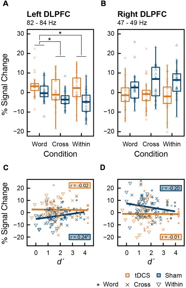
Transcranial direct current stimulation (tDCS) has been shown to have mixed effects on working memory (WM) capacity in healthy individuals. Different stimulation paradigms may account for these discrepancies, with certain features being favored. To determine the effect in the context of anodal tDCS, we investigated whether anodal tDCS induced cortical oscillatory changes during a WM task. Specifically, we tested whether anodal offline tDCS over the left prefrontal cortex (PFC) enhances WM capacity by modulating the oscillatory activity in the left dorsolateral PFC (DLPFC) using magnetoencephalography (MEG). This study employed a double-blind, randomized, crossover design, in which 24 healthy right-handed participants conducted MEG recordings during a 3-back task after administration of 2 mA tDCS or sham stimulation as a placebo. Our results showed that the effect of tDCS did not appear in the behavioral indices-WM accuracy (d') or reaction time (RT). From the results of the time-frequency analysis, significant event-related synchronization (ERS) in the high-gamma band (82-84 Hz) of the left DLPFC was found under the tDCS condition; however, ERS was not correlated with WM capacity. Furthermore, we calculated the modulation index (MI), which indicates the strength of phase-amplitude coupling (PAC). tDCS significantly decreased MI of the left DLPFC, representing the theta-gamma PAC during the n-back task using color names as verbal stimuli. Our results suggest that although tDCS increased the gamma-band oscillation indicating greater neural activity in the left DLPFC, it did not lead to an improvement of WM capacity; this may be due to the inability of gamma-band oscillation to couple with the task-induced theta wave. WM capacity might not increase unless theta-gamma PAC is not enhanced by tDCS.
Keywords: DLPFC; MEG; color; n-back task; phase-amplitude coupling; tDCS; working memory.
Figures


Similar articles
-
Balanced bifrontal transcranial direct current stimulation enhances working memory in adults with high-functioning autism: a sham-controlled crossover study.Mol Autism. 2017 Jul 28;8:40. doi: 10.1186/s13229-017-0152-x. eCollection 2017. Mol Autism. 2017. PMID: 28775825 Free PMC article. Clinical Trial.
-
Transcranial random noise stimulation is more effective than transcranial direct current stimulation for enhancing working memory in healthy individuals: Behavioural and electrophysiological evidence.Brain Stimul. 2020 Sep-Oct;13(5):1370-1380. doi: 10.1016/j.brs.2020.07.001. Epub 2020 Jul 10. Brain Stimul. 2020. PMID: 32659482 Clinical Trial.
-
The effects of offline and online prefrontal vs parietal transcranial direct current stimulation (tDCS) on verbal and spatial working memory.Neurobiol Learn Mem. 2021 Mar;179:107398. doi: 10.1016/j.nlm.2021.107398. Epub 2021 Feb 1. Neurobiol Learn Mem. 2021. PMID: 33540112
-
Does Transcranial Direct Current Stimulation Improve Healthy Working Memory?: A Meta-analytic Review.J Cogn Neurosci. 2016 Aug;28(8):1063-89. doi: 10.1162/jocn_a_00956. Epub 2016 Apr 7. J Cogn Neurosci. 2016. PMID: 27054400 Review.
-
Identifying regions in prefrontal cortex related to working memory improvement: A novel meta-analytic method using electric field modeling.Neurosci Biobehav Rev. 2021 Nov;130:147-161. doi: 10.1016/j.neubiorev.2021.08.017. Epub 2021 Aug 18. Neurosci Biobehav Rev. 2021. PMID: 34418436 Free PMC article. Review.
Cited by
-
A mathematical model and experimental procedure to analyze the cognitive effects of audio frequency magnetic fields.Front Hum Neurosci. 2023 May 12;17:1135511. doi: 10.3389/fnhum.2023.1135511. eCollection 2023. Front Hum Neurosci. 2023. PMID: 37250701 Free PMC article.
-
The Role of the Dorsolateral Prefrontal Cortex for Speech and Language Processing.Front Hum Neurosci. 2021 May 17;15:645209. doi: 10.3389/fnhum.2021.645209. eCollection 2021. Front Hum Neurosci. 2021. PMID: 34079444 Free PMC article.
-
Educational fMRI: From the Lab to the Classroom.Front Psychol. 2019 Dec 6;10:2769. doi: 10.3389/fpsyg.2019.02769. eCollection 2019. Front Psychol. 2019. PMID: 31866920 Free PMC article. Review.
-
[Review of cognitive enhancement techniques based on the combination of cognitive training and transcranial direct current stimulation].Sheng Wu Yi Xue Gong Cheng Xue Za Zhi. 2020 Oct 25;37(5):903-909. doi: 10.7507/1001-5515.201911079. Sheng Wu Yi Xue Gong Cheng Xue Za Zhi. 2020. PMID: 33140616 Free PMC article. Review. Chinese.
-
Dependence of Working Memory on Coordinated Activity Across Brain Areas.Front Syst Neurosci. 2022 Jan 13;15:787316. doi: 10.3389/fnsys.2021.787316. eCollection 2021. Front Syst Neurosci. 2022. PMID: 35095433 Free PMC article. Review.
References
-
- Baddeley A. D., Hitch G. J. (1974). “Working memory,” in Recent Advances in Learning and Motivation, ed Bower G. H. (New York, NY: Academic Press; ), 47–90.
LinkOut - more resources
Full Text Sources
Miscellaneous