. 2019 Apr 26:10:856.
doi: 10.3389/fmicb.2019.00856. eCollection 2019.

Human, Nonhuman Primate, and Bat Cells Are Broadly Susceptible to Tibrovirus Particle Cell Entry

Affiliations

Human, Nonhuman Primate, and Bat Cells Are Broadly Susceptible to Tibrovirus Particle Cell Entry

Yíngyún Caì et al. Front Microbiol. .

Abstract

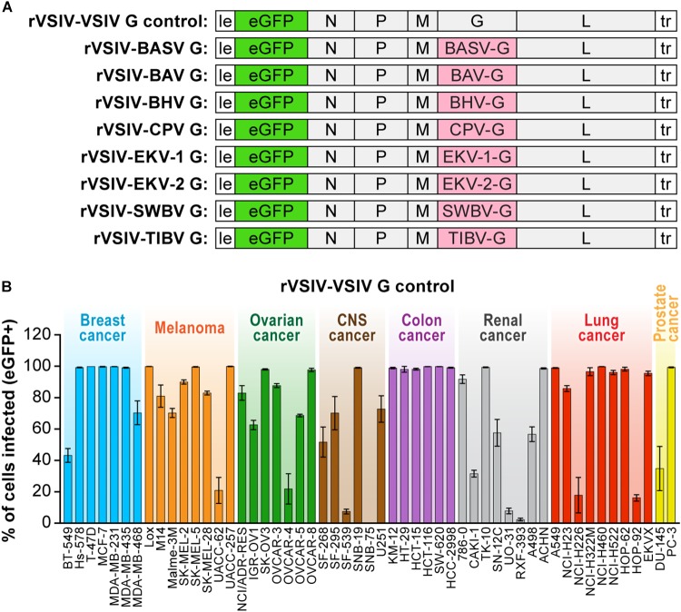
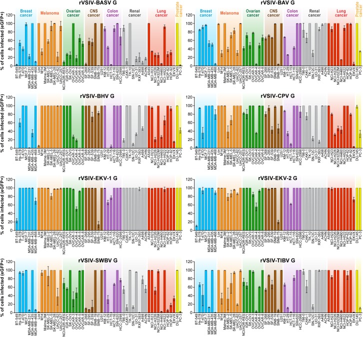
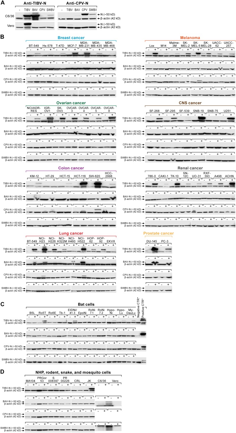
In 2012, the genome of a novel rhabdovirus, Bas-Congo virus (BASV), was discovered in the acute-phase serum of a Congolese patient with presumed viral hemorrhagic fever. In the absence of a replicating virus isolate, fulfilling Koch's postulates to determine whether BASV is indeed a human virus and/or pathogen has been impossible. However, experiments with vesiculoviral particles pseudotyped with Bas-Congo glycoprotein suggested that BASV particles can enter cells from multiple animals, including humans. In 2015, genomes of two related viruses, Ekpoma virus 1 (EKV-1) and Ekpoma virus 2 (EKV-2), were detected in human sera in Nigeria. Isolates could not be obtained. Phylogenetic analyses led to the classification of BASV, EKV-1, and EKV-2 in the same genus, Tibrovirus, together with five biting midge-borne rhabdoviruses [i.e., Beatrice Hill virus (BHV), Bivens Arm virus (BAV), Coastal Plains virus (CPV), Sweetwater Branch virus (SWBV), and Tibrogargan virus (TIBV)] not known to infect humans. Using individual recombinant vesiculoviruses expressing the glycoproteins of all eight known tibroviruses and more than 75 cell lines representing different animal species, we demonstrate that the glycoproteins of all tibroviruses can mediate vesiculovirus particle entry into human, bat, nonhuman primate, cotton rat, boa constrictor, and Asian tiger mosquito cells. Using four of five isolated authentic tibroviruses (i.e., BAV, CPV, SWBV, and TIBV), our experiments indicate that many cell types may be partially resistant to tibrovirus replication after virion cell entry. Consequently, experimental data solely obtained from experiments using tibrovirus surrogate systems (e.g., vesiculoviral pseudotypes, recombinant vesiculoviruses) cannot be used to predict whether BASV, or any other tibrovirus, infects humans.

Keywords: Bas-Congo virus; Mononegavirales; Rhabdoviridae; mononegavirus; rhabdovirus; tibrovirus; tropism; viral hemorrhagic fever.


Figures

FIGURE 1
FIGURE 1
Recombinant vesiculoviruses used in this study. (A) Genome schematic of rVSIV expressing its native G and eGFP (rVSIV–VSIV G control; top row) and rVSIVs created for this study encoding tibrovirus G instead of VSIV G (other rows). (B) Infectivity of rVSIV–VSIV G control (MOI = 3). The percentage of eGFP-expressing NCI-60 human cell panel cell lines was measured by high-content imaging at 24 h post-exposure (“negatives” were confirmed also at 72 h post-exposure, not shown). All experiments were performed in triplicate; error bars show standard deviations. BHV, Beatrice Hill virus; BASV, Bas-Congo virus; BAV, Bivens Arm virus; CNS, central nervous system, CPV; Coastal Plains virus; EKV-1, Ekpoma virus 1; EKV-2, Ekpoma virus 2; MOI, multiplicity of infection; NCI, National Cancer Institute; SWBV, Sweetwater Branch virus; TIBV, Tibrogargan virus. NCI-60 cell lines are listed by their abbreviations and grouped by organ/cancer type.
FIGURE 2
FIGURE 2
Tibrovirus glycoproteins mediate virion entry into a broad range of human cell types. Same experiment as in Figure 1B using rVSIVs expressing diverse tibrovirus G glycoproteins (MOI = 3). The percentage of eGFP-expressing NCI-60 human cell panel cell lines was measured by high-content imaging at 24 h post-exposure. All experiments were performed in triplicates; error bars show standard deviations. BHV, Beatrice Hill virus; BASV, Bas-Congo virus; BAV, Bivens Arm virus; CNS, central nervous system, CPV; Coastal Plains virus; EKV-1, Ekpoma virus 1; EKV-2, Ekpoma virus 2; eGFP, enhanced green fluorescent protein; MOI, multiplicity of infection; NCI, National Cancer Institute; SWBV, Sweetwater Branch virus; TIBV, Tibrogargan virus; rVSIV, recombinant vesicular stomatitis Indiana virus. NCI-60 cell lines are listed by their abbreviations and grouped by organ/cancer type.
FIGURE 3
FIGURE 3
Tibrovirus glycoproteins mediate virion entry into a broad range of animal cell types. Same experiment as in Figure 1B, 2 using different cell types exposed to rVSIV–VSIV G control and rVSIVs expressing diverse tibrovirus glycoproteins (G) (MOI = 3). (A) Bat (PESU-B5L, Ro5T, Ro6E, EidNi/41.3, EpoNi/22.1, RoNi/7.1, RoNi/7.2, HypNi/1.1, HypLu/45.1, Tb1 Lu, MyDauLu/47.1), nonhuman primate (Vero, MA104, RPGor53, S008397, RP00226), hispid cotton rat CRL, and boa constrictor JK cell lines. (B) Asian tiger mosquito C6/36 cells. The percentage of eGFP-expressing cell lines was measured by high-content imaging at 24 h post-exposure (bat, nonhuman primate, hispid cotton rat, and boa constrictor cell lines) or at 24, 48, 72, and 96 h post-exposure (Asian tiger mosquito cells). All experiments were performed in triplicate; error bars show standard deviations. BHV, Beatrice Hill virus; BASV, Bas-Congo virus; BAV, Bivens Arm virus; CPV, Coastal Plains virus; eGFP, enhanced green fluorescent protein; EKV-1, Ekpoma virus 1; EKV-2, Ekpoma virus 2; MOI, multiplicity of infection; SWBV, Sweetwater Branch virus; TIBV, Tibrogargan virus; rVSIV, recombinant vesicular stomatitis Indiana virus.
FIGURE 4
FIGURE 4
Tibrovirus particle host cell entry is dependent on low pH and dynamin but is independent of cholesterol. Tibrovirus G-mediated cell entry occurs via a low-pH-dependent, CME-like pathway. (A) Effects of pretreatment of grivet (Vero) cells with increasing concentrations of endosomal pH modulators on cell entry of rVSIV–VSIV G control and rVSIVs expressing diverse tibrovirus glycoproteins (G). (B) Effect of pretreatment of Vero cells with increasing concentrations of CME inhibitors on cell entry of the same viruses as in A. Cells were pretreated with the indicated concentrations of inhibitors for 30 min and then exposed to rVSIVs (MOI = 0.6) in the presence of inhibitors for 1 h at 37°C, followed by removal of virus inocula. Total expression levels of eGFP were measured using a Tecan microplate reader at 16 h post-exposure. BHV, Beatrice Hill virus; BASV, Bas-Congo virus; BAV, Bivens Arm virus; CME, clathrin-mediated endocytosis; CPV, Coastal Plains virus; eGFP, enhanced green fluorescent protein; EKV-1, Ekpoma virus 1; EKV-2, Ekpoma virus 2; MOI, multiplicity of infection; SWBV, Sweetwater Branch virus; TIBV, Tibrogargan virus; rVSIV, recombinant vesicular stomatitis Indiana virus.
FIGURE 5
FIGURE 5
Authentic tibrovirion cell entry and infections. (A) Asian tiger mosquito (C6/36) and grivet (Vero) cells were exposed to medium-only control (–), BAV, CPV, SWBV, or TIBV particle preparations. Virion entry was detected via western blotting using an anti-TIBV N antibody (here shown to be strongly cross-reactive with BAV in C6/36 cells and weakly cross-reactive with SWBV) and an anti-CPV N antibody (here shown to be cross-reactive with SWBV in C6/36 cells and very weakly cross-reactive with BAV). (B) NCI-60 cells, (C) bat cells, and (D) nonhuman primate (NHP), hispid cotton rat, boa constrictor, and Asian tiger mosquito cells (see also Figure 3) were exposed to medium-only control (–) or BAV, CPV, SWBV, or TIBV (+). Cell lysates were harvested after the appearance of CPE or otherwise at day 10 post-exposure (Table 2). Tibovirion entry was detected via western blotting using the appropriate anti-TIBV, BAV, SWBV, and anti-CPV N antibodies. Protein loading was controlled by detecting β-actin. BAV, Bivens Arm virus; CPV, Coastal Plains virus; N, nucleoprotein; NCI, National Cancer Institute; SWBV, Sweetwater Branch virus; TIBV, Tibrogargan virus.

Similar articles

Cited by

References

    1. Abraham G., Banerjee A. K. (1976). Sequential transcription of the genes of vesicular stomatitis virus. Proc. Natl. Acad. Sci. U.S.A. 73 1504–1508. - PMC - PubMed
    1. Altstiel L. D., Landsberger F. R. (1981). Lipid-protein interactions between the surface glycoprotein of vesicular stomatitis virus and the lipid bilayer. Virology 115 1–9. - PubMed
    1. Babayan S. A., Orton R. J., Streicker D. G. (2018). Predicting reservoir hosts and arthropod vectors from evolutionary signatures in RNA virus genomes. Science 362 577–580. 10.1126/science.aap9072 - DOI - PMC - PubMed
    1. Ball L. A., White C. N. (1976). Order of transcription of genes of vesicular stomatitis virus. Proc. Natl. Acad. Sci. U.S.A. 73 442–446. - PMC - PubMed
    1. Bearzotti M., Delmas B., Lamoureux A., Loustau A.-M., Chilmonczyk S., Bremont M. (1999). Fish rhabdovirus cell entry is mediated by fibronectin. J. Virol. 73 7703–7709. - PMC - PubMed

LinkOut - more resources