. 2019 Apr 15;11(4):2496-2506.
eCollection 2019.

Vitamin D retards intervertebral disc degeneration through inactivation of the NF-κB pathway in mice

Affiliations

Vitamin D retards intervertebral disc degeneration through inactivation of the NF-κB pathway in mice

Hanshui Huang et al. Am J Transl Res. .

Abstract

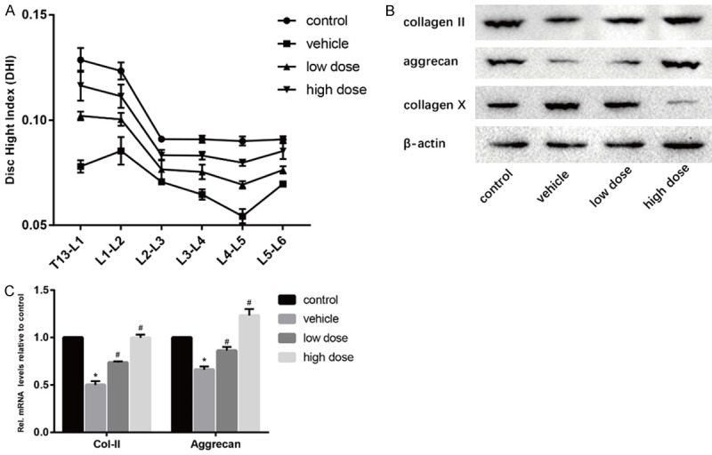
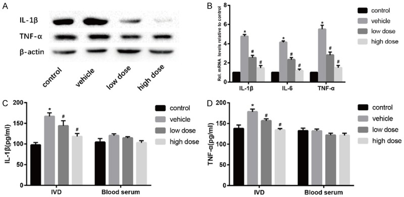
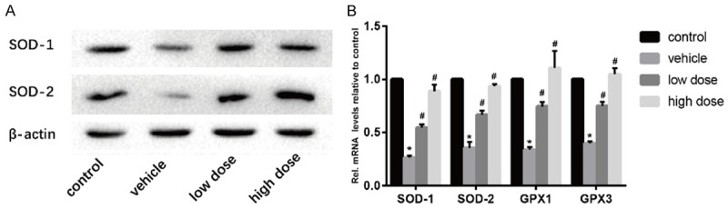
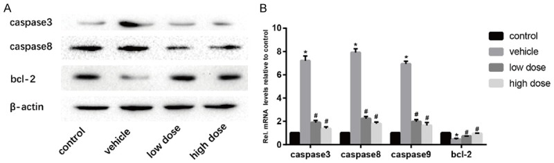
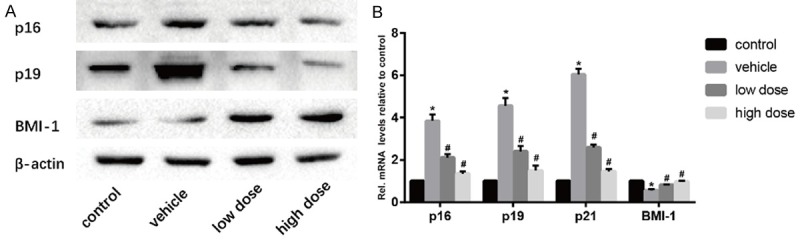
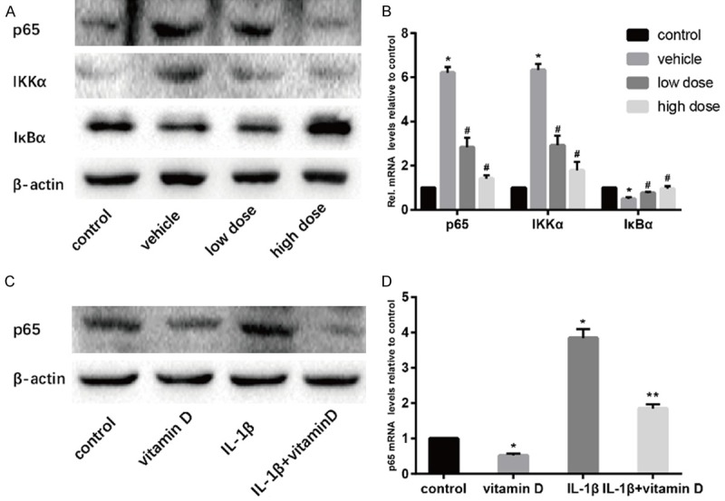
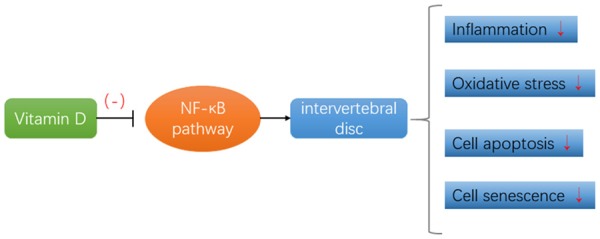
To determine the efficacy and specific mechanism of vitamin D on intervertebral disc degeneration. The model of intervertebral disc degeneration was established in 3-month-old mice. Furthermore, the levels of intervertebral disc degeneration in the vitamin D group and the control group were detected one month later by X-ray, Western boltting, quantitative real-time polymerase chain reaction (qRT-PCR) and enzyme-linked immunosorbent assay (ELISA) et al. In addition, in vitro, we cultured mouse intervertebral disc nucleus pulposus cells to verify the effect of vitamin D on nucleus pulposus cells and study its mechanism. In vivo, compared with the control group, mice in the vitamin D group showed dose-dependent retardation of intervertebral disc degeneration in terms of reducing inflammatory responses, antioxidant stress, inhibiting apoptosis and delaying cell aging. In vitro, compared with the control group, collagen II was increased and collagen X was decreased in mice treated with vitamin D. In vivo and in vitro experiments, the expression of p65 and IκB kinase α was decreased and the expression of inhibitor of NF-κB was increased in the intervertebral disc tissue or nucleus pulposus cells of the vitamin D treatment group, indicating that vitamin D could suppress the NF-κB pathway. Vitamin D retarded intervertebral disc degeneration by inhibiting NF-κB pathway, which may relieve inflammatory reactions, resist oxidative stress, inhibit apoptosis and delay cell senescence.

Keywords: Intervertebral disc degeneration; NF-κB pathway; vitamin D.


Conflict of interest statement

None.

Figures

Figure 1
Figure 1
Effect of vitamin D on intervertebral disc degeneration. A. Results of the difference of DHI in four groups were calculated. B. Results of protein expression of collagen II, aggrecan and collagen X in four groups were determined by Western blotting. β-Actin was used as an internal control. C. Results of mRNA expression of collagen II and aggrecan in four groups were determined by real time PCR. (“*” means there is a statistical difference with the control group and “#” means there is a statistical difference with the vehicle group. N number is all 3. Collagen-II: vehicle-control. P = 0.00002; low dose-vehicle, P = 0.0005; high dose-vehicle, P = 0.00008. Aggrecan, vehicle-control. P = 0.00006; low dose-vehicle, P = 0.0024; high dose-vehicle, P = 0.00019).
Figure 2
Figure 2
Vitamin D reduces inflammation in the intervertebral disc. (A) Results of protein expression of IL-1β and TNF-α in four groups was determined by Western blotting. β-Actin was used as an internal control. (B) Results of mRNA expression of IL-1β, IL-6 and TNF-α in four groups were determined by real time PCR. In addition, The protein expression of IL-1β (C) and TNF-α (D) was measured by Elisa. (“*” means there is a statistical difference with the control group and “#” means there is a statistical difference with the vehicle group. N number is all 3. IL-1β: vehicle-control. P = 0.0003; low dose-vehicle, P = 0.05; high dose-vehicle, P = 0.00156. TNF-α: vehicle-control. P = 0.00285; low dose-vehicle, P = 0.01023; high dose-vehicle, P = 0.00055).
Figure 3
Figure 3
Vitamin D reduces oxidative stress in intervertebral discs. A. Results of protein expression of SOD-1 and SOD-2 in four groups was determined by Western blotting. β-Actin was used as an internal control. B. Results of mRNA expression of SOD-1, SOD-2, GPX1 and GPX3 in four groups were determined by real time PCR. (“*” means there is a statistical difference with the control group and “#” means there is a statistical difference with the vehicle group. N number is all 3. SOD-1: vehicle-control. P = 0.0000007; low dose-vehicle, P = 0.00017; high dose-vehicle, P = 0.00008. SOD-2: vehicle-control. P = 0.00004; low dose-vehicle, P = 0.0014; high dose-vehicle, P = 0.00007. GPX1: vehicle-control. P = 0.0000017; low dose-vehicle, P = 0.00015; high dose-vehicle, P = 0.00124. GPX3: vehicle-control. P = 0.0000007; low dose-vehicle, P = 0.00014; high dose-vehicle, P = 0.00005).
Figure 4
Figure 4
Vitamin D inhibits apoptosis of intervertebral disc cells. A. Results of protein expression of caspase 3, caspase8 and bcl-2 in four groups was determined by Western blotting. β-Actin was used as an internal control. B. Results of mRNA expression of caspase 3, caspase8, caspase9 and bcl-2 in four groups were determined by real time PCR. (“*” means there is a statistical difference with the control group and “#” means there is a statistical difference with the vehicle group. N number is all 3. Caspase3: vehicle-control. P = 0.00001; low dose-vehicle, P = 0.00003; high dose-vehicle, P = 0.00002. Caspase8: vehicle-control. P = 0.000003; low dose-vehicle, P = 0.00001; high dose-vehicle, P = 0.000007. Caspase9: vehicle-control. P = 0.000002; low dose-vehicle, P = 0.000009; high dose-vehicle, P = 0.00001. Bcl-2: vehicle-control. P = 0.00039; low dose-vehicle, P = 0.00568; high dose-vehicle, P = 0.0008).
Figure 5
Figure 5
Vitamin D delays the aging of intervertebral disc cells. A. Results of protein expression of p16, p19, and BMI-1 in four groups was determined by Western blotting. β-Actin was used as an internal control. B. Results of mRNA expression of p16, p19, p21 and BMI-1 in four groups were determined by real time PCR. (“*” means there is a statistical difference with the control group and “#” means there is a statistical difference with the vehicle group. N number is all 3. p16: vehicle-control. P = 0.00009; low dose-vehicle, P = 0.00099; high dose-vehicle, P = 0.00019. p19: vehicle-control. P = 0.00007; low dose-vehicle, P = 0.00113; high dose-vehicle, P = 0.00026. p21: vehicle-control. P = 0.000005; low dose-vehicle, P = 0.000039; high dose-vehicle, P = 0.00001. BMI-1: vehicle-control. P = 0.00017; low dose-vehicle, P = 0.00179; high dose-vehicle, P = 0.00064).
Figure 6
Figure 6
Vitamin D retarded NPCs degeneration in vitro. IL-1β (10 ng/ml) was uesd to promote NPCs degeneration. A. The optimal concentration and time of vitamin D were determined by CCK8 assay. B. Results of protein expression of collagen II and collagen X were determined by Western blotting. C. Results of mRNA expression of collagen II and aggrecan were determined by real time PCR. (“*” means there is a statistical difference with the control group and “**” means there is a statistical difference with the IL-1β group. N number is all 3. Collagen II: vitamin D-control. P = 0.00125; IL-1β+vitamin D-IL-1β, P = 0.0015. Aggrecan: vitamin D-control. P = 0.00012; IL-1β+vitamin D-IL-1β, P = 0.00129).
Figure 7
Figure 7
Vitamin D retarded IVDD by inhibiting NF-κB pathway. Results of protein expression of p65, IKKα and IκBα in vivo was determined by Western blotting (A) and real time PCR (B). Results of expression of p65 in vitro was determined by Western blotting (C) and real time PCR (D). (“*” means there is a statistical difference with the control group, “#” means there is a statistical difference with the vehicle group and “**” means there is a statistical difference with the IL-1β group. N number is all 3. p65: vehicle-control. P = 0.000002; low dose-vehicle, P = 0.00032; high dose-vehicle, P = 0.00001. IKKα: vehicle-control. P = 0.000004; low dose-vehicle, P = 0.00032; high dose-vehicle, P = 0.00008. IκBα: vehicle-control. P = 0.00041; low dose-vehicle, P = 0.01042; high dose-vehicle, P = 0.00526. p65: vitamin D-control. P = 0.0001; IL-1β+vitamin D-IL-1β, P = 0.00024).
Figure 8
Figure 8
Vitamin D inhibits the NF-κB signaling pathway and thus retards IVDD. This role mainly includes relieving inflammatory reactions, resisting oxidative stress, inhibiting apoptosis and delaying cell senescence.

References

    1. Dowdell J, Erwin M, Choma T, Vaccaro A, Iatridis J, Cho SK. Intervertebral disk degeneration and repair. Neurosurgery. 2017;80:S46–S54. - PMC - PubMed
    1. Ji ML, Lu J, Shi PL, Zhang XJ, Wang SZ, Chang Q, Chen H, Wang C. Dysregulated miR-98 contributes to extracellular matrix degradation by targeting IL-6/STAT3 signaling pathway in human intervertebral disc degeneration. J Bone Miner Res. 2016;31:900–909. - PubMed
    1. Shelerud RA. Epidemiology of occupational low back pain. Clin Occup Environ Med. 2006;5:501–528. - PubMed
    1. Xu TT, Liao F, Jin HT, Tong PJ, Xiao LW, Wu CL. Research advance on intervertebral disc degeneration and cell death. Zhongguo Gu Shang. 2015;28:673–678. - PubMed
    1. Podichetty VK. The aging spine: the role of inflammatory mediators in intervertebral disc degeneration. Cell Mol Biol (Noisy-le-grand) 2007;53:4–18. - PubMed

LinkOut - more resources