

Prostate cancer cell growth characteristics in serum and prostate-conditioned media from moderate-intensity exercise-trained healthy and tumor-bearing rats
- PMID: 31105994
- PMCID: PMC6511645
Prostate cancer cell growth characteristics in serum and prostate-conditioned media from moderate-intensity exercise-trained healthy and tumor-bearing rats
Abstract
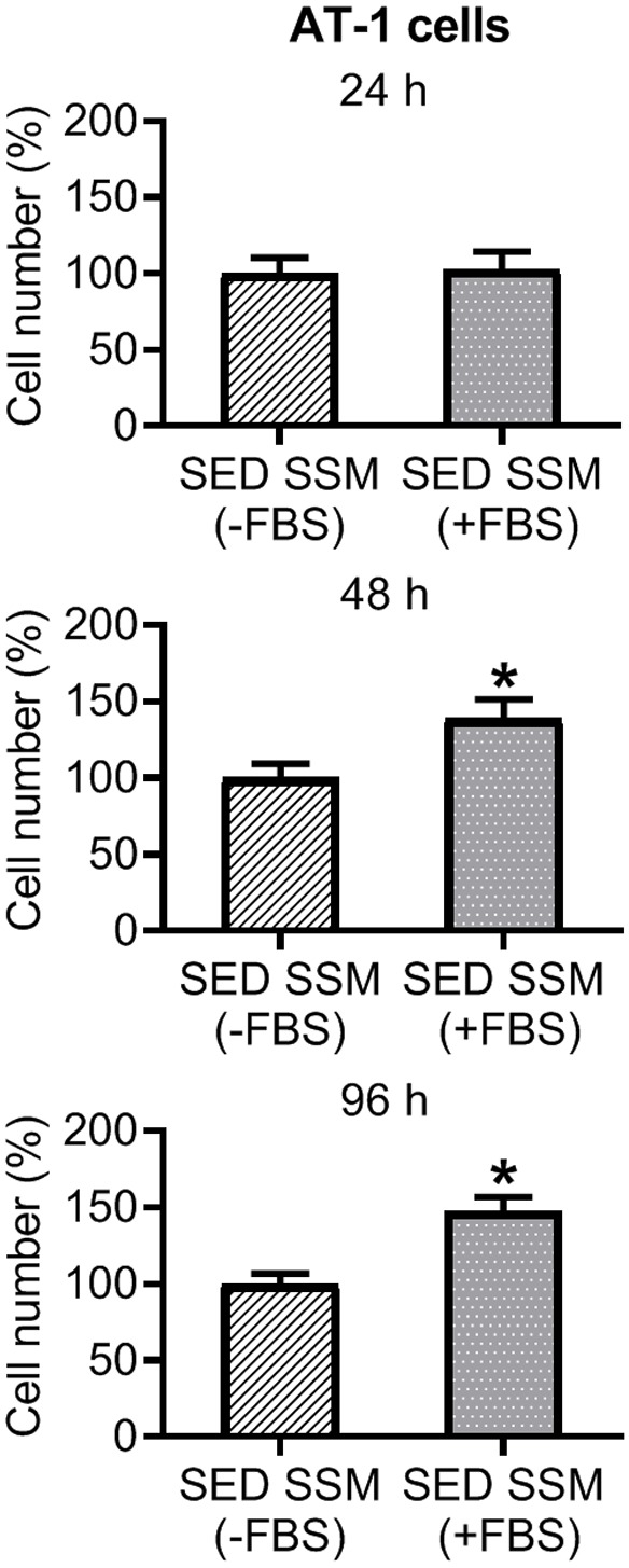
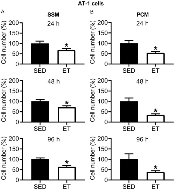
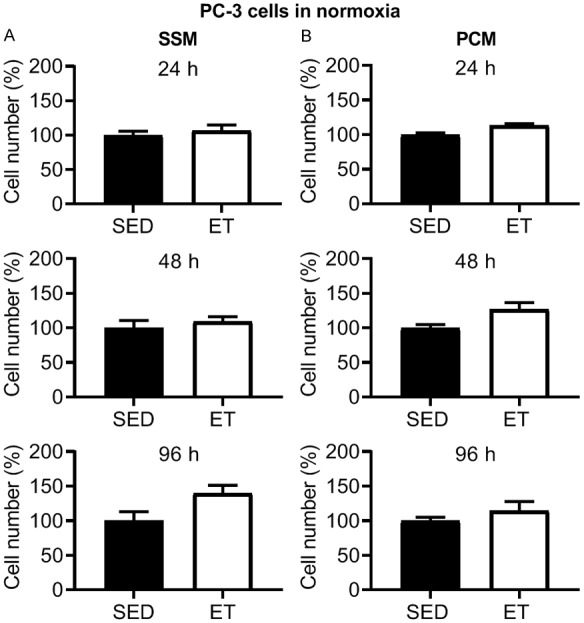
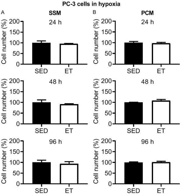
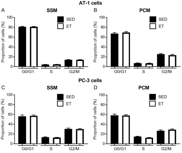
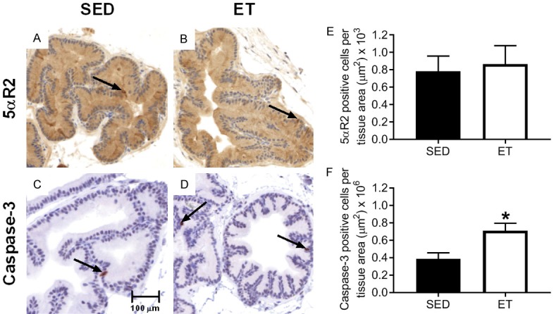
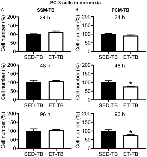
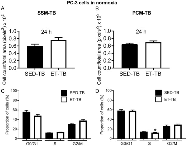
Physical activity is associated with diminished risk of several cancers, and preclinical studies suggest exercise training may alter tumor cell growth in certain tissue(s) (e.g., adipose). From moderate-intensity exercise-trained rats versus sedentary controls, we hypothesized 1) there will be a decreased prostate cancer cell viability and migration in vitro and, within the prostate, a reduced 5α-reductase 2 (5αR2) and increased caspase-3 expression, and 2) that exercise training in tumor-bearing (TB) animals will demonstrate a reduced tumor cell viability in prostate-conditioned media. Serum and prostate were harvested from sedentary or exercise-trained (treadmill running, 10-11 weeks) immune-competent (Copenhagen; n = 20) and -deficient (Nude; n = 18) rats. AT-1 and PC-3 prostate cancer cells were grown in one or more of the following: serum-supplemented media (SSM), SSM from TB rats (SSM-TB), prostate-conditioned media (PCM) or PCM from TB rats (PCM-TB) for 24-96 h under normoxic (18.6% O2) or hypoxic (5% O2) conditions. Under normoxic condition, there was a decreased AT-1 cell viability in SSM and PCM from the exercise-trained (ET) immune-competent rats, but no difference in PC-3 cell viability in SSM and PCM from ET Nude rats versus the sedentary (SED) group, or in SSM-TB from ET-TB Nude rats versus the SED-TB group. However, there was a decreased PC-3 cell viability in the PCM-TB of the ET-TB group versus SED-TB group. PC-3 cell viability in all conditioned media types was not altered between groups with hypoxia. In the prostate, exercise training did not alter 5αR2 expression levels, but increased caspase-3 expression levels. In conclusion, prior exercise status reduced prostate cancer cell viability in the serum and prostate of trained rats but did not modify several other key prostate tumor cell growth characteristics (e.g., migration, cell cycle except in S phase of PC-3 cells in PCM-TB). Importantly, once the tumor was established, exercise training reduced tumor cell viability in the surrounding prostate, which may help explain the reduced severity of the disease in patients that exercise.
Keywords: 5α-reductase 2; Exercise; caspase-3; growth characteristics; hypoxia; normoxia; prostate cancer.
Conflict of interest statement
None.
Figures


Similar articles
-
Effects of exercise training on tumor hypoxia and vascular function in the rodent preclinical orthotopic prostate cancer model.J Appl Physiol (1985). 2013 Dec;115(12):1846-54. doi: 10.1152/japplphysiol.00949.2013. Epub 2013 Oct 31. J Appl Physiol (1985). 2013. PMID: 24177690 Free PMC article.
-
Effects of prostate cancer and exercise training on left ventricular function and cardiac and skeletal muscle mass.J Appl Physiol (1985). 2019 Mar 1;126(3):668-680. doi: 10.1152/japplphysiol.00829.2018. Epub 2018 Dec 20. J Appl Physiol (1985). 2019. PMID: 30571286
-
Exercise delays the hypoxic thermal response in rats.J Appl Physiol (1985). 2003 Jul;95(1):272-8. doi: 10.1152/japplphysiol.00057.2003. Epub 2003 Mar 7. J Appl Physiol (1985). 2003. PMID: 12626482
-
Effects of exercise training on acclimatization to hypoxia: systemic O2 transport during maximal exercise.J Appl Physiol (1985). 2003 Oct;95(4):1531-41. doi: 10.1152/japplphysiol.01220.2001. Epub 2003 Jun 27. J Appl Physiol (1985). 2003. PMID: 12832435
-
Effect of exercise-conditioned human serum on the viability of cancer cell cultures: A systematic review and meta-analysis.Exerc Immunol Rev. 2021;27:24-41. Exerc Immunol Rev. 2021. PMID: 33965899
Cited by
-
Association of non-insulin-based insulin resistance indices, mean platelet volume and prostate cancer: a cross-sectional study.BMC Cancer. 2025 Apr 28;25(1):795. doi: 10.1186/s12885-025-13839-0. BMC Cancer. 2025. PMID: 40295970 Free PMC article.
-
Modulating Tumour Hypoxia in Prostate Cancer Through Exercise: The Impact of Redox Signalling on Radiosensitivity.Sports Med Open. 2022 Apr 8;8(1):48. doi: 10.1186/s40798-022-00436-9. Sports Med Open. 2022. PMID: 35394236 Free PMC article.
-
Anti-carcinogenic effects of exercise-conditioned human serum: evidence, relevance and opportunities.Eur J Appl Physiol. 2021 Aug;121(8):2107-2124. doi: 10.1007/s00421-021-04680-x. Epub 2021 Apr 17. Eur J Appl Physiol. 2021. PMID: 33864493 Free PMC article. Review.
-
Potential Anticarcinogenic Effects From Plasma of Older Adults After Exercise Training: An Exploratory Study.Front Physiol. 2022 Jul 6;13:855133. doi: 10.3389/fphys.2022.855133. eCollection 2022. Front Physiol. 2022. PMID: 35874516 Free PMC article.
-
Effects of high-intensity training on prostate cancer-induced cardiac atrophy.Am J Transl Res. 2021 Jan 15;13(1):197-209. eCollection 2021. Am J Transl Res. 2021. PMID: 33527018 Free PMC article.
References
-
- American Cancer Society. Cancer Facts and Figures. 2019
-
- Ferrara F, Staquicini DI, Driessen WHP, D’Angelo S, Dobroff AS, Barry M, Lomo LC, Staquicini FI, Cardó-Vila M, Soghomonyan S, Alauddin MM, Flores LG, Arap MA, Lauer RC, Mathew P, Efstathiou E, Aparicio AM, Troncoso P, Navone NM, Logothetis CJ, Marchiò S, Gelovani JG, Sidman RL, Pasqualini R, Arap W. Targeted molecular-genetic imaging and ligand-directed therapy in aggressive variant prostate cancer. PNAS. 2016;113:12786–12791. - PMC - PubMed
-
- Wiggins JM, Opoku-Acheampong AB, Baumfalk DR, Siemann DW, Behnke BJ. Exercise and the tumor microenvironment: potential therapeutic implications. Exerc Sport Sci Rev. 2018;46:56–64. - PubMed
-
- Littman AJ, Kristal AR, White E. Recreational physical activity and prostate cancer risk (United States) Cancer Causes Control. 2006;17:831–841. - PubMed
-
- Wiklund F, Lageros YT, Chang E, Bälter K, Johansson J, Adami H, Grönberg H. Lifetime total physical activity and prostate cancer risk: a population-based case-control study in Sweden. Eur J Epidemiol. 2008;23:739–746. - PubMed
LinkOut - more resources
Full Text Sources
Research Materials