

Dermoscopy in the Diagnosis of Cutaneous Leishmaniasis
- PMID: 31106013
- PMCID: PMC6502294
- DOI: 10.5826/dpc.0902a06
Dermoscopy in the Diagnosis of Cutaneous Leishmaniasis
Abstract
Background: Cutaneous leishmaniasis (CL) is a protozoan infectious disease. Dermoscopy is a noninvasive diagnostic tool that has been applied to several skin diseases, including infestations.
Objectives: To determine the dermoscopic patterns of CL lesions and to investigate whether a relationship exists between dermoscopic characteristics and the disease duration, localization, and type of CL lesions.
Methods: Seventy-nine patients (48 male, 31 female) from Hatay, Turkey, were enrolled in the study and a dermoscopic evaluation was performed on 139 lesions. Images of CL lesions were taken via polarized light contact dermoscopy. Chi-square and Fisher exact tests were used for statistical analyses and P values <0.05 were considered significant.
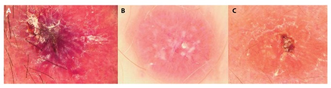
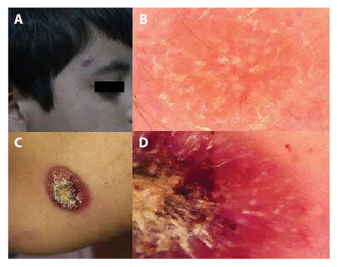
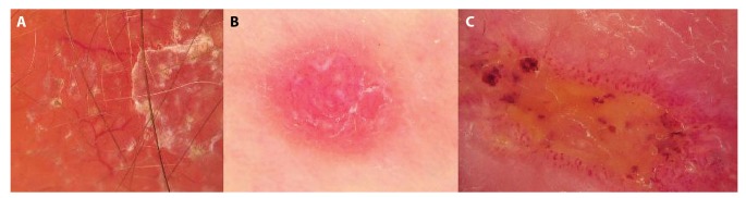
Results: Generalized erythema was seen in all CL lesions. Vascular structures (94.2%), yellow tears (75.5%), and a white starburst-like pattern (58.3%) were the other most common dermoscopic features. Hyperkeratosis (P = 0.001) and white starburst-like pattern (P < 0.001) were more prevalent in the extremities than elsewhere. Among vascular structures, linear irregular (45.8%), hairpin (43.5%), and comma-shaped (25.9%) patterns were the most common dermoscopic findings. Linear irregular (P = 0.023) and arborizing vessels (P = 0.001) were observed in the head-neck region. Dotted (P = 0.009), hairpin (P < 0.001), and glomerular-like (P = 0.016) morphological findings were more prevalent in the extremities. Statistical significances in disease duration were detected in microarborizing (P = 0.027) and arborizing (P = 0.004) vessels and were most prevalent with a disease duration of >6 months. Hairpin vessels were prevalent in the plaque and nodulo-ulcerative type of lesions. Dotted vessels were most commonly seen in the plaque type (47.4%) of lesions.
Conclusions: Generalized erythema, yellow tears, and starburst-like patterns, as well as linear irregular, hairpin, comma-shaped, and arborizing vessels, were the most commonly detected dermoscopic features of CL lesions. We suggest that the presence of these features can be helpful when diagnosing CL lesions by dermoscopy.
Keywords: cutaneous; dermoscopy; leishmaniasis; vascular.
Conflict of interest statement
Competing interests: The authors have no conflicts of interest to disclose.
Figures

Similar articles
-
Dermoscopic evaluation of cutaneous leishmaniasis.Arch Dermatol Res. 2023 Apr;315(3):531-540. doi: 10.1007/s00403-022-02387-3. Epub 2022 Sep 26. Arch Dermatol Res. 2023. PMID: 36163392
-
Dermoscopy of cutaneous leishmaniasis.Br J Dermatol. 2009 Apr;160(4):756-61. doi: 10.1111/j.1365-2133.2008.08986.x. Epub 2008 Dec 16. Br J Dermatol. 2009. PMID: 19120331
-
Cutaneous leishmaniasis: new dermoscopic findings.Int J Dermatol. 2013 Jul;52(7):831-7. doi: 10.1111/j.1365-4632.2012.05815.x. Int J Dermatol. 2013. PMID: 23789601
-
How to diagnose nonpigmented skin tumors: a review of vascular structures seen with dermoscopy: part I. Melanocytic skin tumors.J Am Acad Dermatol. 2010 Sep;63(3):361-74; quiz 375-6. doi: 10.1016/j.jaad.2009.11.698. J Am Acad Dermatol. 2010. PMID: 20708469 Review.
-
Eccrine porocarcinoma shares dermoscopic characteristics with eccrine poroma: A report of three cases and review of the published work.J Dermatol. 2016 Mar;43(3):332-5. doi: 10.1111/1346-8138.13082. Epub 2015 Sep 1. J Dermatol. 2016. PMID: 26333057 Review.
Cited by
-
Entodermoscopy Update: A Contemporary Review on Dermoscopy of Cutaneous Infections and Infestations.Indian Dermatol Online J. 2021 Mar 2;12(2):220-236. doi: 10.4103/idoj.IDOJ_559_20. eCollection 2021 Mar-Apr. Indian Dermatol Online J. 2021. PMID: 33959518 Free PMC article. Review.
-
Dermoscopic evaluation of cutaneous leishmaniasis.Arch Dermatol Res. 2023 Apr;315(3):531-540. doi: 10.1007/s00403-022-02387-3. Epub 2022 Sep 26. Arch Dermatol Res. 2023. PMID: 36163392
-
Update on Dermoscopy and Infectious Skin Diseases.Dermatol Pract Concept. 2019 Dec 31;10(1):e2020003. doi: 10.5826/dpc.1001a03. eCollection 2020. Dermatol Pract Concept. 2019. PMID: 31921490 Free PMC article.
-
Erysipeloid Presentation of Cutaneous Leishmaniasis of the Scalp.Dermatol Pract Concept. 2023 Apr 1;13(2):e2023087. doi: 10.5826/dpc.1302a87. Online ahead of print. Dermatol Pract Concept. 2023. PMID: 37196278 Free PMC article. No abstract available.
-
Dermoscopy of cutaneous granulomatous disorders: A study of 107 cases.Skin Res Technol. 2023 Jan;29(1):e13273. doi: 10.1111/srt.13273. Skin Res Technol. 2023. PMID: 36704887 Free PMC article.
References
-
- Uz OK, Balcıoğlu IC, Taylan Özkan A, Ozensoy S, Ozbel Y. Leishmaniasis in Turkey. Acta Trop. 2002;84(1):43–48. - PubMed
-
- WHO. Leishmaniasis in high-burden countries: an epidemiological update based on data reported in 2014. Wkly Epidemiol Rec. 2016;91(22):287–296. - PubMed
-
- Lallas A, Kyrgidis A, Tzellos TG, et al. Accuracy of dermoscopic criteria for the diagnosis of psoriasis, dermatitis, lichen planus and pityriasis rosea. Br J Dermatol. 2012;166(6):1198–1205. - PubMed
-
- Ayhan E, Ucmak D, Baykara SN, Akkurt ZM, Arica M. Clinical and dermoscopic evaluation of cutaneous leishmaniasis. Int J Dermatol. 2015;54(2):193–201. - PubMed
-
- Yücel A, Günasti S, Denli Y, Uzun S. Cutaneous leishmaniasis: new dermoscopic findings. Int J Dermatol. 2013;52(7):831–837. - PubMed
LinkOut - more resources
Full Text Sources