

Development of an in vitro diagnostic method to determine the genotypic sex of Xenopus laevis
- PMID: 31106075
- PMCID: PMC6500372
- DOI: 10.7717/peerj.6886
Development of an in vitro diagnostic method to determine the genotypic sex of Xenopus laevis
Abstract
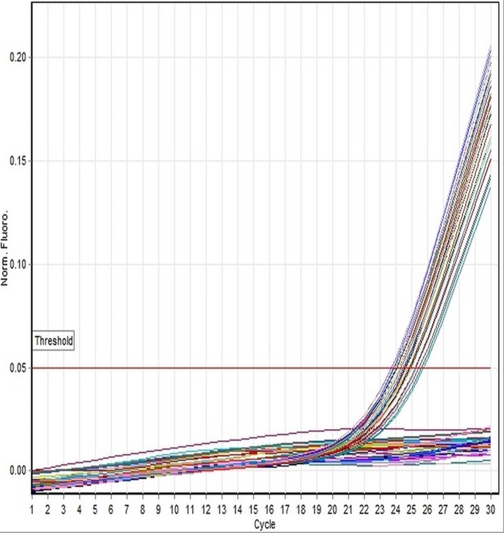
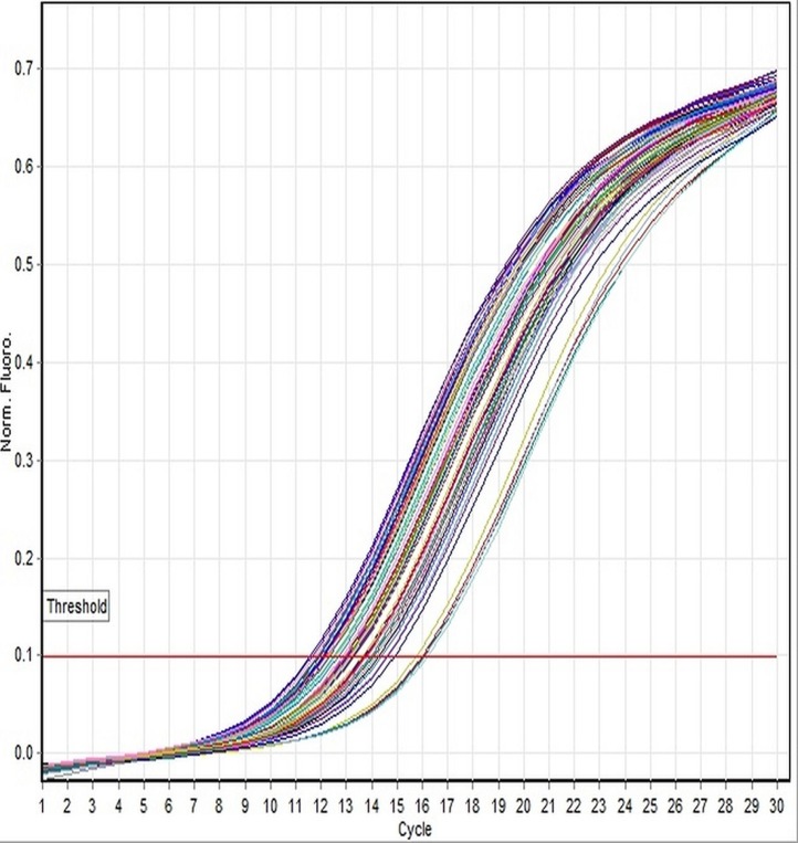
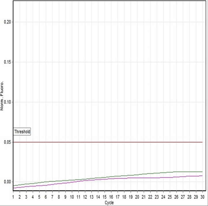
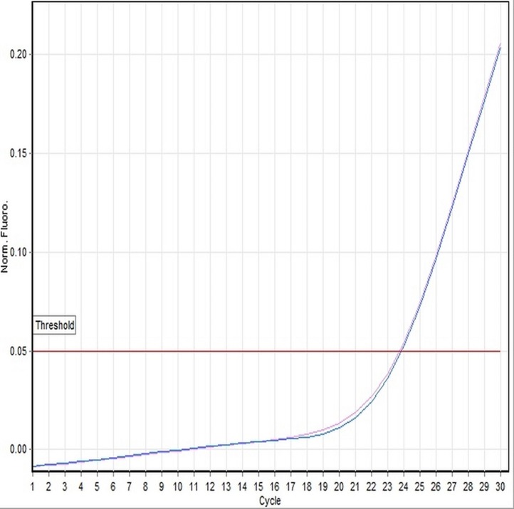
A genotypic sex determination assay provides accurate gender information of individuals with well-developed phenotypic characters as well as those with poorly developed or absent of phenotypic characters. Determination of genetic sex for Xenopus laevis can be used to validate the outcomes of Tier 2 amphibian assays, and is a requirement for conducting the larval amphibian growth and development assay (LAGDA), in the endocrine disruptor screening program (EDSP), test guidelines. The assay we developed uses a dual-labeled TaqMan probe-based real-time polymerase chain reaction (real-time PCR) method to determine the genotypic sex. The reliability of the assay was tested on 37 adult specimens of X. laevis collected from in-house cultures in Eurofins EAG Agroscience, Easton. The newly designed X. laevis-specific primer pair and probe targets the DM domain gene linked-chromosome W as a master female-determining gene. Accuracy of the molecular method was assessed by comparing with phenotypic sex, determined by necropsy and histological examination of gonads for all examined specimens. Genotypic sex assignments were strongly concordant with observed phenotypic sex, confirming that the 19 specimens were male and 18 were female. The results indicate that the TaqMan® assay could be practically used to determine the genetic sex of animals with poorly developed or no phenotypic sex characteristics with 100% precision. Therefore, the TaqMan® assay is confirmed as an efficient and feasible method, providing a diagnostic molecular sex determination approach to be used in the amphibian endocrine disrupting screening programs conducted by regulatory industries. The strength of an EDSP is dependent on a reliable method to determine genetic sex in order to identify reversals of phenotypic sex in animals exposed to endocrine active compounds.
Keywords: Genetic sex determination; OCSPP-LAGDA; TaqMan® real-time PCR; Xenopus laevis.
Conflict of interest statement
Amin Eimanifar is a staff scientist II of the Eurofins EAG Agroscience, John Aufderheide is a senior director, Aquatic, Plant and Invertebrate Toxicology of the Eurofins EAG Agroscience, Suzanne Z. Schneider is a manager of Aquatic Toxicology of the Eurofins EAG Agroscience, Henry Krueger is a VP, Regulatory and Scientific Affairs of the Eurofins EAG Agroscience, and Sean Gallagher is a Scientific Advisor - Ecotoxicology of the Eurofins EAG Agroscience. The authors declare that they have no competing interests.
Figures
Similar articles
-
Development of the Larval Amphibian Growth and Development Assay: Effects of benzophenone-2 exposure in Xenopus laevis from embryo to juvenile.J Appl Toxicol. 2016 Dec;36(12):1651-1661. doi: 10.1002/jat.3336. Epub 2016 May 30. J Appl Toxicol. 2016. PMID: 27241388
-
Development of the Larval Amphibian Growth and Development Assay: effects of chronic 4-tert-octylphenol or 17β-trenbolone exposure in Xenopus laevis from embryo to juvenile.J Appl Toxicol. 2016 Dec;36(12):1639-1650. doi: 10.1002/jat.3330. Epub 2016 May 3. J Appl Toxicol. 2016. PMID: 27143402
-
A ZZ/ZW-type sex determination in Xenopus laevis.FEBS J. 2011 Apr;278(7):1020-6. doi: 10.1111/j.1742-4658.2011.08031.x. Epub 2011 Feb 25. FEBS J. 2011. PMID: 21281450 Review.
-
Impaired gonadal and somatic development corroborate vulnerability differences to the synthetic estrogen ethinylestradiol among deeply diverged anuran lineages.Aquat Toxicol. 2016 Aug;177:503-14. doi: 10.1016/j.aquatox.2016.07.001. Epub 2016 Jul 5. Aquat Toxicol. 2016. PMID: 27434076
-
Evaluation of EPA's Tier 1 Endocrine Screening Battery and recommendations for improving the interpretation of screening results.Regul Toxicol Pharmacol. 2011 Apr;59(3):397-411. doi: 10.1016/j.yrtph.2011.01.003. Epub 2011 Jan 18. Regul Toxicol Pharmacol. 2011. PMID: 21251942 Review.
Cited by
-
CYP19A1 (aromatase) dominates female gonadal differentiation in chicken (Gallus gallus) embryos sexual differentiation.Biosci Rep. 2020 Oct 30;40(10):BSR20201576. doi: 10.1042/BSR20201576. Biosci Rep. 2020. PMID: 32990306 Free PMC article.
-
Telomere dynamics in maturing frogs vary among organs.Biol Lett. 2025 Feb;21(2):20240626. doi: 10.1098/rsbl.2024.0626. Epub 2025 Feb 26. Biol Lett. 2025. PMID: 39999893 Free PMC article.
-
Ageing across the great divide: tissue transformation, organismal growth and temperature shape telomere dynamics through the metamorphic transition.Proc Biol Sci. 2023 Feb 8;290(1992):20222448. doi: 10.1098/rspb.2022.2448. Epub 2023 Feb 8. Proc Biol Sci. 2023. PMID: 36750187 Free PMC article.
References
-
- Babošová M, Vašeková P, Porhajašová JI, Noskovič J. Influence of temperature on reproduction and length of metamorphosis in Xenopus laevis (Amphibia: Anura) European Zoological Journal. 2018;85(1):151–158. doi: 10.1080/24750263.2018.1450456. - DOI
-
- Banaras S, Aman S, Zafar M, Khan M, Abbas S, Shinwari ZK, Shakeel SN. Molecular identification and comparative analysis of novel 18S Ribosomal RNA genomic sequences of a wide range of medicinal plants. Pakistan Journal of Botany. 2012;44(6):2021–2026.
LinkOut - more resources
Full Text Sources
