. 2018 Jul 9;4(7):2319-2329.
doi: 10.1021/acsbiomaterials.8b00304. Epub 2018 Jun 6.

A chemical treatment method for obtaining clean and intact pollen shells of different species

Affiliations

A chemical treatment method for obtaining clean and intact pollen shells of different species

Pedro Gonzalez-Cruz et al. ACS Biomater Sci Eng. .

Abstract

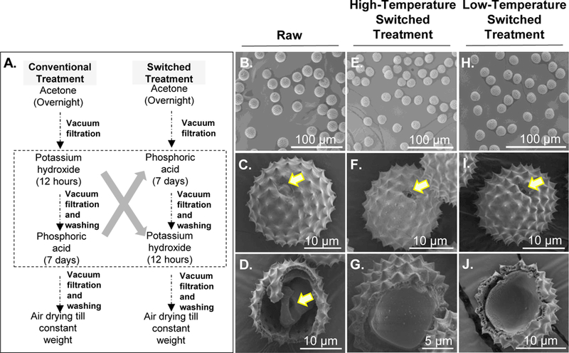
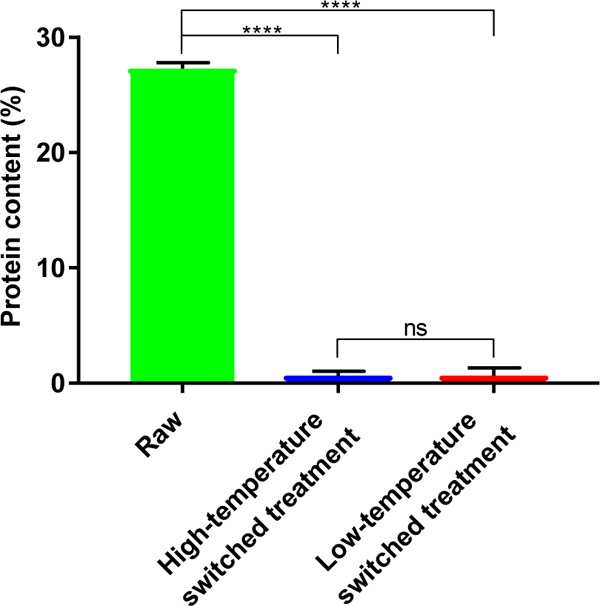
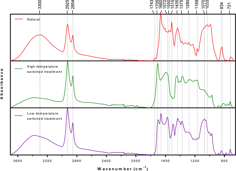
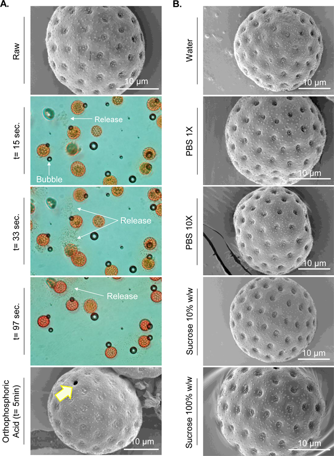
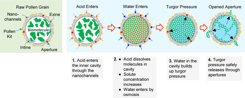
Pollen grains and plant spores have emerged as a novel biomaterial for a broad range of applications including oral drug and vaccine delivery, catalyst support, and removal of heavy metals. However, before pollens can be used, their intrinsic biomolecules, which occupy a large part of the pollen inner cavity must be removed not only to create empty space but because they have potential to cause allergies when used in vivo. These intrinsic materials in the pollen core can be extracted through a chemical treatment to generate clean pollen shells. The commonly used method involves a series of sequential treatments with organic solvents, alkalis, and acids to remove the native pollen biomolecules. This method, though successful for treating lycopodium (Lycopodium clavatum) spores, fails for other species of pollens such as common ragweed (Ambrosia elatior) and thus prevents widespread investigation of different pollens. Herein, we report a new chemical treatment for obtaining clean pollen shells from multiple plant species. This new method involves sequential treatment with acetone, phosphoric acid, and potassium hydroxide. Scanning electron micrographs and protein quantification have shown that the new method can successfully produce clean, intact, and hollow shells from many pollen species including ragweed, sunflower, black alder, and lamb's quarters. These results demonstrate the broad applicability of this method to clean pollens of different species, and paves the way to start investigating them for various applications.

Keywords: Pollen aperture; Pollen treatment; Pollen turgor pressure; Ragweed pollen; Sporopollenin.


Conflict of interest statement

This potential conflict of interest has been disclosed and is managed by Texas Tech University.

Figures

Figure 1.
Figure 1.. Schematic diagram of conventional and modified conventional treatment processes.
Pathway 1 shows processing steps for conventional treatment of pollen grains. Pathways 2 and 3 represent modifications to the conventional treatment and are called modified conventional treatment. Their respective steps are shown. In pathway 2 modification was made at the wash step to compare centrifugation versus vacuum filtration. Pathway 3 shows processing steps after the centrifugation process was selected based on pathway 2 as the more suitable option for washing the pollens.
Figure 2.
Figure 2.. Schematic diagram and images of lycopodium and ragweed pollens processed using the conventional treatment protocol.
A. Schematic diagram of the processing steps of the conventional treatment protocol. B. Lycopodium after processing with conventional treatment and C. zoomed in image of a single lycopodium. Ragweed pollens after 6 hours of KOH treatment: D. photograph of the cake formed on the filter paper after vacuum filtration. E. SEM image of the cake showing pollen entrapped in extraneous matter. F. zoomed in SEM image of the cake showing more details of entrapped pollens. Some entrapped pollens are indicated by arrows.
Figure 3.
Figure 3.. Fourier-transform infrared (FTIR) spectra of extracted materials from ragweed pollen after KOH treatment.
Ragweed pollens were treated with 6% w/v KOH solution for 6 h and extracts were separated from the solution via centrifugation, dried at 120 ºC, and analyzed using FTIR.
Figure 4.
Figure 4.. Images of ragweed pollens processed using conventional treatment and modified conventional treatment protocols.
A. Schematic diagram of the processing steps for figures B to D (vacuum filtration) and E to F (centrifugation). B. Photograph of the cake formed after vacuum filtration. C. SEM image of the cake showing pollens entrapped in extraneous matter. D. Zoomed in SEM image of the cake showing entrapped pollens (indicated by arrows). E. Pollen clumps formed after centrifugation. F. Zoomed in SEM image of the clumps showing more details of clumped pollens. G. Schematic diagram of the processing steps for figures H and I. H. SEM image of pollens clumped together. I. Zoomed in SEM image of the clump showing more details of unclean pollen surfaces.
Figure 5.
Figure 5.. SEM images of ragweed pollens processed using the switched treatment.
A. Comparison diagram of the conventional treatment and switched treatment steps. B. Raw ragweed pollens. C. Zoomed in image of a raw ragweed pollen. D. Interior of raw ragweed pollen showing the presence of natural biological material as indicated by the arrow. E. Ragweed pollen processed with high-temperature switched treatment. F. Zoomed in image of a ragweed pollen after high-temperature switched treatment showing an intact morphology. G. Inside of ragweed pollen after high-temperature switched treatment showing a clean interior. H. Ragweed pollen processed with low-temperature switched treatment. I. Zoomed in image of a ragweed pollen after low-temperature switched treatment showing an intact morphology. J. Inside of ragweed pollen after low-temperature switched treatment showing a clean interior. Arrow in figure C indicates a closed aperture in raw pollen, while arrows in F and I indicate open apertures after switched treatment.
Figure 6.
Figure 6.. Protein content of ragweed shells obtained using the switched treatment protocols.
Ragweed pollens processed by high- and low-temperature switched treatment protocols showed a dramatic reduction in protein content over raw ragweed pollens, indicating success of the switched treatment process in removing native proteinaceous material. Values shown are means ± SD from three independent samples. A two-tailed t-test was used for comparison. (ns = not significant, [****] = p < 0.001)
Figure 7.
Figure 7.
Fourier-transform infrared (FTIR) spectra of ragweed pollen treated using switched treatment protocols.
Figure 8.
Figure 8.. SEM images of different species of pollens processed using the low-temperature switched treatment protocol.
A. Lamb’s quarters pollen. B. Zoomed in image of a lamb’s quarters pollen showing an intact morphology and clean exterior surface. C. Inside of lamb’s quarters pollen showing a clean interior. D. Sunflower pollen. E. Zoomed in image of a sunflower pollen showing an intact morphology and clean exterior surface. F. Inside of sunflower pollen showing a clean interior. G. Black alder pollen. H. Zoomed in image of a black alder pollen showing an intact morphology and clean exterior surface. I. Inside of black alder pollen showing a clean interior. Arrows in B, C, E, F, H, and I indicate open apertures after switched treatment.
Figure 9.
Figure 9.. Images of pollen apertures bursting open.
A. Lamb’s quarters pollen before and at different times after exposure to orthophosphoric acid. Arrows indicate either air bubbles, or release of matter from inside the pollen due to bursting open of apertures. Air bubbles are an artifact and are not related to bursting of apertures. B. SEM images of lamb’s quarters after exposure to other solvents that did not cause aperture to open. It should be noted that due to practical limitations of time needed to apply the cover slip and subsequent lens focusing there is a time gap of about 5 s between addition of acid to lamb’s quarters on the slide and start of video capture. The time label t = 15 s, t = 33 s, and t = 97 s represent time after video capture was started.
Figure 10.
Figure 10.. Schematic showing mechanism of opening of aperture in pollen grains during orthophosphoric acid treatment.
Orthophosphoric acid enters the pollen grains and dissolves the molecules contained in its interior. This causes buildup of an osmotic gradient and allows more water to enter the pollen cavity, which increases turgor pressure and bursts the pollen shell wall open at aperture sites, because they are mechanically weaker than the rest of the pollen wall.

References

    1. Bedinger P, The remarkable biology of pollen. Plant Cell 1992, 4 (8), 879–887. - PMC - PubMed
    1. Borg M; Brownfield L; Twell D, Male gametophyte development: a molecular perspective. J Exp Bot 2009, 60 (5), 1465–1478. 10.1093/jxb/ern355. - DOI - PubMed
    1. Mascarenhas JP, The Male Gametophyte of Flowering Plants. Plant Cell 1989, 1 (7), 657–664. 10.2307/3868955. - DOI - PMC - PubMed
    1. Diego-Taboada A; Beckett ST; Atkin SL; Mackenzie G, Hollow Pollen Shells to Enhance Drug Delivery. Pharmaceutics 2014, 6 (1), 80–96. 10.3390/pharmaceutics6010080. - DOI - PMC - PubMed
    1. Edlund AF; Swanson R; Preuss D, Pollen and stigma structure and function: the role of diversity in pollination. Plant Cell 2004, 16 (Suppl), S84–S97. 10.1105/tpc.015800. - DOI - PMC - PubMed

LinkOut - more resources