

Development and multicentre validation of an agar-based screening method for echinocandin susceptibility testing of Aspergillus species
- PMID: 31106352
- PMCID: PMC6640300
- DOI: 10.1093/jac/dkz154
Development and multicentre validation of an agar-based screening method for echinocandin susceptibility testing of Aspergillus species
Abstract
Background: Reference antifungal susceptibility testing of echinocandins against Aspergillus spp. relies on the determination of the minimal effective concentration, which is difficult to perform, time-consuming and subjective. We developed and evaluated in a multicentre study an agar-based screening method for echinocandin susceptibility testing of Aspergillus spp.
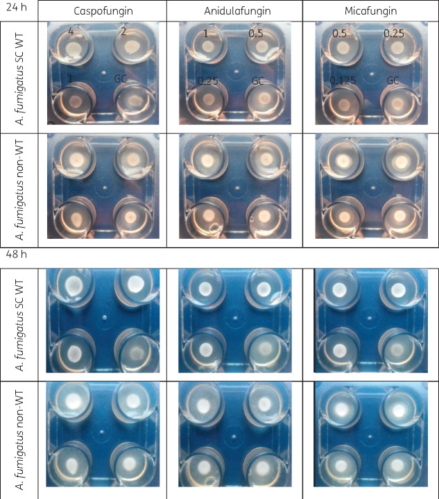
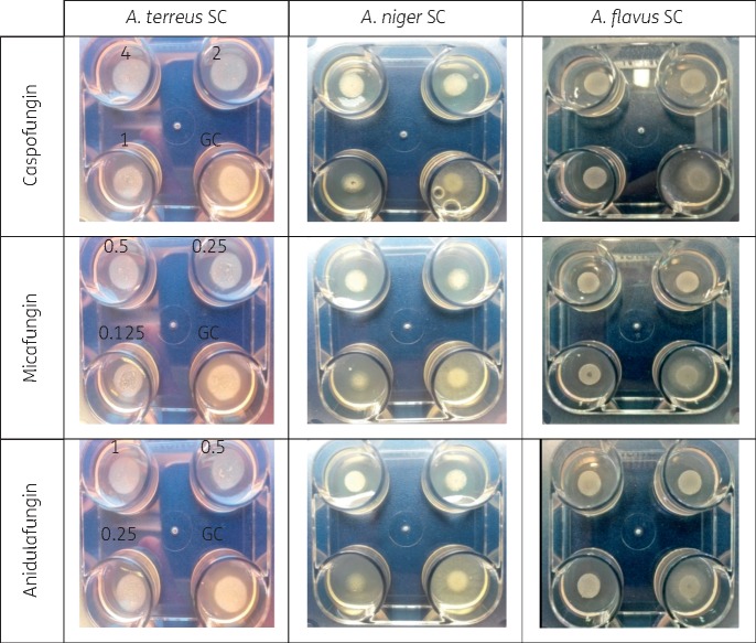
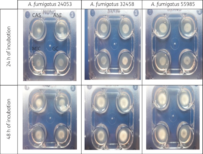
Methods: Forty WT isolates [10 Aspergillus fumigatus species complex (SC), 10 Aspergillus flavus SC, 10 Aspergillus terreus SC and 10 Aspergillus niger SC] and 4 non-WT A. fumigatus isolates with or without known fks alterations were used. The optimal test conditions and stability over time were evaluated in preliminary studies monitoring colony growth. Twenty-microlitre aliquots of 1-2 McFarland inocula in 0.1% Tween 20 aqueous solution were added to each well and plates were incubated for 24/48 h at 35 ± 2°C. Subsequently, all isolates were tested blindly at three centres using four-well screening plates, containing anidulafungin, caspofungin, micafungin or no antifungal in each of the four wells, respectively.
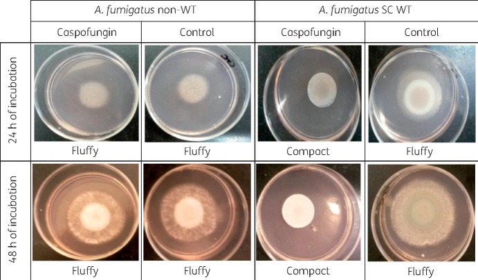
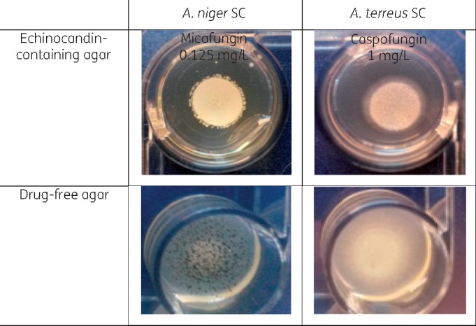
Results: WT isolates produced fluffy colonies on drug-free agar wells only. The non-WT isolates produced fluffy colonies on echinocandin-containing and control agar wells. Using the echinocandin concentrations of 0.25 mg/L anidulafungin, 1 mg/L caspofungin and 0.125 mg/L micafungin, and the compact (non-fluffy) versus fluffy colony morphology endpoint, all centres successfully discriminated non-WT and WT strains even after 24 h. Among the three echinocandins, anidulafungin produced the clearest endpoints.
Conclusions: The four-well plate agar method is suitable for echinocandin susceptibility screening of Aspergillus spp. and can be used to detect echinocandin non-WT isolates.
© The Author(s) 2019. Published by Oxford University Press on behalf of the British Society for Antimicrobial Chemotherapy. All rights reserved. For permissions, please email: journals.permissions@oup.com.
Figures
Similar articles
-
Multicenter Study of Method-Dependent Epidemiological Cutoff Values for Detection of Resistance in Candida spp. and Aspergillus spp. to Amphotericin B and Echinocandins for the Etest Agar Diffusion Method.Antimicrob Agents Chemother. 2016 Dec 27;61(1):e01792-16. doi: 10.1128/AAC.01792-16. Print 2017 Jan. Antimicrob Agents Chemother. 2016. PMID: 27799206 Free PMC article.
-
Evaluation of disk diffusion method compared to broth microdilution for antifungal susceptibility testing of 3 echinocandins against Aspergillus spp.Diagn Microbiol Infect Dis. 2012 May;73(1):53-6. doi: 10.1016/j.diagmicrobio.2012.01.006. Epub 2012 Apr 4. Diagn Microbiol Infect Dis. 2012. PMID: 22480568
-
Echinocandins and Their Activity against Aspergillus terreus Species Complex: a Novel Agar Screening Method.Antimicrob Agents Chemother. 2022 Feb 15;66(2):e0190921. doi: 10.1128/AAC.01909-21. Epub 2021 Dec 13. Antimicrob Agents Chemother. 2022. PMID: 34902268 Free PMC article.
-
Candida and candidaemia. Susceptibility and epidemiology.Dan Med J. 2013 Nov;60(11):B4698. Dan Med J. 2013. PMID: 24192246 Review.
-
[In vitro activity of anidulafungin. Comparison with the activity of other echinocandins].Enferm Infecc Microbiol Clin. 2008 Dec;26 Suppl 14:7-13. doi: 10.1016/s0213-005x(08)76587-x. Enferm Infecc Microbiol Clin. 2008. PMID: 19572429 Review. Spanish.
Cited by
-
Twenty Years in EUCAST Anti-Fungal Susceptibility Testing: Progress & Remaining Challenges.Mycopathologia. 2024 Jul 11;189(4):64. doi: 10.1007/s11046-024-00861-2. Mycopathologia. 2024. PMID: 38990395 Review.
-
A Practical Guide to Antifungal Susceptibility Testing.J Pediatric Infect Dis Soc. 2023 Apr 28;12(4):214-221. doi: 10.1093/jpids/piad014. J Pediatric Infect Dis Soc. 2023. PMID: 36882026 Free PMC article.
-
Drug-Resistant Fungi: An Emerging Challenge Threatening Our Limited Antifungal Armamentarium.Antibiotics (Basel). 2020 Dec 8;9(12):877. doi: 10.3390/antibiotics9120877. Antibiotics (Basel). 2020. PMID: 33302565 Free PMC article. Review.
-
Prevalence of T. rubrum and T. interdigitale Exhibiting High MICs to Terbinafine in Clinical Samples Analyzed in the Portuguese Mycology Reference Laboratory.Pathogens. 2025 Jan 25;14(2):115. doi: 10.3390/pathogens14020115. Pathogens. 2025. PMID: 40005492 Free PMC article.
-
Aspergillus fumigatus and aspergillosis: From basics to clinics.Stud Mycol. 2021 May 10;100:100115. doi: 10.1016/j.simyco.2021.100115. eCollection 2021 Sep. Stud Mycol. 2021. PMID: 34035866 Free PMC article. Review.
References
-
- Bernal-Martínez L, Alastruey-Izquierdo A, Cuenca-Estrella M.. Diagnostics and susceptibility testing in Aspergillus. Future Microbiol 2016; 11: 315–28. - PubMed
-
- Ullmann AJ, Aguado JM, Arikan-Akdagli S. et al. Diagnosis and management of Aspergillus diseases: executive summary of the 2017 ESCMID-ECMM-ERS guideline. Clin Microbiol Infect 2018; 24: e1–38. - PubMed
Publication types
MeSH terms
Substances
Grants and funding
LinkOut - more resources
Full Text Sources
Miscellaneous
