. 2019 Oct 15;200(8):1032-1044.
doi: 10.1164/rccm.201808-1560OC.

Role of B7H3/IL-33 Signaling in Pulmonary Fibrosis-induced Profibrogenic Alterations in Bone Marrow

Affiliations

Role of B7H3/IL-33 Signaling in Pulmonary Fibrosis-induced Profibrogenic Alterations in Bone Marrow

Taku Nakashima et al. Am J Respir Crit Care Med. .

Abstract

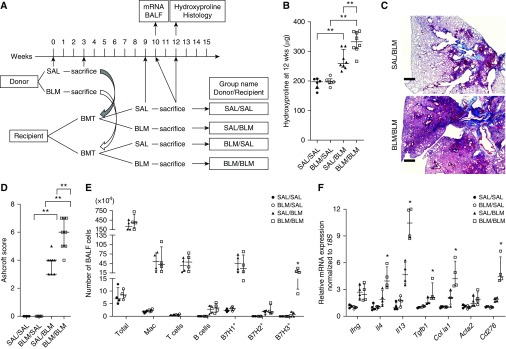
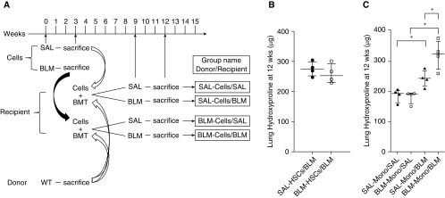
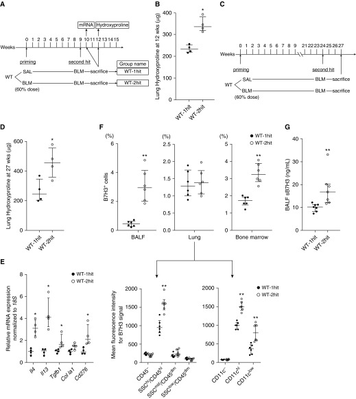
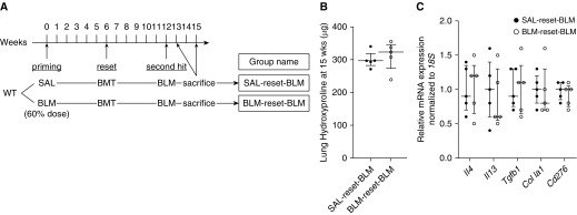
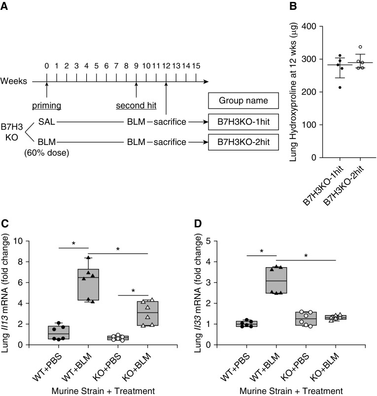
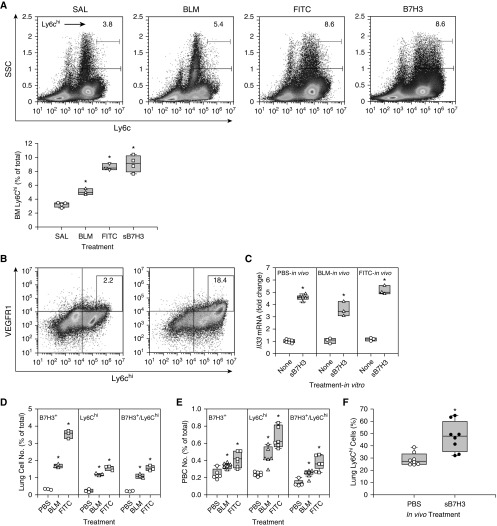
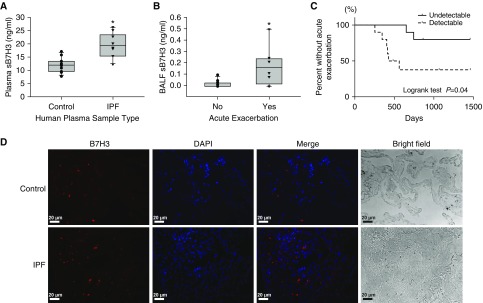
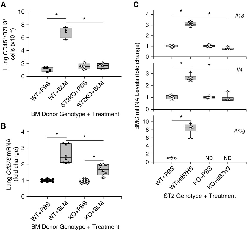
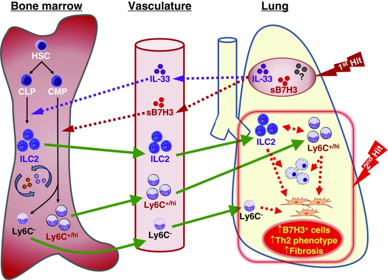
Rationale: The impact of lung insult on the bone marrow (BM) and subsequent disease is unknown.Objectives: To study alterations in the BM in response to lung injury/fibrosis and examine their impact on subsequent lung insult.Methods: BM cells from control or bleomycin-treated donor mice were transplanted into naive mice, which were subsequently evaluated for bleomycin-induced pulmonary fibrosis. In addition, the effect of prior bleomycin treatment on subsequent fibrosis was examined in wild-type and B7H3-knockout mice. Samples from patients with idiopathic pulmonary fibrosis were analyzed for potential clinical relevance of the findings.Measurements and Main Results: Recipient mice transplanted with BM from bleomycin-pretreated donors showed significant exacerbation of subsequent fibrosis with increased B7H3+ cell numbers and a T-helper cell type 2-skewed phenotype. Pretreatment with a minimally fibrogenic/nonfibrogenic dose of bleomycin also caused exacerbation, but not in B7H3-deficient mice. Exacerbation was not observed if the mice received naive BM cell transplant after the initial bleomycin pretreatment. Soluble B7H3 stimulated BM Ly6Chi monocytic cell expansion in vitro and caused similar expansion in the lung in vivo. Notably, soluble B7H3 was elevated in plasma of patients with idiopathic pulmonary fibrosis and in BAL fluid in those with acute exacerbation. Finally, ST2 deficiency diminished the bleomycin-induced B7H3 and IL-13 upregulation, suggesting a role for type 2 innate lymphoid cells.Conclusions: Pulmonary fibrosis caused significant alterations in BM with expansion and activation of monocytic cells, which enhanced fibrosis when transplanted to naive recipients with potential mediation by a novel role for B7H3 in the pathophysiology of pulmonary fibrosis in both mice and humans.

Keywords: bleomycin; bone marrow transplantation; group 2 innate lymphoid cells; idiopathic pulmonary fibrosis; monocytes.


Figures

Figure 1.
Figure 1.
Bleomycin (BLM)-induced pulmonary fibrosis in recipients transplanted with bone marrow (BM) cells from BLM- or saline (SAL)-pretreated donors. (A) Experimental scheme and group naming. Recipients received BM cell transplants from BLM- or SAL-pretreated donors at the indicated time points and evaluated as indicated at the specified time points. (B) Lung hydroxyproline content in each group (n = 6–8/group). **P < 0.01. (C) Masson’s trichrome–stained lung tissue obtained 3 weeks after BLM treatment (or 9 wk after bone marrow transplant) in recipient mice. (D) Ashcroft scores in each group (n = 5–8/group) were obtained from analysis of histological sections as described in C. **P < 0.01. (E) Flow cytometric analysis of cell surface markers in BAL fluid (BALF) cells obtained 1 week after SAL or BLM treatment in recipients (n = 4/group). *P < 0.05 versus SAL/BLM group. (F) Real-time PCR analysis for the indicated lung mRNAs in recipients at 1 week after treatment (n = 4/group). Measured mRNA concentrations were expressed relative to internal control 18S mRNA concentration and normalized to the lowest value in the analyzed groups (which equaled 1). Scale bars, 400 μm. *P < 0.05 versus SAL/BLM group. BMT = bone marrow transplant; Mac = macrophages.
Figure 2.
Figure 2.
Bone marrow (BM) cell transplant (BMT) from bleomycin (BLM)- or saline (SAL)-pretreated mice affects pulmonary fibrosis. (A) Experimental scheme and group naming. Recipients received transplants of normal BM cells (from naive donors) together with the indicated sorted cells obtained from BLM- or SAL-pretreated mice and then analyzed as described at the indicated time points. (B) Lung hydroxyproline content in recipients of transplanted normal BM cells together with sorted hematopoietic stem cells (HSCs) from SAL- or BLM-pretreated donors (n = 4/group). (C) Lung hydroxyproline content in recipients of transplanted normal BM cells together with sorted monocytic cells (Mono) from SAL- or BLM-pretreated donors (n = 4/group). *P < 0.05. WT = wild type.
Figure 3.
Figure 3.
Effect of bleomycin (BLM) pretreatment on subsequent BLM-induced pulmonary fibrosis in wild-type (WT) mice. (A) Experimental scheme and group naming. Mice were pretreated with a reduced dose (60% of normal dose) of BLM at Time 0. After the indicated time intervals, mice were then treated with the normal dose of BLM. (B) Lung hydroxyproline content at 3 weeks after the subsequent full-dose BLM treatment (n = 4/group). *P < 0.05 versus WT one-hit group. (C) Modified experimental scheme with extended time interval after BLM pretreatment. (D) Lung hydroxyproline content at 3 weeks after the subsequent full-dose BLM treatment (n = 4/group). *P < 0.05 versus WT one-hit group. (E) Real-time PCR analysis for indicated lung mRNAs at 1 week after the subsequent full-dose BLM treatment in WT one-hit and WT two-hit groups (n = 4/group). Measured mRNA concentrations were expressed relative to internal control 18S mRNA concentration and normalized to the value in WT one-hit group (which equaled 1). *P < 0.05 versus WT one-hit group. (F) Flow cytometric analysis for B7H3 expression on cells obtained from BAL fluid (BALF), lung tissue, and bone marrow (upper panel) at 3 weeks after the full-dose BLM treatment in WT one-hit and WT two-hit groups (n = 6/group). The percentages of total cells are shown. For lung tissue, total lung cells were divided according to side scatter (SSC) and CD45 or CD11c expression, then further analyzed for each mean fluorescence intensity for B7H3 signal (lower panel). **P < 0.01 versus WT one-hit group. (G) BALF concentrations of soluble B7H3 protein at 3 weeks after the subsequent full-dose BLM treatment in WT one-hit and WT two-hit groups (n = 7/group). **P < 0.01 versus WT one-hit group. SAL = saline.
Figure 4.
Figure 4.
Bone marrow transplant (BMT) using bone marrow (BM) from naive donors abolished exacerbation of fibrosis by bleomycin (BLM) pretreatment. (A) Experimental scheme and group naming. Mice received transplanted BM cells from naive donors (“reset”) after the initial priming BLM treatment (60% of normal dose; control mice received saline [SAL] alone). The recipient mice were subsequently treated with full-dose BLM at the indicated time point. (B) Lung hydroxyproline content at 3 weeks after the full-dose BLM treatment (n = 5/group). (C) Real-time PCR analysis for the indicated lung mRNAs at 1 week after the full-dose BLM treatment in SAL reset BLM and BLM reset BLM groups (n = 5/group). Measured mRNA concentrations were expressed relative to internal control 18S mRNA concentration and normalized to the value in the SAL reset BLM group (which equaled 1). WT = wild type.
Figure 5.
Figure 5.
Effect of bleomycin (BLM) pretreatment on subsequent BLM-induced pulmonary fibrosis in B7H3-knockout (B7H3 KO) mice. (A) Experimental scheme and group naming. The experimental protocol is identical to that in Figure 3, except that B7H3 KO mice were used instead of wild-type (WT) mice. (B) Lung hydroxyproline content at 3 weeks after the subsequent full-dose BLM treatment (n = 5/group). (C) Lung Il13 and (D) Il33 mRNA analysis by real-time PCR at 1 week after saline (SAL) or full-dose BLM treatment in WT or B7H3 KO mice. Results were expressed as fold change relative to the SAL-treated WT group mean value (n = 6/group). Box plots show the median and interquartile range, and the lower and upper bars denote the 10th and 90th percentiles, respectively. PBS = phosphate-buffered saline. *P < 0.05 between the two indicated groups.
Figure 6.
Figure 6.
Effect of soluble B7H3 (sB7H3) on bone marrow (BM) monocytic cells. (A) BM cells isolated from saline (SAL)-treated (control), bleomycin (BLM)-treated, or fluorescein isothiocyanate (FITC)-treated mice and naive BM cells (from untreated mice) treated with 5 μg/ml sB7H3 for 24 hours were analyzed for Ly6C expression by flow cytometry. The quantitative data are shown on the right panel. All box plots show the median and interquartile range, and the lower and upper bars denote the 10th and 90th percentiles, respectively. *P < 0.05 from the respective SAL or PBS controls. (B) sB7H3-treated control BM cells described in A were analyzed for VEGFR1 and Ly6Chi double-positive cells. Representative plots from three separate analyses are shown in A (left panel) and B. (C) SAL BM cells obtained from SAL-, BLM-, or FITC-treated mice were treated with 10 ng/ml granulocyte–macrophage colony–stimulating factor for 5 days and then further treated with or without sB7H3 for another 24 hours. IL-33 mRNA expression was analyzed by real-time PCR (n = 3–6). (D) Whole-lung cell suspension or (E) peripheral blood cells (PBC) obtained from SAL-, BLM-, or FITC-treated mice were analyzed for B7H3+, Ly6Chi, and double-positive cells by flow cytometry (n = 3–6). (F) Mice were treated with sB7H3 by intravenous injection and analyzed for lung Ly6Chi cell population by flow cytometry. Data were expressed as percentage of CD11b+ cells (n = 8). PBS = phosphate-buffered saline; SSC = side scatter. *P < 0.05 compared with respective untreated or PBS controls in CF.
Figure 7.
Figure 7.
B7H3 was induced in human plasma, BAL fluid (BALF), and lung tissue. (A) Plasma concentrations of soluble B7H3 (sB7H3) in healthy control subjects (n = 12) and patients with idiopathic pulmonary fibrosis (IPF) (n = 10). *P < 0.05. (B) BALF concentrations of sB7H3 in patients with IPF who did (n = 8) or did not (n = 12) develop acute exacerbation in their clinical course. *P < 0.05. Box plots show the median and interquartile range, and the lower and upper bars denote the 10th and 90th percentiles, respectively, in A and B. (C) Kaplan-Meier analysis of frequency of acute exacerbation in patients with IPF. The proportion of patients with IPF without acute exacerbations were compared between patients whose BALF concentrations of sB7H3 were detectable (n = 10; dashed line) or undetectable (n = 10; solid line). (D) The immunofluorescence staining of paraffin-embedded human lung tissue sections obtained from control subjects or patients with IPF is shown. B7H3 signals are shown in red and nuclei in purple-blue with DAPI. Representative single and merged images are shown. Scale bars, 20 μm.
Figure 8.
Figure 8.
ST2 effect on B7H3 induction and function. Mice received transplants with wild-type (WT) or ST-knockout (ST2KO) bone marrow (BM), and, after stable engraftment, they were treated with phosphate-buffered saline (PBS) or bleomycin (BLM). Box plots show the median and interquartile range, and the lower and upper bars denote the 10th and 90th percentiles, respectively. (A) The number of lung CD45+/B7H3+ cells was assessed by flow cytometry (n = 4). (B) Lung tissue B7H3 (Cd276) mRNA was analyzed by real-time PCR, and the results are expressed as fold change from the SAL-treated group with WT BM (n = 8/group). (C) Lineage-negative bone marrow cells (BMC) from SAL WT or ST2KO mice were treated with or without 5 μg/ml sB7H3 for 48 hours, and they were then analyzed for the indicated mRNA by real-time PCR. Results are shown as fold changes from their respective untreated controls (n = 6/group). *P < 0.05 between the two indicated groups.
Figure 9.
Figure 9.
Schematic summary of the alteration in bone marrow cells and B7H3/IL-33 signaling in pulmonary fibrosis. Initial injury or first hit in the lung causes elevated concentrations of soluble B7H3 (sB7H3) and other mediators, such as IL-33, etc. (?), to be detected in the lung and plasma. These factors (sB7H3 and IL-33 are shown) are hypothesized to stably influence/alter bone marrow (BM) progenitor cell (e.g., hematopoietic stem cells [HSC], common lymphoid progenitors [CLP], and common myeloid progenitors [CMP]) differentiation (BM priming) (e.g., IL-33–mediated activation of type 2 innate lymphoid cell [ILC2] development from CLP) and sB7H3 promotion of myeloid cell differentiation (e.g., to Ly6C+/hi population and/or other Ly6C cells or progenitors as shown) from CMP (1, 4). These “primed” or preactivated BM cells exhibit enhanced responsiveness for recruitment to the lung upon a second hit or insult, where they may play a paracrine role to promote myeloid differentiation/activation with expression of B7H3, T-helper cell type 2 (Th2) skewing, and fibroblast proliferation and activation, resulting in exacerbation of pulmonary fibrosis. Solid arrows indicate cell differentiation (black) or migration (green), and dotted arrows indicate regulatory effects of mediators (e.g., sB7H3 or IL-33 from the indicated sources as shown). Parts of images were adapted from Motifolio drawing toolkits (www.motifolio.com).

Comment in

  • Remember Me? The Bone Marrow in Pulmonary Fibrosis.
    Rangarajan S, Thannickal VJ. Rangarajan S, et al. Am J Respir Crit Care Med. 2019 Oct 15;200(8):959-960. doi: 10.1164/rccm.201906-1101ED. Am J Respir Crit Care Med. 2019. PMID: 31206314 Free PMC article. No abstract available.

Similar articles

Cited by

References

    1. Nakashima T, Liu T, Yu H, Ding L, Ullenbruch M, Hu B, et al. Lung bone marrow-derived hematopoietic progenitor cells enhance pulmonary fibrosis. Am J Respir Crit Care Med. 2013;188:976–984. - PMC - PubMed
    1. Liu T, Yu H, Ullenbruch M, Jin H, Ito T, Wu Z, et al. The in vivo fibrotic role of FIZZ1 in pulmonary fibrosis. PLoS One. 2014;9:e88362. - PMC - PubMed
    1. Ding L, Liu T, Wu Z, Hu B, Nakashima T, Ullenbruch M, et al. Bone marrow CD11c+ cell-derived amphiregulin promotes pulmonary fibrosis. J Immunol. 2016;197:303–312. - PMC - PubMed
    1. Satoh T, Nakagawa K, Sugihara F, Kuwahara R, Ashihara M, Yamane F, et al. Identification of an atypical monocyte and committed progenitor involved in fibrosis. Nature. 2017;541:96–101. - PubMed
    1. Wang X, Takagawa J, Lam VC, Haddad DJ, Tobler DL, Mok PY, et al. Donor myocardial infarction impairs the therapeutic potential of bone marrow cells by an interleukin-1-mediated inflammatory response. Sci Transl Med. 2011;3:100ra90. - PMC - PubMed

Publication types

MeSH terms