Review
. 2019 Sep;22(5):726-740.
doi: 10.1111/vop.12678. Epub 2019 May 20.

The future of canine glaucoma therapy

Affiliations
Review

The future of canine glaucoma therapy

András M Komáromy et al. Vet Ophthalmol. 2019 Sep.

Abstract

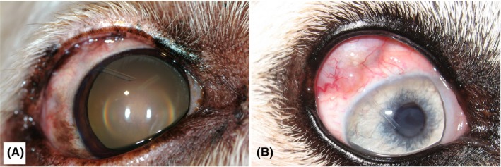
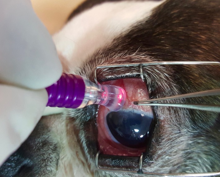
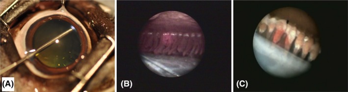
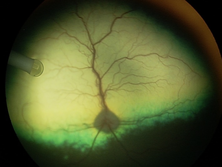
Canine glaucoma is a group of disorders that are generally associated with increased intraocular pressure (IOP) resulting in a characteristic optic neuropathy. Glaucoma is a leading cause of irreversible vision loss in dogs and may be either primary or secondary. Despite the growing spectrum of medical and surgical therapies, there is no cure, and many affected dogs go blind. Often eyes are enucleated because of painfully high, uncontrollable IOP. While progressive vision loss due to primary glaucoma is considered preventable in some humans, this is mostly not true for dogs. There is an urgent need for more effective, affordable treatment options. Because newly developed glaucoma medications are emerging at a very slow rate and may not be effective in dogs, work toward improving surgical options may be the most rewarding approach in the near term. This Viewpoint Article summarizes the discussions and recommended research strategies of both a Think Tank and a Consortium focused on the development of more effective therapies for canine glaucoma; both were organized and funded by the American College of Veterinary Ophthalmologists Vision for Animals Foundation (ACVO-VAF). The recommendations consist of (a) better understanding of disease mechanisms, (b) early glaucoma diagnosis and disease staging, (c) optimization of IOP-lowering medical treatment, (d) new surgical therapies to control IOP, and (e) novel treatment strategies, such as gene and stem cell therapies, neuroprotection, and neuroregeneration. In order to address these needs, increases in research funding specifically focused on canine glaucoma are necessary.

Keywords: aqueous humor; canine; glaucoma; intraocular pressure; optic nerve; surgery.


Conflict of interest statement

The authors have the following potential conflicts of interest (in alphabetic order): Aerie Pharmaceutical (RLF and SEM: research funding, clinical trial grant), Allergan (SEM: research funding, clinical trial grant), Bausch and Lomb (RLF: research funding), Beaver‐Visitec (RLF: consultant), Cara Life (DWE: previous consultant), Elsevier (PEM: book royalties), Ivantis (CBT: research funding), Nicox (CBT: research funding), OSOD (PEM: consultant), PolyActiva (AMK: research funding), Santen (CBT: research funding), and Wolters Kluwer (SEM: book royalties). The opinions expressed in this article are the authors' own and do not necessarily reflect the view of the United States Food and Drug Administration (FDA) or the United States Government. Special thanks go to Ms. Jen Gazdacko (ACVO‐VAF) for her technical support.

Figures

Figure 1
Figure 1
Cross‐sectional anatomy (A) and aqueous humor drainage routes (B) in the canine eye. Once through the trabecular meshwork, the aqueous can pass into the angular aqueous plexus and is directed either anteriorly into the more superficial episcleral venules (1) or posteriorly into the scleral venous plexus and the vortex venous system (2). An alternative aqueous humor drainage pathway (3) is the diffusion through the ciliary muscle interstitium to the suprachoroidal space and through the sclera (ie, uveoscleral flow). Abbreviation: AVAs, arteriovenous anastomosis (from Tsai et al14; with permission)
Figure 2
Figure 2
Optical coherence tomography (OCT) images of the canine eye taken with the Spectralis® (Heidelberg Engineering GmbH, Heidelberg, Germany). A, Iridocorneal angle of a 2.5‐year old, female Beagle with POAG (IOP during imaging: 23 mm Hg). OCT often provides higher resolution than routine high‐resolution ultrasonography (Figure 3), but there are still limits when imaging deeper tissues, such as the aqueous humor outflow pathways (*). B, ONH images of a normal (B1; 6.5‐years old female) and POAG‐affected (B2; 9.5‐years old female) Beagle. While the nondegenerated, well‐myelinated normal canine ONH bulges into the vitreous (B1; IOP during imaging: 15 mm Hg), the chronically glaucomatous ONH appears cupped (B2; IOP during imaging: 19 mm Hg). The white arrows indicate the location of the lamina cribrosa. Unless there is extensive degeneration, the canine lamina cribrosa is difficult to visualize, even with the current enhanced depth imaging (EDI) technology, due to the thick, myelinated prelaminar ONH. AC, anterior chamber; C, cornea; S, sclera; V, vitreous
Figure 3
Figure 3
High‐resolution ultrasound (HRUS) images of the canine iridocorneal angle. Compared to a normal eye with physiologic IOP (A) with flat iris and open ciliary cleft (white arrows), the iris has a sigmoidal shape with increased corneal contact (black arrow) and a collapsed ciliary cleft (white arrow) in an eye with acute PACG and IOP of 55 mm Hg (B). AC, anterior chamber; C, cornea; CB, ciliary body; I, iris. (From Miller143; with permission)
Figure 4
Figure 4
The canine ciliary cleft may collapse following lens removal by phacoemulsification. Tissue cross‐sections of the iridocorneal angle in normal Bouin's fixed globes show that compared to the normal, unoperated eye (A) the ciliary cleft is severely reduced in an eye 24 h after phacoemulsification (B). The IOP in this eye reached 52 mm Hg 3 h after surgery and decreased to 15 mm Hg at 24 h. Despite the normalization of IOP, the ciliary cleft remained reduced. The arrows denote the approximate boundaries of the ciliary cleft. Bars = 0.2 mm. (from Miller et al49; with permission)
Figure 5
Figure 5
Tube positioning of an Ahmed VS‐2 valved drainage implant (New World Medical Inc, Rancho Cucomonga, CA, USA) in the anterior chamber of two dogs (A and B). B, Subconjunctival filtering bleb is shown underneath the upper eyelid
Figure 6
Figure 6
Positioning of the MicroPulse® Cyclo G6 probe (Iridex) 3 mm posterior to the limbus of a dog during transscleral cyclophotocoagulation (TSCP)
Figure 7
Figure 7
Endolaser cyclophotocoagulation (ECP) in the canine eye. A, The laser endoscope is inserted through a limbal incision and the pupil to access the ciliary processes. The endoscopic view shows the red aiming beam on the ciliary processes before (B) and following laser treatment when they appear white and shrunken (C). The lens capsule is shown on the bottom and the posterior iris surface on the top (B and C)
Figure 8
Figure 8
Sustained intraocular delivery of ciliary neurotrophic factor (CNTF) by encapsulated cell technology (ECT) in a canine eye. The NT‐501 implant containing CNTF‐secreting human cells (Neurotech Pharmaceuticals, Inc) is located within the vitreous and anchored to the pars plana of the ciliary body

References

    1. Gelatt KN, MacKay EO. Prevalence of the breed‐related glaucomas in pure‐bred dogs in North America. Vet Ophthalmol. 2004;7:97‐111. - PubMed
    1. Gelatt KN, MacKay EO. Secondary glaucomas in the dog in North America. Vet Ophthalmol. 2004;7:245‐259. - PubMed
    1. Quigley HA, Broman AT. The number of people with glaucoma worldwide in 2010 and 2020. Br J Ophthalmol. 2006;90:262‐267. - PMC - PubMed
    1. Tham YC, Li X, Wong TY, et al. Global prevalence of glaucoma and projections of glaucoma burden through 2040: a systematic review and meta‐analysis. Ophthalmology. 2014;121:2081‐2090. - PubMed
    1. Newbold GM, Kelch WJ, Chen T, et al. Phacoemulsification outcomes in Boston terriers as compared to non‐Boston terriers: a retrospective study (2002–2015). Vet Ophthalmol. 2018;21:353‐361. - PubMed