

Corneal Perforation After Corneal Cross-Linking in Keratoconus Associated With Potentially Pathogenic ZNF469 Mutations
- PMID: 31107761
- PMCID: PMC6612572
- DOI: 10.1097/ICO.0000000000002002
Corneal Perforation After Corneal Cross-Linking in Keratoconus Associated With Potentially Pathogenic ZNF469 Mutations
Abstract
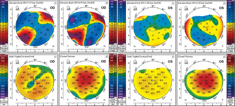
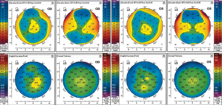
Purpose: To report a case of bilateral and repetitive corneal perforations after corneal cross-linking (CXL) for keratoconus in a woman harboring potentially pathogenic variants in the ZNF469 gene and to characterize the keratoconus phenotype in this woman and her daughter who shared the same ZNF469 mutations.
Methods: Clinical characterization of the proband and her daughter followed by sequencing of the genes associated with brittle cornea syndrome, ZNF469 and PRDM5, in both individuals.
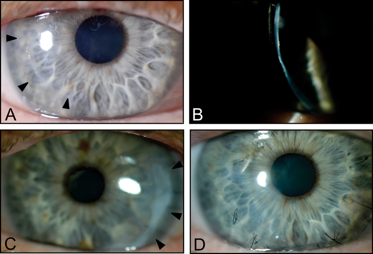
Results: An Ashkenazi Jewish woman in her sixth decade presented with diffuse corneal thinning and progressive steepening consistent with keratoconus. After CXL, epithelium-off in the first eye and epithelium-on in the second, she developed spontaneous corneal perforations in each eye. Her daughter in her fourth decade demonstrated a similar pattern of diffuse corneal thinning and progressive corneal steepening but did not undergo CXL and did not develop corneal perforation. Screening of the ZNF469 and PRDM5 genes revealed 3 missense ZNF469 variants (c.2035G>A, c.10244G>C, and c.11119A>G) in cis arrangement on 1 allele of ZNF469 in both proband and her daughter. Although the 3 variants share low (<0.01) global minor allele frequencies, each has significantly higher minor allele frequencies (0.01-0.03) in the Ashkenazi Jewish population, leading to uncertainty regarding a pathogenic role for the identified variants.
Conclusions: CXL may be associated with the development of corneal perforation in particular at-risk individuals with keratoconus. Identifying clinical and genetic risk factors, including screening of ZNF469 and PRDM5, may be useful in the prevention of significant complications after CXL.
Conflict of interest statement
Conflicts of interest: None
Figures
References
-
- Ziaei M, Barsam A, Shamie N, et al. Reshaping procedures for the surgical management of corneal ectasia. J Cataract Refract Surg 2015;41:842–872. - PubMed
-
- Al-Hussain H, Zeisberger SM, Huber PR, et al. Brittle cornea syndrome and its delineation from the kyphoscoliotic type of Ehlers–Danlos syndrome (EDS VI): Report on 23 patients and review of the literature. Am J Med Genet A 2004;124A:28–34. - PubMed
-
- Wollensak G Crosslinking treatment of progressive keratoconus: new hope. Curr Opin Ophthalmol 2006;17:356–360. - PubMed
Publication types
MeSH terms
Substances
Grants and funding
LinkOut - more resources
Full Text Sources
