. 2019 May 20;14(5):e0216527.
doi: 10.1371/journal.pone.0216527. eCollection 2019.

The evolution and multi-molecular properties of NF1 cutaneous neurofibromas originating from C-fiber sensory endings and terminal Schwann cells at normal sites of sensory terminations in the skin

Affiliations

The evolution and multi-molecular properties of NF1 cutaneous neurofibromas originating from C-fiber sensory endings and terminal Schwann cells at normal sites of sensory terminations in the skin

Frank L Rice et al. PLoS One. .

Abstract

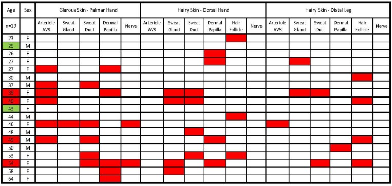
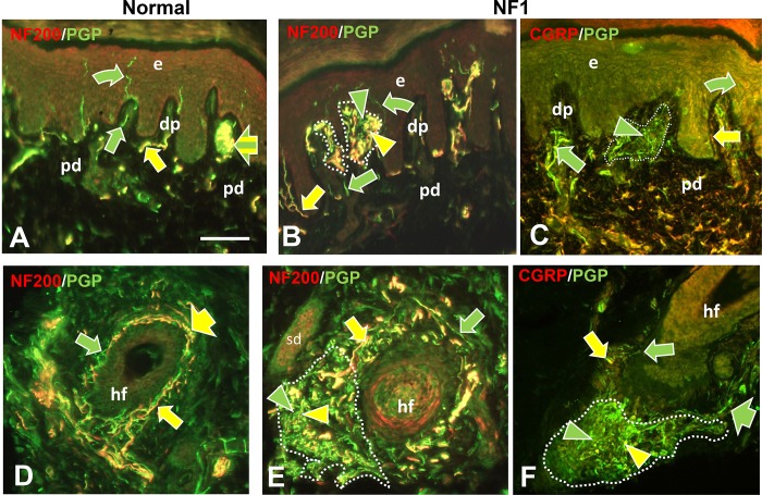
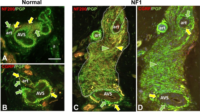
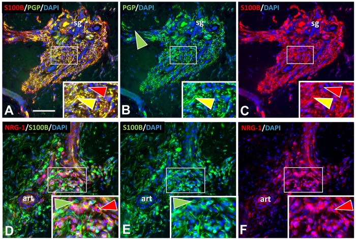
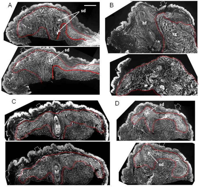
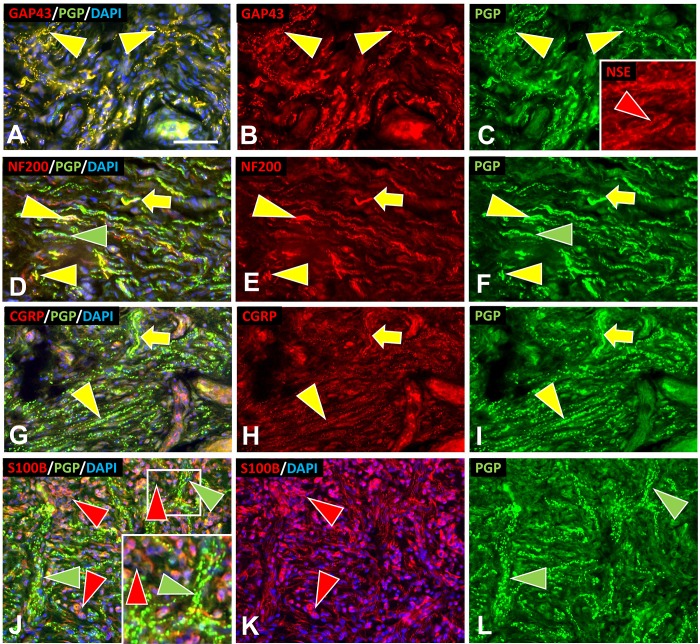
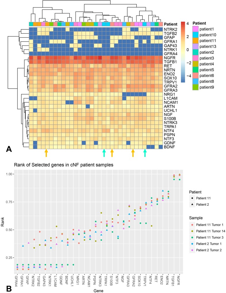
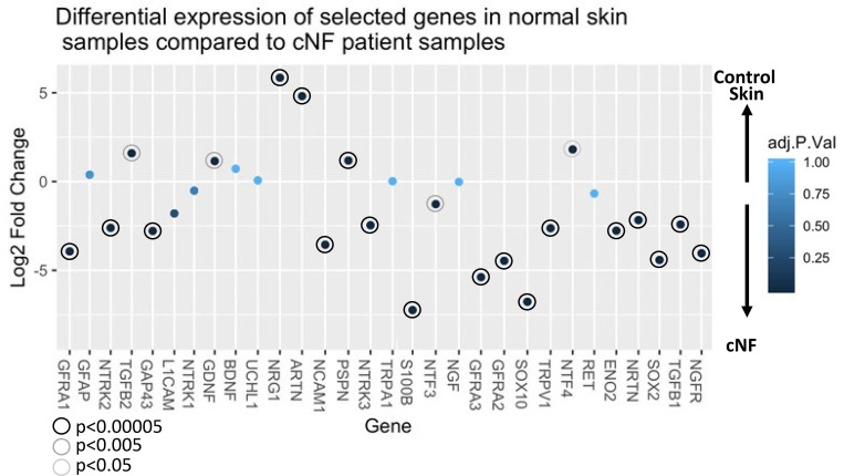
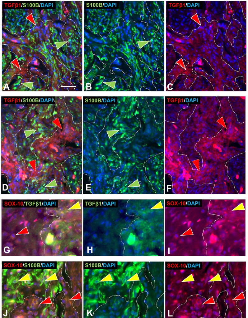
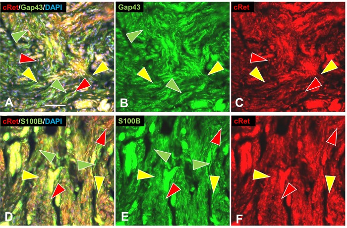
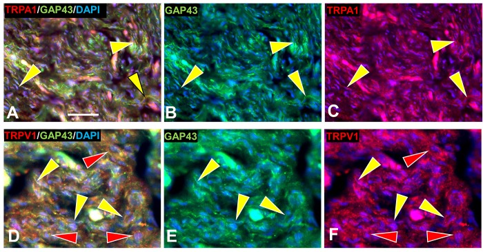
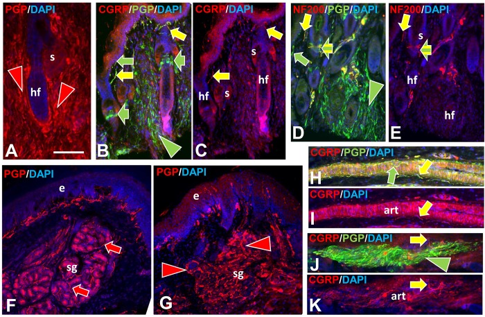
In addition to large plexiform neurofibromas (pNF), NF1 patients are frequently disfigured by cutaneous neurofibromas (cNF) and are often afflicted with chronic pain and itch even from seemingly normal skin areas. Both pNFs and cNF consist primarily of benign hyperproliferating nonmyelinating Schwann cells (nSC). While pNF clearly arise within deep nerves and plexuses, the role of cutaneous innervation in the origin of cNF and in chronic itch and pain is unknown. First, we conducted a comprehensive, multi-molecular, immunofluorescence (IF) analyses on 3mm punch biopsies from three separate locations in normal appearing, cNF-free skin in 19 NF1 patients and skin of 16 normal subjects. At least one biopsy in 17 NF1 patients had previously undescribed micro-lesions consisting of a small, dense cluster of nonpeptidergic C-fiber endings and the affiliated nSC consistently adjoining adnexal structures-dermal papillae, hair follicles, sweat glands, sweat ducts, and arterioles-where C-fiber endings normally terminate. Similar micro-lesions were detected in hind paw skin of mice with conditionally-induced SC Nf1-/- mutations. Hypothesizing that these microlesions were pre-cNF origins of cNF, we subsequently analyzed numerous overt, small cNF (s-cNF, 3-6 mm) and discovered that each had an adnexal structure at the epicenter of vastly increased nonpeptidergic C-fiber terminals, accompanied by excessive nSC. The IF and functional genomics assays indicated that neurturin (NTRN) and artemin (ARTN) signaling through cRET kinase and GFRα2 and GFRα3 co-receptors on the aberrant C-fiber endings and nSC may mutually promote the onset of pre-cNF and their evolution to s-cNF. Moreover, TrpA1 and TrpV1 receptors may, respectively, mediate symptoms of chronic itch and pain. These newly discovered molecular characteristics might be targeted to suppress the development of cNF and to treat chronic itch and pain symptoms in NF1 patients.


Conflict of interest statement

FLR and PJA are the co-founders and co-owners of INTiDYN, which is a for-profit contract and private equity research and consulting organization. GH and MD are research employees of INTiDYN. INTiDYN has no other commercial affiliations but does conduct contract research for pharmaceutical companies and academic collaborators unrelated to, and having no conflict of interest in this research. This does not alter our adherence to PLOS ONE policies on sharing data and materials.

Figures

Fig 1
Fig 1. Summary of the incidence and locations of the pre-cNF among the biopsies of 19 NF1 patients.
The three skin types and biopsy locations are listed in the first row. The sites where sensory endings terminate in each biopsy site are listed in the second row. Except for the first column, red boxes indicate where a pre-cNF was detected among the biopsies for each patient. In column 1, red boxes indicate those patients who had at least one pre-cNF in each biopsy. Green boxes indicate those patients who had no detectable pre-cNF.
Fig 2
Fig 2. Immunofluorescence profile of pre-cNF among dermal papilla and hair follicles.
Immunolabeling of NF1 patient skin biopsies revealed pre-cNF in dermal papillae (dp in B, C) that are invaginations of papillary dermis (pd) into the epidermis (e), and adjacent to hair follicles (hf in E, F). A, D: Innervation of dp and hf in normal subjects. Normally, dp and hf are innervated by a mix of large and small-caliber fibers, all of which label for PGP with the larger caliber fibers co-labeling for NF200 (A, B, D, E; yellow arrows) and a contingent of small-caliber fibers co-labeling for CGRP (C, F; yellow arrow). Other small caliber fibers only labeled with PGP (green arrows). In some cases, such as a Meissner’s corpuscle (A; broad yellow and green striped arrow), endings of large-caliber NF200-positive and small-caliber NF200-negative terminals are intermingled. Arrowheads indicate abnormal extremely fine-caliber, dense innervation within focal pre-cNF circumscribed by dotted outlines. In pre-cNF, the densely packed, extremely small-caliber fibers labeled for PGP. A high proportion co-labeled with NF200 (B, E; yellow compared to green arrowheads). Very few co-labeled for CGRP (C, F). Scale bar = 50μm.
Fig 3
Fig 3. Immunofluorescence profile of pre-cNF among sweat ducts and glands.
Immunolabeling of NF1 patient skin biopsies revealed pre-cNF engulfing sweat ducts (sd in B, C) and infiltrating sweat glands (sg in E, F). A, D: Innervation of sd and sg in normal subjects. The sweat ducts normally have an extremely sparse innervation with NF200-positive and NF200-negative fibers (A, B; yellow and green arrows) and virtually no fibers with CGRP (C: green arrow). Normal sweat gland tubules are each surrounded by a loose tangle of fibers of which nearly all revealed by PGP labeling (D-F; green arrows) are cholinergic sympathetic with a sparse contingent of sensory fibers that co-label for NF200 or CGRP (D-F; yellow arrows). In pre-cNF engulfing sweat ducts or embedded within sweat glands (dotted outlines), the densely packed, extremely small-caliber fibers labeled for PGP. A high proportion co-labeled with NF200 (E; yellow compared to green arrowheads). Very few co-labeled for CGRP (F; yellow arrowhead). Scale bar = 50μm.
Fig 4
Fig 4. Immunofluorescence profiling of pre-cNF among arterioles and AVS.
Immunolabeling of NF1 patient skin biopsies revealed pre-cNF (C, D) occurring at the border of arterioles (art) and arteriole venule shunts (AVS). A, B: Innervation of art and AVS in normal subjects. The perimeters of the arterioles and especially the AVS normally have a dense PGP-labeled, small-caliber innervation that consists of numerous sensory endings of which nearly all co-label for CGRP (B, D; yellow arrows) as well as most co-labeling for NF200 (A, C; yellow arrows). The remainder of the innervation labels only for PGP (green arrows) consists of some sensory fibers and mostly noradrenergic sympathetic fibers. In the pre-cNF (dotted outlines) located at the perimeter of art and AVS, the exceptionally small-caliber and extremely dense innervation has a unique and very high proportion that co-label with NF200 (C; yellow arrowhead), but hardly any that co-label for CGRP. Scale bar = 50μm.
Fig 5
Fig 5. SC and other cells in pre-cNF.
Immunolabeling revealed the presence in pre-cNF of abnormal concentrations of SC immunolabeled for S100B (A-C) and another non-neuronal cell type immunolabeled for NRG-1 (D-F). Inserts are 2X enlargements of sites in the small white rectangles. A-C: In this pre-cNF located at the edge of a sweat gland (sg), dense aberrant axons labeled for PGP are intimately lined with SC processes labeled with S100B (yellow arrowheads). S100B is expressed in SC bodies (red arrowheads). D-F: In this pre-cNF located at the edge of a small arteriole (art), S100B-labeled SC bodies and their processes (green arrowheads) are completely distinct from clusters of other cells with few processes that label for NRG-1 (red arrowheads). Scale bar = 50μm.
Fig 6
Fig 6. Structural organization of s-cNF around an adnexal core (perpendicular).
Immunolabeling reveals the adnexal core of four s-cNF as seen in each of two of the serial sections each cut perpendicular to the s-cNF surface and immunolabeled for PGP. Concentrated areas of aberrant dense innervation are outlined in red and typically consist of fibers ascending adjacent to the adnexal core structure and spreading out under and parallel to, but not penetrating the papillary dermis (between broad arrows). In A and D, the core structure is a sweat duct; in C, a hair follicle. In B, a hair follicle is pushed to the side by excessive nerve fibers originating from a deeper sweat gland. Scale bar = 500μm.
Fig 7
Fig 7. Structural organization of an s-cNF around an adnexal core (parallel).
Immunolabeling reveals the adnexal core of a 4mm s-cNF sectioned parallel to the epidermal surface and co-labeled for GAP-43 (green), S100B (red), and DAPI (blue). Small blood vessels filled with albumen autofluorescence (yellow arrow) A-C: Complete montages of 3 sections at successively deeper levels. The red contours outline the perimeter of a dense concentration of small-caliber fibers oriented circumferentially around a core. The area in the white square of A is shown at 2X magnification in D-F. D-F: Increased magnification reveals a sweat duct (white arrow) surrounded by small-caliber fibers cut in cross-section at the core. The fibers shift to a circumferential orientation around this core. S100B-labeled SC (E) intimately match the orientations and concentrations of the fibers labeled for GAP-43. The area in the white square of D is shown at still a 2X higher magnification in G-I. G-I: An increased magnification of the sweat duct and associated small-caliber fibers cut in cross-section at the core of the s-cNF. Insets in G-H are a further 2X magnification of the broken line area in G that show the intimate association of SC and their processes (red arrowheads) with the aberrant small-caliber fibers (green arrowheads). Scale bar = 500μm.
Fig 8
Fig 8. Multi-molecular immunofluorescence characteristics of s-cNF aberrant innervation.
Multi-molecular immunolableing double-labeled with combinations for PGP together with either GAP-43, NSE, S100B, NF200, or CGRP revealed aberrations among s-cNF. A-C: Virtually all of the aberrant innervation co-labels for PGP and GAP-43 (yellow arrowhead). NSE is more widely expressed likely on innervation and SC (red arrowhead). D-F. A high proportion of the aberrant fibers co-label for NF200 (yellow arrowheads). Others only label for PGP (green arrowheads). Yellow arrows indicate what normal NF200 innervation should look like. G-I: Yellow arrows indicate fibers that are typical of normal CGRP and PGP co-labeling. Most of the aberrant fibers have faint tiny punctate labeling for CGRP (yellow arrowheads). Yellow arrows indicate what normal CGRP innervation should look like. J-L: Aberrant concentrations of PGP-labeled fibers (green arrowheads) are intermingled with SC (red arrowheads). Inset is a 2X of the small square. Scale bar = 25μm.
Fig 9
Fig 9. mRNA expression for most of immunochemically assessed proteins in this study.
A: Shows the absolute levels in multiple s-cNF from 13 different patients color coded by Z-score, with blue representing poorly expressed genes and red representing highly expressed genes in each biopsy from each patient. Patient key is on the right. The s-cNF biopsies for the ITD-CMA are from Patient 2 (turquoise arrows) and Patient 11 (orange arrows). B: Shows the relative mRNA expression levels for only two s-cNF from Patient 2 and three s-cNF from Patient 11.
Fig 10
Fig 10. Differential expression values of selected genes.
Comparisons were performed to determine differential expression values of selected genes in Fig 9 compared to 33 normal skin controls. Negative values indicate up-regulation in s-cNF while positive values confer down-regulation. Corrected P-value is indicated by color of the point. Significantly different data points are encircled.
Fig 11
Fig 11. Partitioning of s-cNF into zones.
Multi-molecular immunolabeling revealed a partitioning of s-cNF into zones (broken lines) of aberrant innervation and affiliated SC interdigitated with aberrant intercalated zones of densely packed clusters of another cell type that had cytoplasmic labeling for TGFβ1 (red arrowheads). Immunofluorescence double labeling for TGFβ1 (red) and S100B (green) revealed that pre-cNF (A-C) and s-cNF both have partitioning of SC immunolabeled for S100B. G-L. The SOX10 immunolabeling is co-express on TGFβ1-positive/S100 cells as well as likely definitive S100B labeled SC in pre-cNF (G-I, yellow and red arrowheads respectively) and in s-cNF (J-L, red and yellow arrowheads respectively). Scale bar = 25μm.
Fig 12
Fig 12. Multi-molecular immunolabeling with c-Ret.
Immunolabeling for c-Ret is expressed on most of the aberrant innervation labeled for GAP-43 (A, B) and accompanying SC labeled for S100B (D, F) in s-cNF. A-C: Some nerve fibers labeled only for GAP-43 (green arrowheads), whereas others were double-labeled for both GAP-43 and c-Ret (yellow arrowheads). Other profiles labeled only for c-Ret (red arrowheads). D-F: Profiles were extensively co-labeled for c-Ret and S100B (yellow arrowheads). Fewer are labeled only for c-Ret (red arrowheads) or only for S100B (green arrowheads). Scale bar = 25μm.
Fig 13
Fig 13. Aberrant innervation of s-cNF express co-receptors of GDNF family ligands.
s-cNF aberrant innervation expresses immunolabeling for c-Ret co-receptors GFRα2 and GFRα3. A-F: No co-labeling was detected for GFRα1 and GFRα4 on the aberrant innervation labeled for GAP-43 (green arrowheads). G-L: Labeling for GFRα2 was widely co-expressed on or associated with most, but not all GAP-43-labeled aberrant innervation (yellow and green arrowheads). Co-labeling for S100B revealed some GFRα2 on SC as well as the innervation. M-O: GFRα3 is expressed on some, but not all of the GAP-43-labeled innervation (yellow and green arrowheads). GFRα3 was not obviously expressed on SC. Scale bar = 25μm.
Fig 14
Fig 14. Immunolabeling for TrpA1 and TrpV1 in s-cNF.
A. Immunolabeling in s-cNF of TrpA1 co-localized with GAP-43 on extensive aberrant innervation (yellow arrowheads). B. Immunolabeling in s-cNF of TrpV1 on at least some of the aberrant innervation (yellow arrowheads) but also on SC (red arrowheads). Scale bar = 25μm.
Fig 15
Fig 15. Immunolabeling among human-like pre-cNF in mice with SC-conditional Nf1/Nf1 mutations.
A-E: Hair follicles (hf) from the dorsum of the foot–some are engulfed in dense fine-caliber aberrant innervation (arrowheads). S = sebaceous gland. A. PGP labeling (red arrowheads) of innervation fully engulfing a follicle. B, C. Some follicles with normal innervation are shown to the left and a partially engulfed follicle to the right. Normal innervation with and without CGRP co-labeling of PGP is present in the upper dermis and neck of the hair follicle (yellow and green arrowheads), as well as normal pilo-neural innervation (broad arrows) located just below the follicle sebaceous glands. D, E. Some follicles with normal innervation are shown to the left and two follicles to the right partially in dense aberrant innervation (arrow head). Normal innervation is seen with and without NF200 (yellow and green arrows). The pilo-neural complexes have a mix of endings with and without NF200 (yellow and green striped arrows). F. Sweat gland with normal PGP-labeled innervation (red arrows). G. Nf1/Nf1 sweat gland with dense aberrant innervation (red arrowheads). H, I. An arteriole with normal innervation labeled only for PGP (green arrow) and double-labeled for CGRP and PGP (yellow arrows). J.K. An Nf1/Nf1 arteriole engulfed in dense aberrant innervation (green arrowhead), but with some normal CGRP innervation (yellow arrows). Scale bar = 50μm.

References

    1. Longo JF, Weber SM, Turner-Ivey BP, Carroll SL. Recent Advances in the Diagnosis and Pathogenesis of Neurofibromatosis Type 1 (NF1)-associated Peripheral Nervous System Neoplasms. Adv Anat Pathol. 2018. Epub 2018/05/16. 10.1097/PAP.0000000000000197 . - DOI - PMC - PubMed
    1. Riccardi VM. Neurofibromatosis: Phenotype, Natural History and Pathogenesis. Baltimore: Johns Hopkins University Press; 1992. 1–498 p.
    1. Riccardi VM. Translational Genetics and Genomics: The Fundamental Nature of NF1 Neurofibromas. JTranslGenetGenom. 2017;1(1):1–12. Epub 2/123/2017.
    1. Le LQ, Parada LF. Tumor microenvironment and neurofibromatosis type I: connecting the GAPs. Oncogene. 2007;26(32):4609–16. Epub 2007/02/14. 1210261 [pii] 10.1038/sj.onc.1210261 - DOI - PMC - PubMed
    1. Tucker T, Riccardi VM, Brown C, Fee J, Sutcliffe M, Vielkind J, et al. S100B and neurofibromin immunostaining and X-inactivation patterns of laser microdissected cells indicate a multicellular origin of some NF1-associated neurofibromas. Journal of Neuroscience Research. 2011;89:1451–60. 10.1002/jnr.22654 - DOI - PubMed

Publication types

MeSH terms