. 2019 May 20;15(5):e1007773.
doi: 10.1371/journal.ppat.1007773. eCollection 2019 May.

Triggering NETosis via protease-activated receptor (PAR)-2 signaling as a mechanism of hijacking neutrophils function for pathogen benefits

Affiliations

Triggering NETosis via protease-activated receptor (PAR)-2 signaling as a mechanism of hijacking neutrophils function for pathogen benefits

Danuta Bryzek et al. PLoS Pathog. .

Abstract

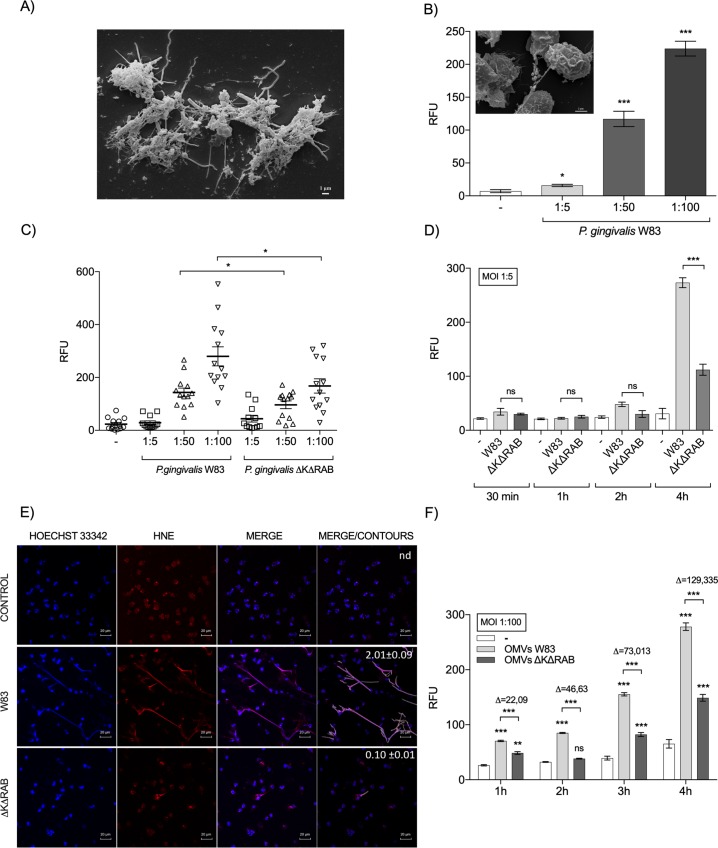
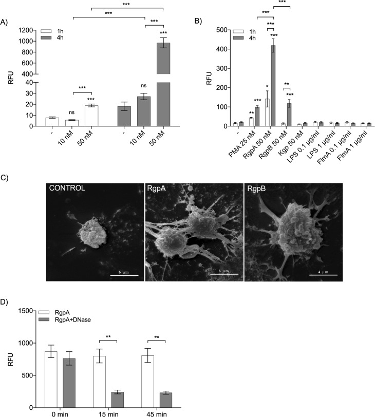
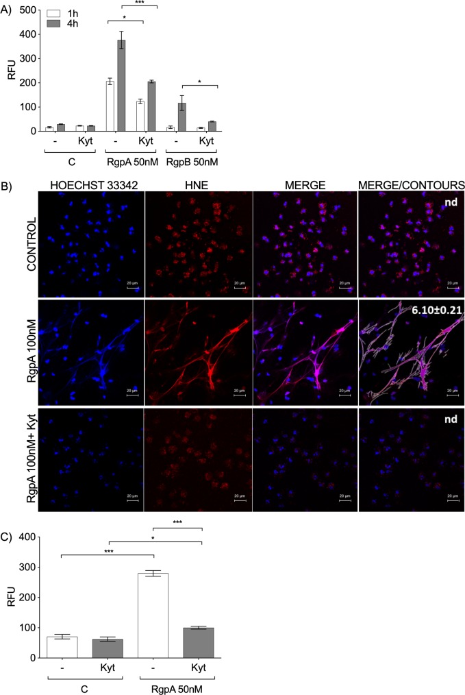
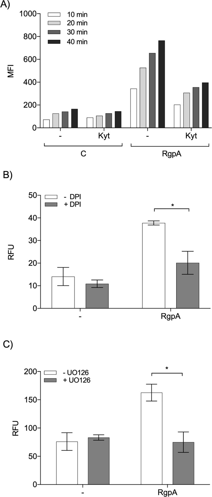
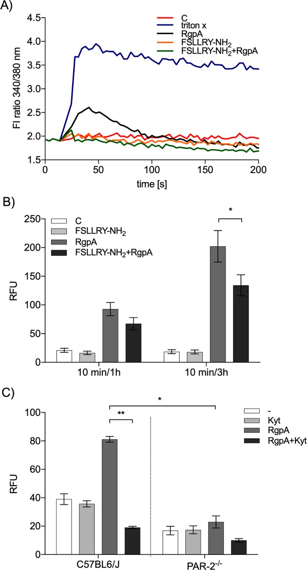
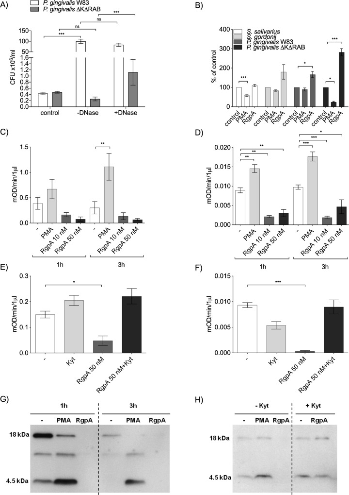
Neutrophil-derived networks of DNA-composed extracellular fibers covered with antimicrobial molecules, referred to as neutrophil extracellular traps (NETs), are recognized as a physiological microbicidal mechanism of innate immunity. The formation of NETs is also classified as a model of a cell death called NETosis. Despite intensive research on the NETs formation in response to pathogens, the role of specific bacteria-derived virulence factors in this process, although postulated, is still poorly understood. The aim of our study was to determine the role of gingipains, cysteine proteases responsible for the virulence of P. gingivalis, on the NETosis process induced by this major periodontopathogen. We showed that NETosis triggered by P. gingivalis is gingipain dependent since in the stark contrast to the wild-type strain (W83) the gingipain-null mutant strain only slightly induced the NETs formation. Furthermore, the direct effect of proteases on NETosis was documented using purified gingipains. Notably, the induction of NETosis was dependent on the catalytic activity of gingipains, since proteolytically inactive forms of enzymes showed reduced ability to trigger the NETs formation. Mechanistically, gingipain-induced NETosis was dependent on proteolytic activation of protease-activated receptor-2 (PAR-2). Intriguingly, both P. gingivalis and purified Arg-specific gingipains (Rgp) induced NETs that not only lacked bactericidal activity but instead stimulated the growth of bacteria species otherwise susceptible to killing in NETs. This protection was executed by proteolysis of bactericidal components of NETs. Taken together, gingipains play a dual role in NETosis: they are the potent direct inducers of NETs formation but in the same time, their activity prevents P. gingivalis entrapment and subsequent killing. This may explain a paradox that despite the massive accumulation of neutrophils and NETs formation in periodontal pockets periodontal pathogens and associated pathobionts thrive in this environment.


Conflict of interest statement

The authors have declared that no competing interests exist.

Figures

Fig 1
Fig 1. The generation of NETs by P. gingivalis is gingipain-dependent.
(A) NETs visualized by SEM in GCF from patients with chronic periodontitis. (B) The generation of NETs by P. gingivalis W83 (MOI 1:5, 1:50, 1:100). The level of extracellular DNA released by neutrophils 1 h post-bacterial exposure was estimated by QPG. SEM visualization of P. gingivalis entrapped in NET structures induced by pathogens (W83) in neutrophils from healthy donors (insert). (C, D) Neutrophils were infected with P. gingivalis strains diametrically differing in the expression of gingipains (WT W83 and the gingipain-null ΔKΔRAB mutant) at MOIs of 1:5, 1:50, and 1:100 for 1 h (C), or at a MOI of 1:5 from 30 min to 4 h (D). The level of extracellular DNA was estimated by QPG. (E) Visualization of NETs by confocal laser scanning microscopy. DNA is shown in blue (Hoechst 33342) and human neutrophil elastase (HNE) is shown in red. Bars represent 20 μm. Quantitative analysis of NETs images was performed by merging blue and red channels (merge/contours). Percentage of the NET area in relation to the area of an image is presented as the mean value (± SEM) from three independent images; n.d.–NETs not detected. (F) OMVs isolated from W83 and the ΔKΔRAB mutant strains were incubated with neutrophils from 1 h to 4 h. The level of NETs was determined by QPG. Statistical significance was evaluated by unpaired t-test (B), two-way (C) and one-way (D, F) ANOVA, followed by Bonferroni’s multiple comparisons posttest. Mean data (± SEM) from 13 (C) or 3 (B, D, F) independent experiments using neutrophils from different healthy donors are shown. *P < 0.05, **P < 0.01, and ***P < 0.001; ns, non-significant.
Fig 2
Fig 2. Purified gingipains promote NET generation.
(A) The level of NETs induced by gingipain cocktails containing each enzyme at 10 or 50 nM after 1 or 4 h of incubation, as determined by QPG. (B) Isolated neutrophils were stimulated with different gingipains (RgpA, RgpB, or Kgp; 50 nM), LPS and FimA (each at 0.1 or 1 μg/ml), or 25 nM PMA, as a control for NET generation. The level of NETs was determined by QPG. (C) NET structures visualized by SEM after 4 h of incubation with 10 nM Arg-X gingipains (RgpA and RgpB). (D) Degradation of the DNA backbone of the NETs induced for 4 h with 50 nM RgpA. Collected NETs were incubated with DNase I (50 μg/ml) for 0, 15, or 45 min. (A, B, D) Statistical significance was evaluated by two-way ANOVA, followed by Bonferroni’s multiple comparisons posttest. Data are the mean (± SEM) from three separate experiments. *P < 0.05, **P < 0.01, and ***P < 0.001; ns, non-significant.
Fig 3
Fig 3. The role of the proteolytic activity of gingipains in NET formation.
Human peripheral blood neutrophils (A) and neutrophils isolated from mouse bone marrow (C) were stimulated with 50 nM RgpA and/or RgpB in the presence or absence of Kyt-1 at a final concentration of 1 μM. The level of NETs was estimated by QPG at 1 h (A) and 4 h (A, C) after enzyme exposure. (B) Confocal laser scanning microscopy of NETs generated by human neutrophils, DNA is shown in blue (Hoechst 33342) and human neutrophil elastase (HNE) expression is shown in red. Bars represent 20 μm. Quantitative analysis of NETs images was performed by merging blue and red channels (merge/contours). Percentage of the NET area in relation to the area of an image is presented as the mean value (± SEM) from three independent images; n.d.–NETs not detected. (A, C) Statistical significance was evaluated by one-way ANOVA, followed by Bonferroni’s multiple comparisons posttest. Mean data (± SEM) from three independent experiments are shown. *P < 0.05 and ***P < 0.001.
Fig 4
Fig 4. The signal transduction pathway triggered by gingipains.
(A) Induction of respiratory burst by active and inactive (Kyt-treated) gingipains. Neutrophils were pretreated with 20 μM DCFH-DA for 10 min, then 50 nM RgpA was added after pretreatment with Kyt-1 or a vehicle control (1 μM). Data represent the mean fluorescence intensity (MFI) of the DCF-positive cells measured at 10, 20, 30, and 40 min after stimulation with RgpA. A representative result from three independent experiments is shown. (B, C) Cells were pretreated with 5 μM DPI (NADPH inhibitor) (B) or 10 μM UO126 (ERK inhibitor) (C) for 30 min. Then, neutrophils were exposed to 10 nM (B) or 50 nM (C) RgpA for 4 h. The level of NETs was determined by QPG. Statistical significance was evaluated by two-way ANOVA, followed by Bonferroni’s multiple comparisons posttest. Mean data (± SEM) from two independent experiments are shown. *P < 0.05; ns, non-significant.
Fig 5
Fig 5. Activation of PAR-2 in NETosis induced by gingipains.
(A) Neutrophils were loaded with Fura-2, then exposed to 100 μM FSLLRY-NH2, followed by 200 nM RgpA. Triton-X was used as a positive control for cellular calcium influx. The cytoplasmic concentration of calcium in a representative experiment is shown. (B) Neutrophils were stimulated for 1 and 3 h with active or inactive RgpA (50 nM) after preincubation with 100 μM FSLLRY-NH2 for 10 min. Statistical significance was evaluated by one-way ANOVA, followed by Bonferroni’s multiple comparisons posttest. Mean data (± SEM) from three independent experiments are shown. *P < 0.05. (C) Peritoneal neutrophils from WT C57BL6/J and PAR-2-/- mice were stimulated for 4 h with 50 nM RgpA with or without pretreatment with Kyt-1 (1 μM). The level of extracellular DNA was estimated by QPG. Statistical significance was evaluated by two-way ANOVA, followed by Bonferroni’s multiple comparisons posttest. Mean data (± SEM) from one experiment using neutrophils from six mice per group are shown. *P < 0.05, and **P < 0.01.
Fig 6
Fig 6. Bactericidal activity of NETs induced by gingipains.
(A) Neutrophils in serum-free DMEM were infected with P. gingivalis W83 and/or ΔKΔRAB (MOI 1:10) in the presence or absence of DNase I. In parallel, bacteria were inoculated into the same medium but without neutrophils. After 3 h incubation mixtures of bacteria with neutrophils (with or without DNase) or bacteria alone in medium (control) were plated and CFUs were determined. (B) Selected bacterial species (MOI 1:5) were added to PMA (25 nM)- or RgpA (50 nM)-derived NETs or serum-free DMEM alone. After 2 h of incubation, bacteria were plated, and CFUs were determined. For each bacterium CFU in the control (bacteria in medium) was taken as 100% and bacterial survival after exposure to differently induced NETs was calculated as percent of the appropriate control. (A, B) Statistical significance was evaluated by one-way ANOVA, followed by Bonferroni’s multiple comparisons posttest. Mean data (± SEM) from three separate experiments are shown. *P < 0.05 and ***P < 0.001; ns, non-significant. (C–F) Enzymatic activity of human NE (C, E) and cat G (D, F) in NETs generated by PMA (25 nM) and/or RgpA in the presence or absence of a specific protease inhibitor (1 μM Kyt-1). Statistical significance was evaluated by unpaired t-test. Data represent the mean ± SEM of three independent experiments. *P < 0.05, **P < 0.01, and ***P < 0.001. (G, H) The presence of LL-37 within NETs generated by PMA (25 nM) or RgpA (50 nM) was visualized by immunoblot analysis at 1 h (G, H) and 3 h post-stimulation (G), in the presence of 1 μM Kyt-1 (H). A representative immunoblot from three separate experiments using neutrophils derived from different donors is shown.

Similar articles

Cited by

References

    1. Brinkmann V, Reichard U, Goosmann C, Fauler B, Uhlemann Y, Weiss DS, et al. Neutrophil extracellular traps kill bacteria. Science. 2004;303(5663):1532–5. Epub 2004/03/06. 10.1126/science.1092385 . - DOI - PubMed
    1. Pilsczek FH, Salina D, Poon KK, Fahey C, Yipp BG, Sibley CD, et al. A novel mechanism of rapid nuclear neutrophil extracellular trap formation in response to Staphylococcus aureus. J Immunol. 2010;185(12):7413–25. Epub 2010/11/26. 10.4049/jimmunol.1000675 . - DOI - PubMed
    1. Floyd M, Winn M, Cullen C, Sil P, Chassaing B, Yoo DG, et al. Swimming Motility Mediates the Formation of Neutrophil Extracellular Traps Induced by Flagellated Pseudomonas aeruginosa. PLoS Pathog. 2016;12(11):e1005987 Epub 2016/11/18. 10.1371/journal.ppat.1005987 - DOI - PMC - PubMed
    1. Pieterse E, Rother N, Yanginlar C, Hilbrands LB, van der Vlag J. Neutrophils Discriminate between Lipopolysaccharides of Different Bacterial Sources and Selectively Release Neutrophil Extracellular Traps. Front Immunol. 2016;7:484 Epub 2016/11/22. 10.3389/fimmu.2016.00484 - DOI - PMC - PubMed
    1. Liu S, Su X, Pan P, Zhang L, Hu Y, Tan H, et al. Neutrophil extracellular traps are indirectly triggered by lipopolysaccharide and contribute to acute lung injury. Sci Rep. 2016;6:37252 Epub 2016/11/17. 10.1038/srep37252 - DOI - PMC - PubMed

Publication types

MeSH terms